Copyright
©The Author(s) 2015.
World J Ophthalmol. May 12, 2015; 5(2): 73-79
Published online May 12, 2015. doi: 10.5318/wjo.v5.i2.73
Published online May 12, 2015. doi: 10.5318/wjo.v5.i2.73
Figure 1 Previous 24 wk infant post menstral age of 32 wk presents with stage 2, zone 1 with preplus which rapidly progresses to aggressive posterior retinopathy of prematurity within 1 wk (image not shown).
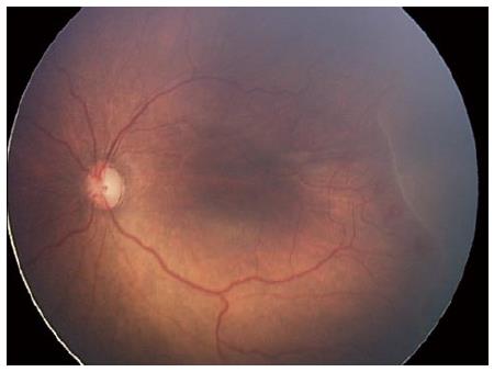
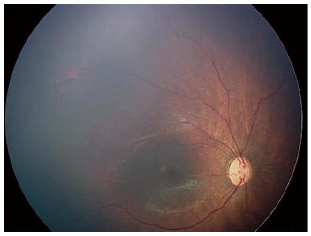
Figure 2 Previous 24 wk infant status post bevacizumab injection a for aggressive posterior retinopathy of prematurity.
RetCam imaging of the right eye reveals regressed retinopathy of prematurity with vascularization into zone 2. Plus disease is no longer present. The patient is post menstral age of 55 wk.
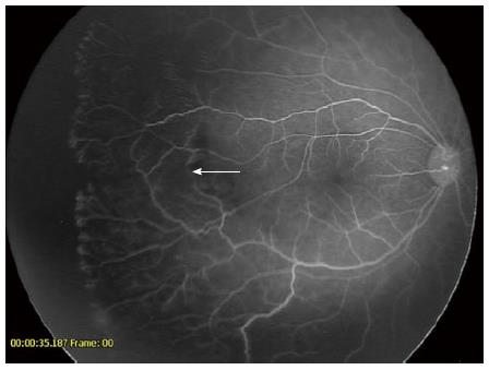
Figure 3 Fluorescein angiogram of the right eye reveals extensive areas of avascular retina.
There is a well demarcated line of advancing vessels with areas of neovascularization present. The arrow depicts the original area of the stage 3 ridge in zone 1 at the time of bevacizumab injection.
- Citation: Pulido CM, Quiram PA. Current understanding and management of aggressive posterior retinopathy of prematurity. World J Ophthalmol 2015; 5(2): 73-79
- URL: https://www.wjgnet.com/2218-6239/full/v5/i2/73.htm
- DOI: https://dx.doi.org/10.5318/wjo.v5.i2.73


