Copyright
©2014 Baishideng Publishing Group Inc.
World J Ophthalmol. Aug 12, 2014; 4(3): 52-55
Published online Aug 12, 2014. doi: 10.5318/wjo.v4.i3.52
Published online Aug 12, 2014. doi: 10.5318/wjo.v4.i3.52
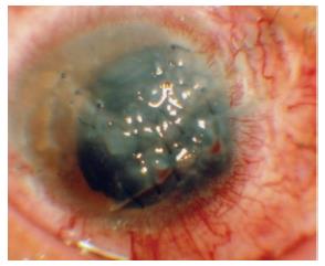
Figure 1 Anterior segment of the eye with severe penetrating corneal injury[6].
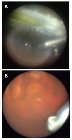
Figure 2 Intraoperative endoscopic view.
A: A bubble of silicone oil and yellow IOL can be observed; B: A tiny retinal tear was identified[5].
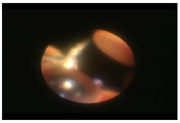
Figure 3 The direction should be arranged properly by projecting the fingers of the right hand outside of the eye.
- Citation: Kita M. Endoscope-assisted vitrectomy. World J Ophthalmol 2014; 4(3): 52-55
- URL: https://www.wjgnet.com/2218-6239/full/v4/i3/52.htm
- DOI: https://dx.doi.org/10.5318/wjo.v4.i3.52


