Copyright
©2013 Baishideng Publishing Group Co.
World J Ophthalmol. Aug 12, 2013; 3(3): 20-31
Published online Aug 12, 2013. doi: 10.5318/wjo.v3.i3.20
Published online Aug 12, 2013. doi: 10.5318/wjo.v3.i3.20
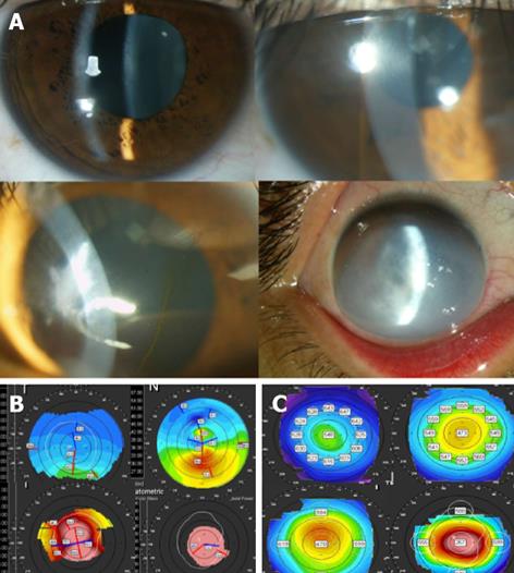
Figure 1 Keratoconus clinical and topographic variation examples.
A: Several clinical presentations and severity of keratoconus cases; B: Different keratometric stages of keratoconus; C: Pachymetric maps showing different grades of KC cases.
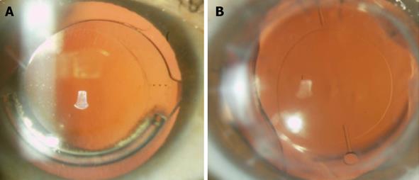
Figure 2 Combined procedures.
A: Combination of intrastromal ring segments and pseudophakic toric intraocular lens; B: Pseudophakic plate toric intraocular lens following penetrating keratoplasty.
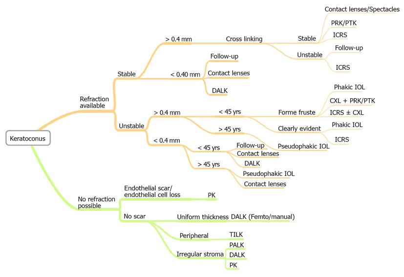
Figure 3 Proposed algorithm for keratoconus treatment.
PRK: Photorefractive keratectomy; PTK: Phototherapeutic keratectomy; ICRS: Intrastromal corneal ring segments; DALK: Deep anterior lamellar keratoplasty; CXL: Corneal collagen cross-linking; IOL: Intraocular lenses; PK: Penetrating keratoplasty; TILK: “Tuck-In” lamellar keratoplasty; PALK: DALK assited by pachymetry.
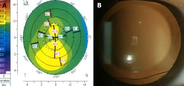
Figure 4 Phakic toric intraocular lens implantation in (A) forme fruste keratoconus case, (B) notice the rhomboidal marks of the lens toricity axis.
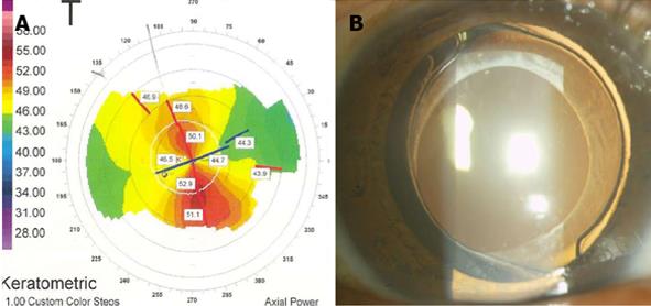
Figure 5 Pseudophakic toric intraocular lens in (A) frank keratoconus, (B) notice the three dot marks for toric intraocular lenses alignment.
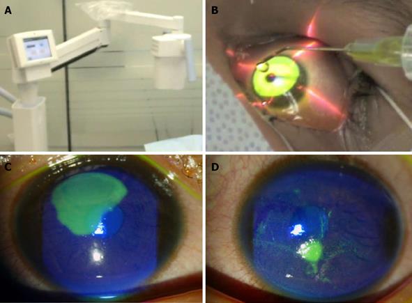
Figure 6 Collagen cross-linking.
A, B: Accelerated corneal collagen cross-linking (A) equipment (B) and riboflavin instillation, collagen cross-linking (CXL) treatment could be decreased to 3 min with Ultraviolet-light intensity of 30 mW/cm2 achieving the same energy on cornea of conventional CXL of 5 J/cm2; C: Right eye three days after accelerated cross-linking showing corneal epithelium recovery; D: Left eye also after three days following accelerated CXL.
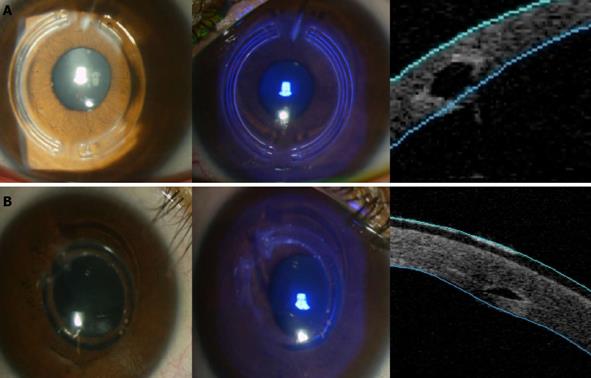
Figure 7 Different intrastromal ring segments models (A) clinical and optical coherence tomography showing hexagonal shape and (B) another design with triangular shape.
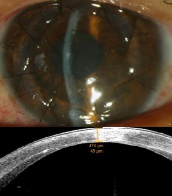
Figure 8 Femtosecond anterior lamellar keratoplasty, upper image showing the clinical photograph and lower image optical coherence tomography showing the residual stromal and endothelial tissue of around 50 microns.
- Citation: Jaimes M, Ramirez-Miranda A, Graue-Hernández EO, Navas A. Keratoconus therapeutics advances. World J Ophthalmol 2013; 3(3): 20-31
- URL: https://www.wjgnet.com/2218-6239/full/v3/i3/20.htm
- DOI: https://dx.doi.org/10.5318/wjo.v3.i3.20


