Copyright
©2013 Baishideng.
World J Ophthalmol. Feb 12, 2013; 3(1): 1-15
Published online Feb 12, 2013. doi: 10.5318/wjo.v3.i1.1
Published online Feb 12, 2013. doi: 10.5318/wjo.v3.i1.1
Figure 1 The treatment addresses the irregularity of the cornea and the steepness of the meridian.
A: Represents the angular circumference of the cornea; B: Illustrates one possible calculation and location for the paired arcuate incisions; C: Illustrates the combination of the arcuate keratotomy and modified form of circular keratotomy; D: The yellow arc marks the area of low corneal power from 210° to 330° and indicates an area of 90% incision depth, while the incision in the red area would be at a depth of 70%.
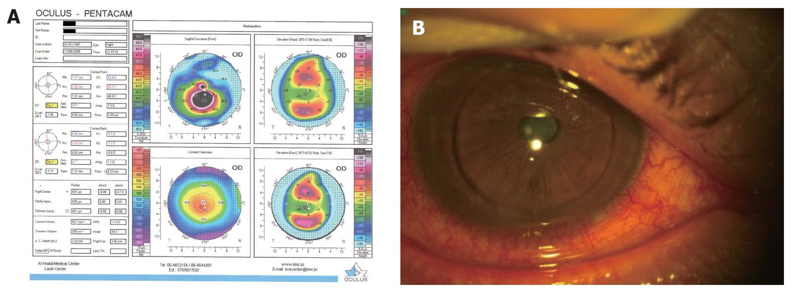
Figure 2 Pre-op Oculus Pentacam tomography.
A: Pre-op Oculus Pentacam tomography for the patient’s right eye; B: The patient’s eye on the day of the surgery immediately after the operation.
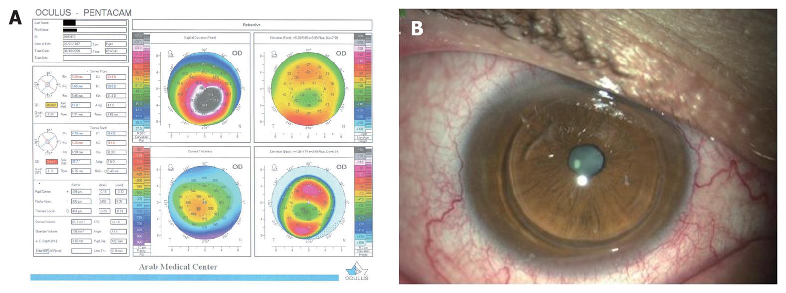
Figure 3 Post-op Pentacam tomography 13 mo after surgery.
A: Post-op Pentacam tomography of the same patient’s eye almost 13 mo after surgery; B: The same patient’s eye 13 mo after surgery.
- Citation: Quawasmi SA. Paired arcuate and modified circular keratotomy in keratoconus. World J Ophthalmol 2013; 3(1): 1-15
- URL: https://www.wjgnet.com/2218-6239/full/v3/i1/1.htm
- DOI: https://dx.doi.org/10.5318/wjo.v3.i1.1


