Published online Feb 10, 2016. doi: 10.5317/wjog.v5.i1.118
Peer-review started: April 29, 2015
First decision: June 9, 2015
Revised: October 14, 2015
Accepted: November 10, 2015
Article in press: November 11, 2015
Published online: February 10, 2016
Processing time: 282 Days and 2.6 Hours
AIM: To investigate the expression patterns of different adrenoceptor isoforms in ovarian cancer and their association with survival and tumor recurrence.
METHODS: The protein expression levels of α1B, α2C and β2 adrenoceptor were assessed in unselected ovarian cancer using immunohistochemistry on microarrayed archival tissue samples. A database containing clinical and pathology parameters and follow-up was used to investigate the association between adrenoceptor isoform expression with ovarian specific survival and tumor recurrence, using univariate and multivariate statistical analysis.
RESULTS: Expression of α1B showed an association with reduced ovarian specific survival (P = 0.05; CI: 1.00-1.49) and increased tumor recurrence (P = 0.021, CI: 1.04-1.69) in the whole patient group. On sub-analysis the expression of α1B in endometrioid cancers (χ2 = 5.867, P = 0.015) was found to predict reduced ovarian specific survival and increased tumor recurrence independently of tumor grade, clinical stage and chemotherapy. An association with clinical outcome was not seen for α2C or β2 AR.
CONCLUSION: Alpha1B adrenoceptor protein was found to predict increased risk of tumor recurrence and reduced mortality in patients with endometrioid type ovarian cancer and should be investigated as a biomarker for identifying patients at increased risk of disease progression. Furthermore, α adrenergic receptor antagonists with α1B selectivity should be investigated as a possible adjuvant therapy for treating patients with endometrioid cancer. Proof of principle could be tested in a retrospective population study.
Core tip: Epidemiological studies suggest that β-blockers might have a role in reducing metastatic spread and tumor recurrence and thereby prolong patient survival in some cancer types. In this novel study we found α1B adrenoceptor is a biomarker of tumor recurrence in endometrioid ovarian cancer. Further studies are needed to test if selective α1 adrenergic receptor antagonists inhibit tumor recurrence and prolong survival in patients with this type of cancer.
- Citation: Deutsch D, Deen S, Entschladen F, Coveney C, Rees R, Zänker KS, Powe DG. Alpha1B adrenoceptor expression is a marker of reduced survival and increased tumor recurrence in patients with endometrioid ovarian cancer. World J Obstet Gynecol 2016; 5(1): 118-126
- URL: https://www.wjgnet.com/2218-6220/full/v5/i1/118.htm
- DOI: https://dx.doi.org/10.5317/wjog.v5.i1.118
Ovarian cancer is the fifth leading cause of cancer related death amongst women in the United Kingdom and the second most common gynaecological malignancy. The lifetime risk of developing ovarian cancer is estimated at 1 in 68 for women in the United States[1]. It arises with non-specific symptoms, thus about 70% of patients are diagnosed with late stage disease with a 5-year survival rate of less than 30%. In contrast, patients diagnosed with early-stage disease have a > 90% 5-year survival rate[2]. Endometrioid type tumors account for 10%-20% ovarian cancers and compared to other forms have a relatively better 5-year survival rate. Patients would benefit from earlier cancer detection improved therapies for preventing metastasis and disease recurrence.
Increased levels of catecholamine hormones are linked with poor prognosis in ovarian cancer[3-5] possibly explained by their ability at promoting cell invasion and proliferation via activation of adrenergic receptors (adrenoceptors, AR)[5,6]. Recent laboratory studies suggest that beta adrenergic receptor antagonists (β-blockers) abrogate cancer cell migration[7-9], an essential element of metastasis, and disrupt the stress/inflammatory/cancer pathway interactions[10]. Sloan et al[10] showed that epinephrine-induced beta2 (β2) adrenoceptor activated cancer cells produce mediators that recruit tumor associated macrophages, and the process is inhibited by the β-blocker propranolol[11].
There are 9 adrenoceptor subtypes classified in two major classes, α and β, belonging to the G-protein coupled receptor family[12]. Promiscuous AR coupling results in the activation of multiple cancer cell signalling pathways[13]. Included among these, β2 activation induces phosphokinase A (PKA) and ERK[14] cell signalling in response to upregulation of cyclic AMP (cAMP)[15], leading to migration in some cancer cell types. ERK1/2 phosphorylation may occur following α1B adrenoceptor activation in ovarian cancer cells[15-17] but opposing this, α2C adrenoceptor can preferentially inhibit cAMP and PKA gene transcription[17]. For this reason it can be hypothesised that activation of α2C adrenoceptors may have an anti-cancer regulatory role compared to α1B and β2 receptors.
A small number of conflicting population studies have investigated the impact of β-blocker usage on survival in ovarian cancer patients. These have either shown potential benefits[7,18] or no effectiveness[19,20]. Such differences could possibly be explained by AR expression heterogeneity and so the current study is designed to assess the distribution and pattern of α1B, α2C and β2 AR proteins expressed in ovarian tumors and association with clinical outcome. Knowledge of this information will improve the study design when assessing the feasibility of using adrenergic receptor antagonists for targeted adjuvant cancer therapeutics. It was found that α1B rather than β2 adrenoceptor expression correlated with poor survival and tumor recurrence in epithelioid tumors.
The protein expression of α1B, α2C and β2 was characterised in formalin-fixed paraffin-embedded tissue microarrays of ovarian cancer from unselected patients attending Nottingham University Hospitals Trust. Damaged tissue cores and those that did not contain invasive carcinoma were censored. The study was approved by the Derbyshire ethics committee (07/H0401/156).
Following a diagnosis of ovarian cancer, patients were selected for chemotherapy treatment according to the East Midlands Cancer Network ovarian cancer treatment algorithm (http://www.eastmidlandscancernetwork.nhs.uk/Library/OvarianTreatmentAlgorithm.pdf). Tissue microarrays were produced by incorporating cores of archival formalin fixed ovarian tumor tissue from Nottingham patients presenting in 1991-2006. The presence of cancer was confirmed by a pathologist. Clinicopathological data was available for patients up to 240 mo post-diagnosis and was categorised as poor (< 60 mo) or better (> 60 mo) prognosis.
Four micron thick TMA sections had immunohistochemistry performed using a linked streptavidin peroxidase/biotinylated AB technique in accordance to the supplier’s recommendations (DAKO, Cambs, United Kingdom). Microwave antigen retrieval was performed in 0.01 mol/mL citrate buffer (pH6). The primary antibodies were previously optimized in full face breast cancer tissues as previously described[21] and in ovarian TMA sections with negative controls. Primary rabbit polyclonal antibodies against α1B (ab13297, Abcam, Cambs, United Kingdom), α2C (ab46536, Abcam), and β2 (ab13163, Abcam) AR were used diluted at 1:50, 1:750 and 1:450, respectively.
Statistical analysis was performed using statistical software, SPSS 20.0 (SPSS Inc., Chicago, IL, United States). Levels of adrenoceptor protein expression were microscopically assessed for staining intensity in malignant epithelium only and patients categorised into negative (negative or weak intensity) verse positive (moderate to strong intensity). Patients with missing clinical data were censored. Association between AR expression and different clinicopathology factors was evaluated using the non-parametric χ2 test. Survival and tumor recurrence analysis was modelled using the Kaplan-Meier method with a univariate log rank test to assess significance. Patients that died due to causes other than ovarian cancer were censored during survival analysis. Multivariate Cox proportional hazard regression (95%CI) was used to evaluate the independence of adrenoceptors for predicting survival and tumor recurrence compared to other clinical variables. A P value of < 0.05 was considered to indicate statistical significance.
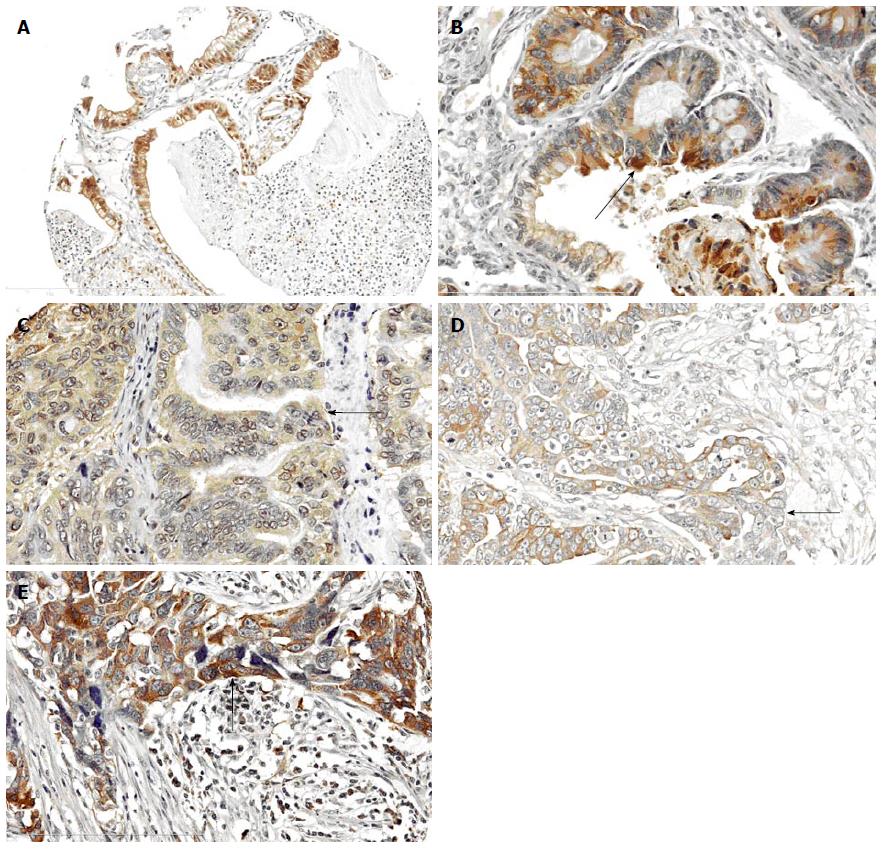
Tissue cores were available for assessment in 168 patients stained for α1B and α2C expression and 146 for β2 adrenoceptor protein. The reduced number of cores available for β2 assessment was due to detachment during immunohistochemistry processing. Adrenoceptor protein appeared localised in the cytoplasm of malignant ovarian tissue (Figure 1).
The median age at cancer diagnosis was 61 years (range 31-87) and the proportion of different cancer subtypes is shown in Table 1. Just over half (53%) of the patient cohort investigated in this study were categorised with poor survival (less than 5 years post-diagnosis) and the remainder survived 5-20 years.
| Adrenergic receptor | Absent | Present | χ2 | P | ||
| Age at diagnosis | < 49 | α-1B | 25 (66) | 13 (34) | 1.076 | 0.584 |
| 50-69 | 55 (59) | 38 (41) | ||||
| > 70 | 24 (54) | 20 (46) | ||||
| < 49 | α-2C | 11 (29) | 27 (71) | 2.600 | 0.273 | |
| 50-69 | 38 (42) | 52 (58) | ||||
| > 70 | 19 (45) | 23 (55) | ||||
| < 49 | β2 | 9 (31) | 20 (69) | 1.956 | 0.376 | |
| 50-69 | 36 (43) | 47 (57) | ||||
| > 70 | 13 (33) | 26 (67) | ||||
| Tumour type | High grade serous | α-1B | 49 (55) | 40 (45) | 14.648 | 0.005 |
| Low grade serous | 4 (67) | 2 (33) | ||||
| Endometrioid | 25 (60) | 17 (40) | ||||
| Clear cell | 16 (94) | 1 (6) | ||||
| Mucinous | 4 (29) | 10 (71) | ||||
| High grade serous | α-2C | 44 (48) | 47 (52) | 11.038 | 0.026 | |
| Low grade serous | 4 (67) | 2 (33) | ||||
| Endometrioid | 8 (21) | 31 (79) | ||||
| Clear cell | 6 (35) | 11 (65) | ||||
| Mucinous | 5 (33) | 10 (67) | ||||
| High grade serous | β2 | 25 (32) | 54 (68) | 13.559 | 0.009 | |
| Low grade serous | 1 (17) | 5 (83) | ||||
| Endometrioid | 20 (54) | 17 (46) | ||||
| Clear cell | 8 (61) | 5 (39) | ||||
| Mucinous | 1 (9) | 10 (91) | ||||
| Tumour grade | 1 | α-1B | 11 (42) | 15 (58) | 3.472 | 0.176 |
| 2 | 6 (55) | 5 (45) | ||||
| 3 | 81 (62) | 50 (38) | ||||
| 1 | α-2C | 9 (35) | 17 (65) | 0.456 | 0.796 | |
| 2 | 4 (36) | 7 (64) | ||||
| 3 | 54 (41) | 77 (59) | ||||
| 1 | β2 | 4 (18) | 18 (82) | 4.236 | 0.12 | |
| 2 | 4 (44) | 5 (56) | ||||
| 3 | 47 (41) | 68 (59) | ||||
| Mortality (5 yr) | No | α-1B | 55 (69) | 25 (31) | 4.821 | 0.028 |
| Yes | 47 (52) | 43 (48) | ||||
| < 5 yr | α-2C | 31 (39) | 48 (61) | 0.001 | 0.969 | |
| > 5 yr | 34 (39) | 52 (61) | ||||
| < 5 yr | β2 | 25 (36) | 44 (64) | 0.284 | 0.594 | |
| > 5 yr | 32 (41) | 47 (59) |
The distribution of adrenoceptor expression differed by tumor type with 41%, 60%, 62% of the full cohort showing expression of α1B, α2C and β2 respectively (Table 1). Increased α1B expression was more frequently seen in mucinous cancers but in contrast was reduced in low grade serous and clear cell tumors. Levels of α2C were increased in endometrioid, clear cell and mucinous tumors. β2 expression was more frequently increased in high and low grade serous tumors and mucinous cancers (Table 1). No association was found between individual adrenoceptor types and tumor grade or clinical stage (Table 1). Patients with tumors expressing α2C adrenoceptor showed an association with low stage clinical disease (Table 2).
| Adrenergic receptor | Absent | Present | χ2 | P | ||
| Clinical stage | 1 | α-1B | 39 (65) | 21 (35) | 2.486 | 0.478 |
| 2 | 12 (50) | 12 (50) | ||||
| 3 | 47 (59) | 33 (41) | ||||
| 4 | 4 (44) | 5 (56) | ||||
| 1 | α-2C | 17 (29) | 41 (71) | 6.014 | 0.111 | |
| 2 | 10 (42) | 14 (58) | ||||
| 3 | 38 (49) | 40 (51) | ||||
| 4 | 2 (25) | 6 (75) | ||||
| 1 | β2 | 18 (37) | 31 (63) | 6.508 | 0.089 | |
| 2 | 4 (20) | 16 (80) | ||||
| 3 | 31 (42) | 43 (58) | ||||
| 4 | 5 (71) | 2 (29) | ||||
| Stage 1 vs other stages | Stage 1 | α-1B | 37 (63) | 22 (37) | 0.894 | 0.344 |
| Other stages | 59 (55) | 48 (45) | ||||
| Stage 1 | α-2C | 17 (29) | 42 (71) | 4.376 | 0.036 | |
| Other stages | 49 (45) | 59 (55) | ||||
| Stage 1 | β2 | 18 (36) | 32 (64) | 0.090 | 0.764 | |
| Other stages | 37 (39) | 59 (61) | ||||
| Chemotherapy regimen | Before surgery | α-1B | 11 (58) | 8 (42) | 0.734 | 0.693 |
| After surgery | 84 (57) | 62 (43) | ||||
| Before surgery | α-2C | 8 (42) | 11 (58) | 0.702 | 0.704 | |
| After surgery | 58 (40) | 87 (60) | ||||
| Before surgery | β2 | 8 (47) | 9 (53) | 1.316 | 0.518 | |
| After surgery | 46 (36) | 80 (64) | ||||
| Chemotherapy resistance | Refractory | α-1B | 8 (80) | 2 (20) | 2.647 | 0.266 |
| Resistant within 6 mo | 4 (44) | 5 (56) | ||||
| Responsive | 72 (58) | 52 (42) | ||||
| Refractory | α-2C | 2 (25) | 6 (75) | 2.342 | 0.310 | |
| Resistant within 6 mo | 6 (60) | 4 (40) | ||||
| Responsive | 50 (41) | 73 (59) | ||||
| Refractory | β2 | 5 (56) | 4 (44) | 1.010 | 0.604 | |
| Resistant within 6 mo | 4 (40) | 6 (60) | ||||
| Responsive | 40 (38) | 64 (62) |
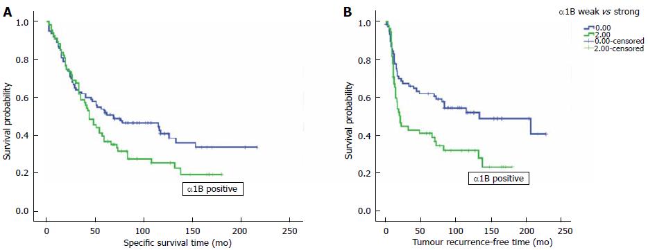
A Kaplan-Meier technique with a log rank test was used to model the independence of adrenoceptor protein expression in predicting ovarian cancer specific survival and tumor recurrence in the full patient cohort. High α1B protein expression was associated with reduced survival across the full patient cohort due to ovarian cancer specific mortality (P = 0.05; 95%CI: 1.00-1.49), resulting in a reduction in the median survival time from 63.5 to 44 mo (Figure 2 and Table 3). Similarly, α1B adrenoceptor protein expression was associated with increased tumor recurrence (P = 0.021, 95%CI: 1.04-1.69). A subanalysis of survival (χ2 = 3.907, P = 0.048) and tumor recurrence (χ2 = 5.867, P = 0.015) in the different cancer subclasses showed an association with endometrioid type cancer. No association was found for α2C or β2 AR with survival or tumor recurrence.
| HR | 95%CI | P value | ||
| Cancer specific survival | ||||
| Alpha1B expression | 1.221 | 1.000 | 1.493 | 0.050 |
| Tumor grade | 1.353 | 0.992 | 1.844 | 0.056 |
| Clinical stage | 1.922 | 1.529 | 2.416 | < 0.001 |
| Chemotherapy | 0.556 | 0.283 | 1.090 | 0.087 |
| Tumor recurrence | ||||
| Alpha1B expression | 1.326 | 1.043 | 1.687 | 0.021 |
| Tumor grade | 1.390 | 0.949 | 2.035 | 0.091 |
| Clinical stage | 2.469 | 1.825 | 3.340 | < 0.001 |
| Chemotherapy | 0.614 | 0.257 | 1.467 | 0.272 |
In the full patient cohort, tumor adrenoceptor protein expression showed no association with chemoresistance or responsiveness. A subanalysis of patients with endometrioid tumors showed similar results.
Relative expression ofα1B andα2C adrenoceptor proteins: Association with survival
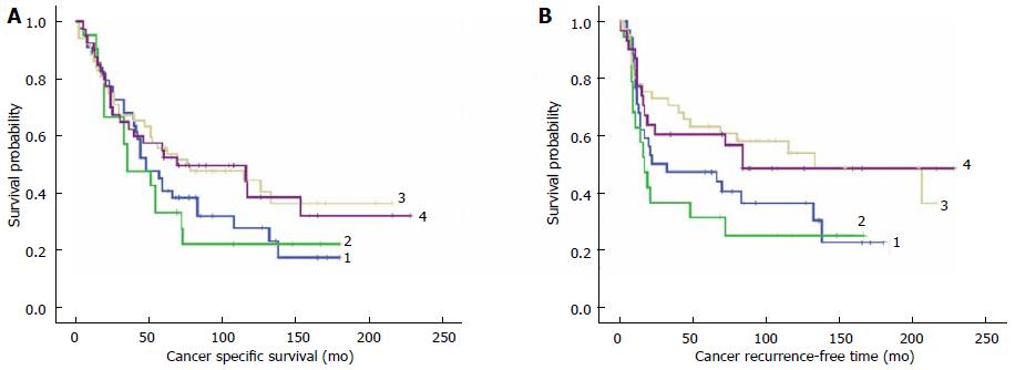
To test the hypothesis that patient clinical outcome might be influenced by the balance of Gs-adrenoceptor proteins (α1B and β2 are proposed biomarkers of disease progression) compared to those with Gi-protein affinity (α2C is proposed as a biomarker of good prognosis), patients were categorised into different groups according to their relative expression levels of tumor-stimulating α1B and tumor-inhibitory α2C. Four patient groups were defined comprising: Group 1: α1Bpositive/αα2Cpositive; Group 2: α1Bpositive/α2Cnegative; Group 3: α1Bnegative/α2Cpositive; and Group 4: α1Bnegative/α2Cnegative.
No significant difference was identified between the four adrenoceptor groups with ovarian cancer specific survival (χ2 = 4.211, P = 0.240) or tumor recurrence (χ2 = 7.361, P = 0.061). For tumor recurrence, the best separation between plots was achieved between the singly positive Group 2 (α1Bpositive/α2Cnegative) and Group 3 (α1Bnegative/α2Cpositive) (χ2 = 5.136, P = 0.023). Groups showing co-expression of α1B/α2C showed intermediate risk of tumor recurrence suggesting the presence of α2C expression has a modifying effect on α1B expression population studies (Figure 3).
Similarly, the relationship involving relative expression between β2 and α2C was tested by defining 4 patient subgroups: Group 1: β2positive/α2Cpositive; Group 2: β2positive/α2Cnegative; Group 3: β2negative/α2Cpositive; and Group 4: β2negative/α2Cnegative.
The effect of clinical outcome was assessed using Kaplan-Meier with a log rank test. No significant difference was seen between the patient subgroups when considering survival (χ2 = 2.253, P = 0.689) or tumor recurrence-free time (χ2 = 0.463, P = 0.927).
α1B is an independent prognostic biomarker
Multivariate Cox regression hazard analysis was used to test the independence of α1B as a prognostic biomarker for predicting ovarian cancer specific survival and tumor recurrence in patients with ovarian cancer. Tumor grade, clinical stage and systemic chemotherapy were included in the model. α1B adrenoceptor expression was found to contribute significant prediction ability concerning survival (HR = 1.221, P = 0.05, 95%CI: 1.000-1.493) and tumor recurrence (HR = 1.326, P = 0.021, 95%CI: 1.043-1.687) over and above the routinely used clinical parameters included in the model (Table 3).
Ovarian cancer has a complex pathogenesis but recent studies have focused on the association between cancer progression and stress[4,22,23] involving the catecholamine hormones epinephrine and norepinephrine, and activation of β2 adrenoceptors. A recent paradigm proposed for a mouse model of breast cancer suggests cross-talk between cancer cells and macrophages triggering pro-metastasis cell signalling[10]. This paradigm might also extend to ovarian cancer because macrophages are implicated in ovarian metastasis, immunosuppression, angiogenesis and poor clinical outcome[24,25]. Consequently, it has been proposed that blockade of adrenoceptors using β-blockers could inhibit tumor progression in a number of cancer types including ovary[7], breast[26-28], prostate[29] and skin[30,31]. In addition, there is increasing evidence from patient[21] and in vitro[32] studies that alpha adrenoceptors are also implicated in breast cancer progression and for this reason we sought to identify the distribution and pattern of different adrenoceptor types in ovarian cancer patients.
The pattern of adrenoceptor expression was altered in different ovarian tumor types but overall, no association was found with tumor grade. Serous and endometrioid tumors generally differ in their prognostic outlook with the endometrioid type having better prognosis. A significant difference in pro-(β2) and anti-migratory (α2C) adrenoceptor expression patterns was identified. Compared to serous tumors, endometrioid cancers less frequently expressed β2 receptors (46% vs 68%) and were more likely to show α2 receptor expression (79% vs 52%). But a subset of patients (40%) with endometrioid tumors expressed high levels of α1B protein and this correlated with poor prognosis due to a significantly shortened survival time and reduced tumor recurrence-free interval, independently of chemotherapy, tumor grade and clinical stage. The pathogenesis of endometrioid tumors is thought to be associated with endometriosis[33] and notable gene mutations in the phosphoinositide-3-kinase (PI3K) cell signalling pathway including PIK3CA and PTEN genes[34] in endometrial-derived cancer[35]. Activation of adrenoceptors provide a route to PI3K upregulation via the intermediary cAMP. Our findings suggest that α1B is a candidate biomarker and here it identified 17% of patients with endometrioid type cancer that require more intensive therapy and follow-up surveillance. Moreover, our findings suggest adrenoceptor antagonists and PI3K inhibitors provide potential for a targeted adjuvant therapy approach to complement existing therapies. In considering candidate anti-α adrenergic receptor drugs consideration has to be given to their selectivity and adverse effects. Alpha AR antagonists are used in the treatment of benign prostatic hyperplasia (e.g., Prazosin, Doxazosin), urinary tract symptoms and hypertension. The non-selective drugs phenoxybenzamine and phentolamine would not be advocated, whereas some current tricyclic antidepressants could be considered but a recent study found amitriptyline, nortriptyline and imipramine are relatively weak α1B antagonists[36]. More promising is the recent development of a new family of 8-OMe benzodioxane analogues of the research drug WB4101 which has been shown to have high affinity for α1B AR[37]. The side effects of α1 antagonists including postural hypotension (Prazosin, Doxazosin), arrhythmia and CNS disturbances (tricyclic antidepressants) can be reduced by careful titration and active monitoring.
Cancer cell line studies have shown that norepinephrine activates α AR resulting in HIF1α dependent vascular endothelial growth factor transcription, required for angiogenesis[38]. Interestingly, Park et al[38] found that the α1 adrenoceptor inhibitor prazosin blocks the angiogenic pathway in the epithelial-to-mesenchymal type MDA-MB-231 breast cancer cell line, but not in liver (SK-Hep1) or prostate (PC3) cancer cells[38]. To translate this to ovarian cancer, we tested the proposal that α1B and 2C adrenoceptors might have an opposing promoting and inhibitory affect respectively on cancer progression and survival. To do this, patients were sub-classified according to α/β adrenoceptor phenotype, by comparing survival in patients with tumors expressing only one adrenoceptor (α1B, α2C or β2 positive) to those with co-expression of α2C. Although Kaplan-Meier models suggest α2C expression improves tumor recurrence-free times the finding was insignificant. Further studies are needed to better stratify patients for assessing possible therapeutic response to adrenoceptor antagonists.
No significant association between β2 protein expression levels and clinical outcome was found in this study. Recent laboratory studies show a significant pathologic role for neouroendocrine-induced progression in ovarian cancer (reviewed by Kang et al[39]), mediated by the β adrenoceptor activated cAMP - PKA cell signalling pathway. Increased cAMP activates Rap guanine-nucleotide-exchange-factor 3 (EPAC) leading to increased cell: Matrix adhesion needed for cancer cell implantation. Cancer growth is maintained due to enhanced cell survival resulting from γSrc-FAK signalling and STAT3 induced angiogenesis. These mechanisms explain the murine in vivo observation that the β2 antagonist propranolol inhibits ovarian cell growth[40]. However, other in vitro studies suggest that β-blockers would not be effective. In some instances, β2 agonists have been found to reduce cell proliferation[32,41,42] and migration[43]. In the latter case, it is proposed that β blockers could actually increase ovarian disease progression by promoting cell migration. Another explanation is that β blockers induce increased cell proliferation leaving unopposed α2C AR activity[42] in contrast to our findings suggesting that α1B is moderated by α2C in ovarian cancer. Recent proof-of-concept population studies of β-blocker users among ovarian cancer patients have produced conflicting findings. One study showed an association between increased progression-free survival (PFS) in users (n = 23) compared to non-users[7]. In contrast, a more recent study found no benefit in PFS or overall survival in platinum-sensitive patients prescribed β-blockers (n = 8)[5]. Clearly, larger studies are needed allowing for possible confounders and tumor receptor typing. Although previous studies have focused on the use of β-blockers to retard disease progression, the results presented here and in a recent breast cancer study[21] suggest that the possible therapeutic benefits of alpha adrenoceptor antagonists should be investigated.
We thank Brett Blackbourn and Claire Boag for photographic assistance.
Many cancers are treatable by surgery, radio-/chemotherapy, or targeted drug treatments, or any combination of these. In some instances a cancer can spread (metastasise) to tissues distant from the original site. This process can place a patient at increased risk of disease progression and demise and in addition, it can present clinicians with more challenging medical management of a patient’s disease. Knowledge about the biological process involved in cancer spread is increasing but there remains an unmet need to develop new treatment approaches to prevent it. Being able to identify patients that are at increased risk of metastasis can rationalise clinical management by focusing extensive treatment on those that will best benefit. Laboratory experiments have shown that some cancer cells are stimulated to migrate when adrenergic receptor proteins (stress receptors) are activated by stress hormones. Drugs are available that inhibit adrenergic receptor function and could be used to neutralise certain cancer cell functions.
Identifying the expression pattern of adrenergic receptors (AR) in different cancer types and their association with disease progression and survival could provide insight into using AR inhibitor drugs for targeted anti-cancer treatment.
The expression pattern of 3 AR proteins (αB1, α2C and β2) was investigated and its significance statistically related to survival and metastasis outcome in patients with different types of ovarian cancer. Only αB1 was found to predict shortened survival and increased risk of tumor recurrence, especially in patients with endometrioid type cancer, independently of tumour grade, clinical stage and chemotherapy treatment.
The results suggest that adrenergic receptor antagonists with anti-α1B selectivity could be used to limit disease progression in patients with endometrioid type tumors expressing α1B AR.
Metastasis development is a major cause of mortality in patients with cancer and involves a multistep biological pathway resulting in tumor cells leaving the primary cancer and disseminating to distant body tissues. AR belong to a family of G protein-coupled receptors comprising 9 members.
This is a well written paper.
P- Reviewer: Rovas L, Zafrakas M, Zhang XQ S- Editor: Ji FF L- Editor: A E- Editor: Wu HL
| 1. | Copeland LJ. Chapter 11 - Epithelial Ovarian Cancer. In Philip JDM, William T. Creasman MD (eds): Clinical Gynecologic Oncology (Seventh Edition). Edinburgh: Mosby 2007; 313-367. |
| 2. | Howlader N, Noone AM, Krapcho M, Neyman N, Aminou R (Eds): SEER Cancer statistics review, 1975-2009 (Vintage 2009 Population). MD: National Cancer Institute, Bethesda, 2012. Available from: http://seer.cancer.gov/csr/1975_2009_pops09/. |
| 3. | Lee JW, Shahzad MM, Lin YG, Armaiz-Pena G, Mangala LS, Han HD, Kim HS, Nam EJ, Jennings NB, Halder J. Surgical stress promotes tumor growth in ovarian carcinoma. Clin Cancer Res. 2009;15:2695-2702. [RCA] [PubMed] [DOI] [Full Text] [Full Text (PDF)] [Cited by in Crossref: 165] [Cited by in RCA: 173] [Article Influence: 10.8] [Reference Citation Analysis (0)] |
| 4. | Sood AK, Bhatty R, Kamat AA, Landen CN, Han L, Thaker PH, Li Y, Gershenson DM, Lutgendorf S, Cole SW. Stress hormone-mediated invasion of ovarian cancer cells. Clin Cancer Res. 2006;12:369-375. [RCA] [PubMed] [DOI] [Full Text] [Full Text (PDF)] [Cited by in Crossref: 379] [Cited by in RCA: 381] [Article Influence: 20.1] [Reference Citation Analysis (0)] |
| 5. | Thaker PH, Lutgendorf SK, Sood AK. The neuroendocrine impact of chronic stress on cancer. Cell Cycle. 2007;6:430-433. [RCA] [PubMed] [DOI] [Full Text] [Cited by in Crossref: 89] [Cited by in RCA: 91] [Article Influence: 5.1] [Reference Citation Analysis (0)] |
| 6. | Armaiz-Pena GN, Lutgendorf SK, Cole SW, Sood AK. Neuroendocrine modulation of cancer progression. Brain Behav Immun. 2009;23:10-15. [RCA] [PubMed] [DOI] [Full Text] [Full Text (PDF)] [Cited by in Crossref: 104] [Cited by in RCA: 97] [Article Influence: 6.1] [Reference Citation Analysis (0)] |
| 7. | Diaz ES, Karlan BY, Li AJ. Impact of beta blockers on epithelial ovarian cancer survival. Gynecol Oncol. 2012;127:375-378. [RCA] [PubMed] [DOI] [Full Text] [Cited by in Crossref: 112] [Cited by in RCA: 121] [Article Influence: 9.3] [Reference Citation Analysis (0)] |
| 8. | Sood AK, Armaiz-Pena GN, Halder J, Nick AM, Stone RL, Hu W, Carroll AR, Spannuth WA, Deavers MT, Allen JK. Adrenergic modulation of focal adhesion kinase protects human ovarian cancer cells from anoikis. J Clin Invest. 2010;120:1515-1523. [RCA] [PubMed] [DOI] [Full Text] [Full Text (PDF)] [Cited by in Crossref: 222] [Cited by in RCA: 223] [Article Influence: 14.9] [Reference Citation Analysis (0)] |
| 9. | Lutgendorf SK, DeGeest K, Dahmoush L, Farley D, Penedo F, Bender D, Goodheart M, Buekers TE, Mendez L, Krueger G. Social isolation is associated with elevated tumor norepinephrine in ovarian carcinoma patients. Brain Behav Immun. 2011;25:250-255. [RCA] [PubMed] [DOI] [Full Text] [Full Text (PDF)] [Cited by in Crossref: 151] [Cited by in RCA: 137] [Article Influence: 9.8] [Reference Citation Analysis (0)] |
| 10. | Sloan EK, Priceman SJ, Cox BF, Yu S, Pimentel MA, Tangkanangnukul V, Arevalo JM, Morizono K, Karanikolas BD, Wu L. The sympathetic nervous system induces a metastatic switch in primary breast cancer. Cancer Res. 2010;70:7042-7052. [RCA] [PubMed] [DOI] [Full Text] [Full Text (PDF)] [Cited by in Crossref: 652] [Cited by in RCA: 634] [Article Influence: 42.3] [Reference Citation Analysis (0)] |
| 11. | Wei W, Mok SC, Oliva E, Kim SH, Mohapatra G, Birrer MJ. FGF18 as a prognostic and therapeutic biomarker in ovarian cancer. J Clin Invest. 2013;123:4435-4448. [RCA] [PubMed] [DOI] [Full Text] [Cited by in Crossref: 54] [Cited by in RCA: 80] [Article Influence: 6.7] [Reference Citation Analysis (0)] |
| 12. | Entschladen F, Zänker KS, Powe DG. Heterotrimeric G protein signaling in cancer cells with regard to metastasis formation. Cell Cycle. 2011;10:1086-1091. [RCA] [PubMed] [DOI] [Full Text] [Cited by in Crossref: 19] [Cited by in RCA: 17] [Article Influence: 1.2] [Reference Citation Analysis (0)] |
| 13. | Evans BA, Broxton N, Merlin J, Sato M, Hutchinson DS, Christopoulos A, Summers RJ. Quantification of functional selectivity at the human α(1A)-adrenoceptor. Mol Pharmacol. 2011;79:298-307. [RCA] [PubMed] [DOI] [Full Text] [Cited by in Crossref: 67] [Cited by in RCA: 64] [Article Influence: 4.6] [Reference Citation Analysis (0)] |
| 14. | Enserink JM, Price LS, Methi T, Mahic M, Sonnenberg A, Bos JL, Taskén K. The cAMP-Epac-Rap1 pathway regulates cell spreading and cell adhesion to laminin-5 through the alpha3beta1 integrin but not the alpha6beta4 integrin. J Biol Chem. 2004;279:44889-44896. [RCA] [PubMed] [DOI] [Full Text] [Cited by in Crossref: 101] [Cited by in RCA: 103] [Article Influence: 4.9] [Reference Citation Analysis (0)] |
| 15. | Fredriksson JM, Lindquist JM, Bronnikov GE, Nedergaard J. Norepinephrine induces vascular endothelial growth factor gene expression in brown adipocytes through a beta -adrenoreceptor/cAMP/protein kinase A pathway involving Src but independently of Erk1/2. J Biol Chem. 2000;275:13802-13811. [RCA] [PubMed] [DOI] [Full Text] [Cited by in Crossref: 137] [Cited by in RCA: 136] [Article Influence: 5.4] [Reference Citation Analysis (0)] |
| 16. | Buffin-Meyer B, Crassous PA, Delage C, Denis C, Schaak S, Paris H. EGF receptor transactivation and PI3-kinase mediate stimulation of ERK by alpha(2A)-adrenoreceptor in intestinal epithelial cells: a role in wound healing. Eur J Pharmacol. 2007;574:85-93. [RCA] [PubMed] [DOI] [Full Text] [Cited by in Crossref: 28] [Cited by in RCA: 27] [Article Influence: 1.5] [Reference Citation Analysis (0)] |
| 17. | Karkoulias G, Mastrogianni O, Lymperopoulos A, Paris H, Flordellis C. alpha(2)-Adrenergic receptors activate MAPK and Akt through a pathway involving arachidonic acid metabolism by cytochrome P450-dependent epoxygenase, matrix metalloproteinase activation and subtype-specific transactivation of EGFR. Cell Signal. 2006;18:729-739. [RCA] [PubMed] [DOI] [Full Text] [Cited by in Crossref: 33] [Cited by in RCA: 36] [Article Influence: 1.9] [Reference Citation Analysis (0)] |
| 18. | Diaz ES, Karlan BY, Li AJ. Impact of Beta Blockers on Epithelial Ovarian Cancer Survival. Obstet Gynecol Surv. 2013;68:109-110. [RCA] [DOI] [Full Text] [Cited by in Crossref: 1] [Cited by in RCA: 2] [Article Influence: 0.2] [Reference Citation Analysis (0)] |
| 19. | Eskander R, Bessonova L, Chiu C, Ward K, Culver H, Harrison T, Randall L. Beta blocker use and ovarian cancer survival: A retrospective cohort study. Gynecol Oncol. 2012;127:S21. [RCA] [DOI] [Full Text] [Cited by in Crossref: 13] [Cited by in RCA: 13] [Article Influence: 1.0] [Reference Citation Analysis (0)] |
| 20. | Johannesdottir SA, Schmidt M, Phillips G, Glaser R, Yang EV, Blumenfeld M, Lemeshow S. Use of ß-blockers and mortality following ovarian cancer diagnosis: a population-based cohort study. BMC Cancer. 2013;13:85. [RCA] [PubMed] [DOI] [Full Text] [Full Text (PDF)] [Cited by in Crossref: 38] [Cited by in RCA: 48] [Article Influence: 4.0] [Reference Citation Analysis (0)] |
| 21. | Powe DG, Voss MJ, Habashy HO, Zänker KS, Green AR, Ellis IO, Entschladen F. Alpha- and beta-adrenergic receptor (AR) protein expression is associated with poor clinical outcome in breast cancer: an immunohistochemical study. Breast Cancer Res Treat. 2011;130:457-463. [RCA] [PubMed] [DOI] [Full Text] [Cited by in Crossref: 66] [Cited by in RCA: 74] [Article Influence: 5.3] [Reference Citation Analysis (0)] |
| 22. | Lutgendorf SK, Cole S, Costanzo E, Bradley S, Coffin J, Jabbari S, Rainwater K, Ritchie JM, Yang M, Sood AK. Stress-related mediators stimulate vascular endothelial growth factor secretion by two ovarian cancer cell lines. Clin Cancer Res. 2003;9:4514-4521. [PubMed] |
| 23. | Thaker PH, Han LY, Kamat AA, Arevalo JM, Takahashi R, Lu C, Jennings NB, Armaiz-Pena G, Bankson JA, Ravoori M. Chronic stress promotes tumor growth and angiogenesis in a mouse model of ovarian carcinoma. Nat Med. 2006;12:939-944. [RCA] [PubMed] [DOI] [Full Text] [Cited by in Crossref: 875] [Cited by in RCA: 946] [Article Influence: 49.8] [Reference Citation Analysis (0)] |
| 24. | Cole SW, Sood AK. Molecular pathways: beta-adrenergic signaling in cancer. Clin Cancer Res. 2012;18:1201-1206. [RCA] [PubMed] [DOI] [Full Text] [Cited by in Crossref: 387] [Cited by in RCA: 497] [Article Influence: 35.5] [Reference Citation Analysis (0)] |
| 25. | Robinson-Smith TM, Isaacsohn I, Mercer CA, Zhou M, Van Rooijen N, Husseinzadeh N, McFarland-Mancini MM, Drew AF. Macrophages mediate inflammation-enhanced metastasis of ovarian tumors in mice. Cancer Res. 2007;67:5708-5716. [RCA] [PubMed] [DOI] [Full Text] [Cited by in Crossref: 153] [Cited by in RCA: 161] [Article Influence: 8.9] [Reference Citation Analysis (0)] |
| 26. | Barron TI, Connolly RM, Sharp L, Bennett K, Visvanathan K. Beta blockers and breast cancer mortality: a population- based study. J Clin Oncol. 2011;29:2635-2644. [RCA] [PubMed] [DOI] [Full Text] [Cited by in Crossref: 370] [Cited by in RCA: 423] [Article Influence: 30.2] [Reference Citation Analysis (0)] |
| 27. | Melhem-Bertrandt A, Chavez-Macgregor M, Lei X, Brown EN, Lee RT, Meric-Bernstam F, Sood AK, Conzen SD, Hortobagyi GN, Gonzalez-Angulo AM. Beta-blocker use is associated with improved relapse-free survival in patients with triple-negative breast cancer. J Clin Oncol. 2011;29:2645-2652. [RCA] [PubMed] [DOI] [Full Text] [Cited by in Crossref: 304] [Cited by in RCA: 382] [Article Influence: 27.3] [Reference Citation Analysis (0)] |
| 28. | Powe DG, Voss MJ, Zänker KS, Habashy HO, Green AR, Ellis IO, Entschladen F. Beta-blocker drug therapy reduces secondary cancer formation in breast cancer and improves cancer specific survival. Oncotarget. 2010;1:628-638. [PubMed] |
| 29. | Grytli HH, Fagerland MW, Fosså SD, Taskén KA, Håheim LL. Use of β-blockers is associated with prostate cancer-specific survival in prostate cancer patients on androgen deprivation therapy. Prostate. 2013;73:250-260. [PubMed] |
| 30. | De Giorgi V, Grazzini M, Gandini S, Benemei S, Lotti T, Marchionni N, Geppetti P. Treatment with β-blockers and reduced disease progression in patients with thick melanoma. Arch Intern Med. 2011;171:779-781. [RCA] [PubMed] [DOI] [Full Text] [Cited by in Crossref: 94] [Cited by in RCA: 129] [Article Influence: 9.2] [Reference Citation Analysis (0)] |
| 31. | Lemeshow S, Sørensen HT, Phillips G, Yang EV, Antonsen S, Riis AH, Lesinski GB, Jackson R, Glaser R. β-Blockers and survival among Danish patients with malignant melanoma: a population-based cohort study. Cancer Epidemiol Biomarkers Prev. 2011;20:2273-2279. [RCA] [PubMed] [DOI] [Full Text] [Cited by in Crossref: 158] [Cited by in RCA: 176] [Article Influence: 12.6] [Reference Citation Analysis (0)] |
| 32. | Gargiulo L, Copsel S, Rivero EM, Galés C, Sénard JM, Lüthy IA, Davio C, Bruzzone A. Differential β₂-adrenergic receptor expression defines the phenotype of non-tumorigenic and malignant human breast cell lines. Oncotarget. 2014;5:10058-10069. [PubMed] |
| 33. | Melin A, Sparén P, Bergqvist A. The risk of cancer and the role of parity among women with endometriosis. Hum Reprod. 2007;22:3021-3026. [RCA] [PubMed] [DOI] [Full Text] [Cited by in Crossref: 101] [Cited by in RCA: 98] [Article Influence: 5.4] [Reference Citation Analysis (0)] |
| 34. | Oda K, Stokoe D, Taketani Y, McCormick F. High frequency of coexistent mutations of PIK3CA and PTEN genes in endometrial carcinoma. Cancer Res. 2005;65:10669-10673. [RCA] [PubMed] [DOI] [Full Text] [Cited by in Crossref: 360] [Cited by in RCA: 348] [Article Influence: 18.3] [Reference Citation Analysis (0)] |
| 35. | Weigelt B, Warne PH, Lambros MB, Reis-Filho JS, Downward J. PI3K pathway dependencies in endometrioid endometrial cancer cell lines. Clin Cancer Res. 2013;19:3533-3544. [RCA] [PubMed] [DOI] [Full Text] [Cited by in Crossref: 107] [Cited by in RCA: 109] [Article Influence: 9.1] [Reference Citation Analysis (0)] |
| 36. | Nojimoto FD, Mueller A, Hebeler-Barbosa F, Akinaga J, Lima V, Kiguti LR, Pupo AS. The tricyclic antidepressants amitriptyline, nortriptyline and imipramine are weak antagonists of human and rat alpha1B-adrenoceptors. Neuropharmacology. 2010;59:49-57. [RCA] [PubMed] [DOI] [Full Text] [Cited by in Crossref: 16] [Cited by in RCA: 19] [Article Influence: 1.3] [Reference Citation Analysis (0)] |
| 37. | Fumagalli L, Pallavicini M, Budriesi R, Bolchi C, Canovi M, Chiarini A, Chiodini G, Gobbi M, Laurino P, Micucci M. 6-methoxy-7-benzofuranoxy and 6-methoxy-7-indolyloxy analogues of 2-[2-(2,6-Dimethoxyphenoxy)ethyl]aminomethyl-1,4-benzodioxane (WB4101): 1 discovery of a potent and selective α1D-adrenoceptor antagonist. J Med Chem. 2013;56:6402-6412. [RCA] [PubMed] [DOI] [Full Text] [Cited by in Crossref: 23] [Cited by in RCA: 24] [Article Influence: 2.0] [Reference Citation Analysis (0)] |
| 38. | Park SY, Kang JH, Jeong KJ, Lee J, Han JW, Choi WS, Kim YK, Kang J, Park CG, Lee HY. Norepinephrine induces VEGF expression and angiogenesis by a hypoxia-inducible factor-1α protein-dependent mechanism. Int J Cancer. 2011;128:2306-2316. [RCA] [PubMed] [DOI] [Full Text] [Cited by in Crossref: 102] [Cited by in RCA: 113] [Article Influence: 7.5] [Reference Citation Analysis (0)] |
| 39. | Kang Y, Lutgendorf S, Hu W, Cole SW, Sood AK. Stress related neuroendocrine influences in ovarian cancer. Current Cancer Therapy Reviews. 2012;8:100-109. [RCA] [DOI] [Full Text] [Cited by in Crossref: 1] [Cited by in RCA: 2] [Article Influence: 0.2] [Reference Citation Analysis (0)] |
| 40. | Landen CN, Lin YG, Armaiz Pena GN, Das PD, Arevalo JM, Kamat AA, Han LY, Jennings NB, Spannuth WA, Thaker PH. Neuroendocrine modulation of signal transducer and activator of transcription-3 in ovarian cancer. Cancer Res. 2007;67:10389-10396. [RCA] [PubMed] [DOI] [Full Text] [Cited by in Crossref: 102] [Cited by in RCA: 115] [Article Influence: 6.4] [Reference Citation Analysis (0)] |
| 41. | Carie AE, Sebti SM. A chemical biology approach identifies a beta-2 adrenergic receptor agonist that causes human tumor regression by blocking the Raf-1/Mek-1/Erk1/2 pathway. Oncogene. 2007;26:3777-3788. [RCA] [PubMed] [DOI] [Full Text] [Cited by in Crossref: 51] [Cited by in RCA: 61] [Article Influence: 3.4] [Reference Citation Analysis (0)] |
| 42. | Pérez Piñero C, Bruzzone A, Sarappa MG, Castillo LF, Lüthy IA. Involvement of α2- and β2-adrenoceptors on breast cancer cell proliferation and tumour growth regulation. Br J Pharmacol. 2012;166:721-736. [RCA] [PubMed] [DOI] [Full Text] [Cited by in Crossref: 78] [Cited by in RCA: 100] [Article Influence: 7.7] [Reference Citation Analysis (0)] |
| 43. | Bastian P, Balcarek A, Altanis C, Strell C, Niggemann B, Zaenker KS, Entschladen F. The inhibitory effect of norepinephrine on the migration of ES-2 ovarian carcinoma cells involves a Rap1-dependent pathway. Cancer Lett. 2009;274:218-224. [RCA] [PubMed] [DOI] [Full Text] [Cited by in Crossref: 20] [Cited by in RCA: 25] [Article Influence: 1.5] [Reference Citation Analysis (0)] |


