Copyright
©The Author(s) 2015.
World J Obstet Gynecol. Aug 10, 2015; 4(3): 58-63
Published online Aug 10, 2015. doi: 10.5317/wjog.v4.i3.58
Published online Aug 10, 2015. doi: 10.5317/wjog.v4.i3.58
Figure 1 Transvaginal ultrasound image of a gestational sac consistent with an early intrauterine pregnancy.
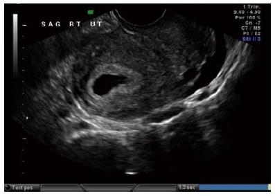
Figure 2 Transvaginal ultrasound image of a fluid collection within the endometrial cavity, a pseudosac, consistent with an extrauterine pregnancy.
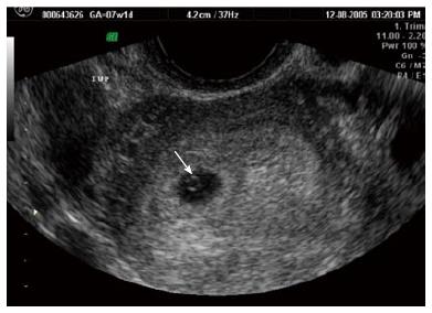
Figure 3 Transvaginal ultrasound image of an intrauterine true gestational sac containing a yolk sac.
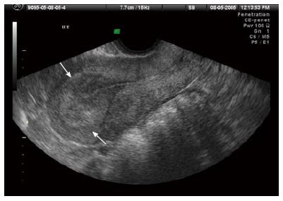
Figure 4 Transvaginal ultrasound image of a thickened endometrial echo before identification of what will be an intrauterine pregnancy.
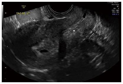
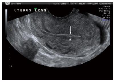
Figure 5 Transvaginal ultrasound image of a thin endometrial echo associated with and ectopic pregnancy.
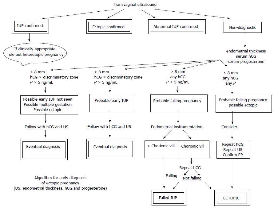
Figure 6 Non-surgical algorithm for the early diagnosis of ectopic pregnancy utilizing vaginal ultrasound, including endometrial echo thickness, serum human chorionic gonadotrophin and serum progesterone.
hCG: Human chorionic gonadotropin; US: Ultrasound; EP: Ectopic pregnancy; IUP: Inverted urothelial papilloma.
- Citation: Fylstra DL. Avoiding misdiagnosing an early intrauterine pregnancy as an ectopic pregnancy. World J Obstet Gynecol 2015; 4(3): 58-63
- URL: https://www.wjgnet.com/2218-6220/full/v4/i3/58.htm
- DOI: https://dx.doi.org/10.5317/wjog.v4.i3.58


