Copyright
©The Author(s) 2023.
World J Obstet Gynecol. Mar 3, 2023; 12(2): 11-16
Published online Mar 3, 2023. doi: 10.5317/wjog.v12.i2.11
Published online Mar 3, 2023. doi: 10.5317/wjog.v12.i2.11
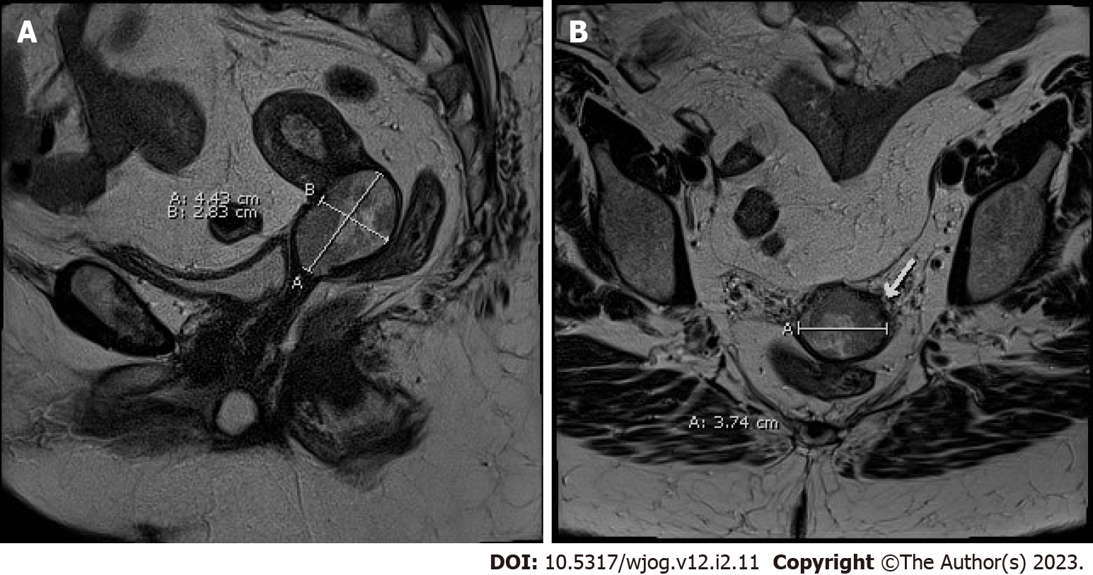
Figure 1 Magnetic resonance imaging showing a 4.
4 cm × 3.7 cm × 2.8 cm lesion in the cervix with extension to the lower third of the endometrium. A: Sagittal view; B: Transverse view.
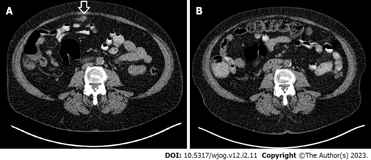
Figure 2 Comparison of computed tomography-scans of the abdomen.
A: A suspicious lesion is seen in front of transverse colon (arrow) before surgery; B: The lesion is not seen 4 mo after surgery.
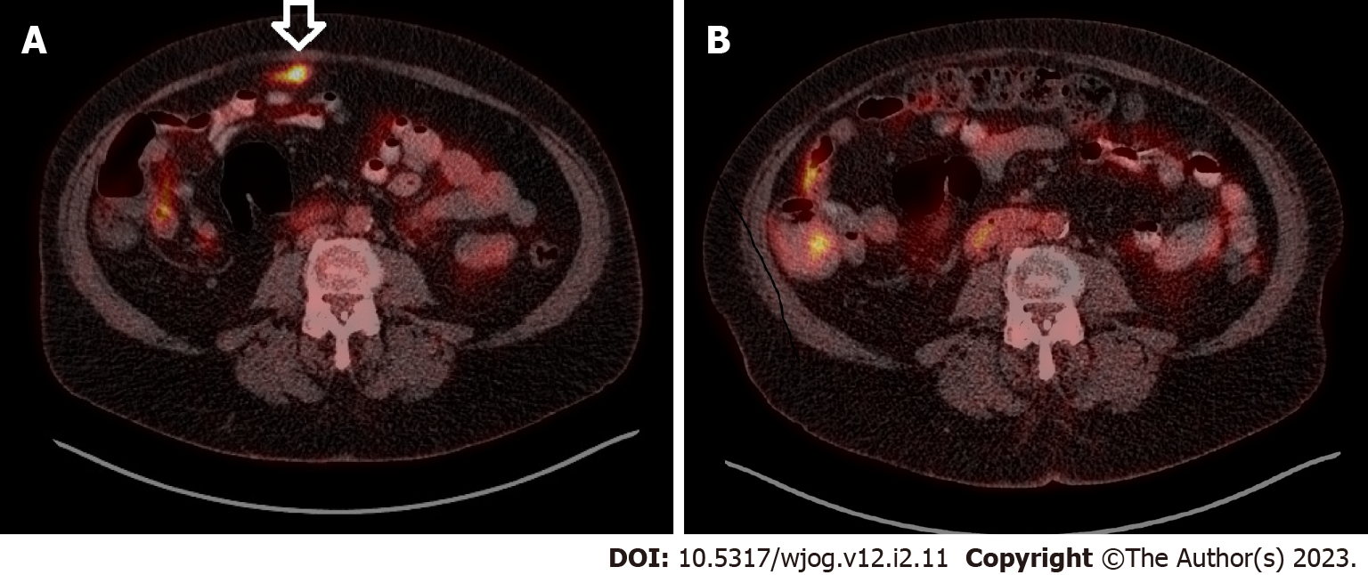
Figure 3 Comparison of fluorine 18 fluorodeoxyglucose position emission tomography with computed tomography.
A: A hypermetabolic intraabdominal lesion is demonstrated (arrow); B: The lesion has disappeared 4 mo after surgery.
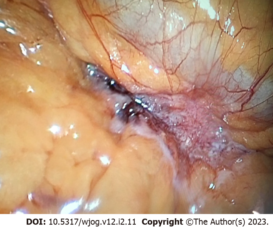
Figure 4 Image of the intraperitoneal lesion besides the transverse colon during laparoscopic exploration.
- Citation: Chan KL, Lord M, McNamara D, Désilets É, Bergeron E. Spilled gallstone mimicking metastasis from cervix cancer on positron emission tomography – computed tomography. World J Obstet Gynecol 2023; 12(2): 11-16
- URL: https://www.wjgnet.com/2218-6220/full/v12/i2/11.htm
- DOI: https://dx.doi.org/10.5317/wjog.v12.i2.11


