Copyright
©The Author(s) 2022.
World J Obstet Gynecol. Jun 29, 2022; 11(3): 33-39
Published online Jun 29, 2022. doi: 10.5317/wjog.v11.i3.33
Published online Jun 29, 2022. doi: 10.5317/wjog.v11.i3.33
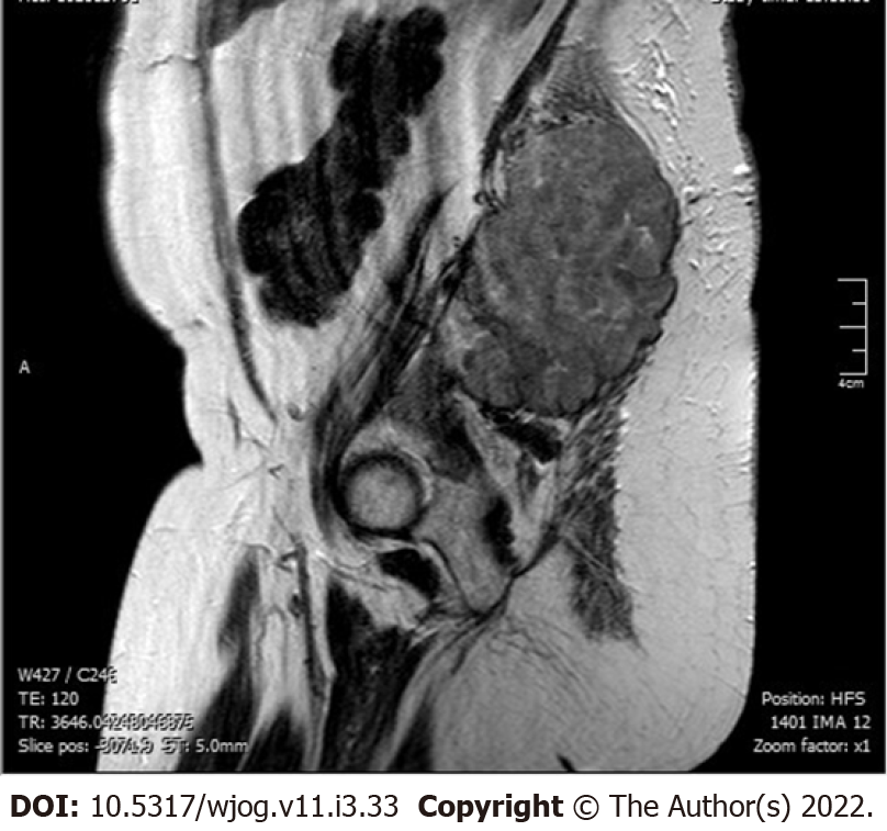
Figure 1 Magnetic resonance imaging of the lower abdomen and pelvis.
Solid formation with osteolytic involvement of the right sacrum, of the sacroiliac synchondrosis and of the contiguous iliac bone was observed.
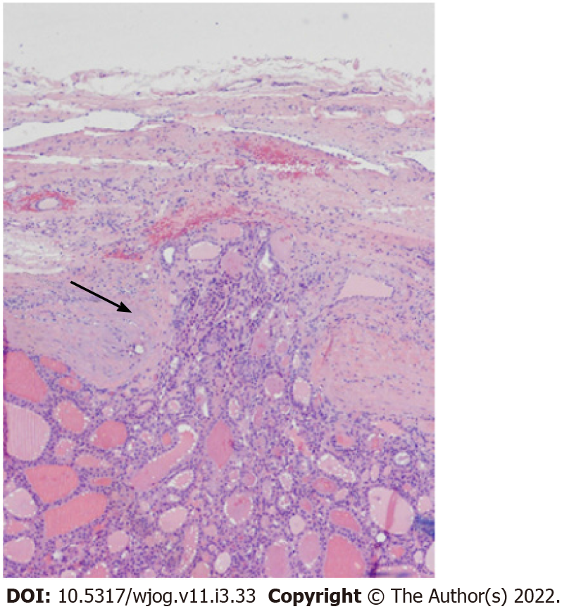
Figure 2 Follicular thyroid carcinoma.
Fibrous capsule invasion (black arrow). The growth pattern is typically micro and/or macrofollicular. No cytonuclear atypia were present. Original magnification for the panel, × 40.
- Citation: Spinelli C, Sanna B, Ghionzoli M, Micelli E. Therapeutic challenges in metastatic follicular thyroid cancer occurring in pregnancy: A case report. World J Obstet Gynecol 2022; 11(3): 33-39
- URL: https://www.wjgnet.com/2218-6220/full/v11/i3/33.htm
- DOI: https://dx.doi.org/10.5317/wjog.v11.i3.33


