Copyright
©2013 Baishideng.
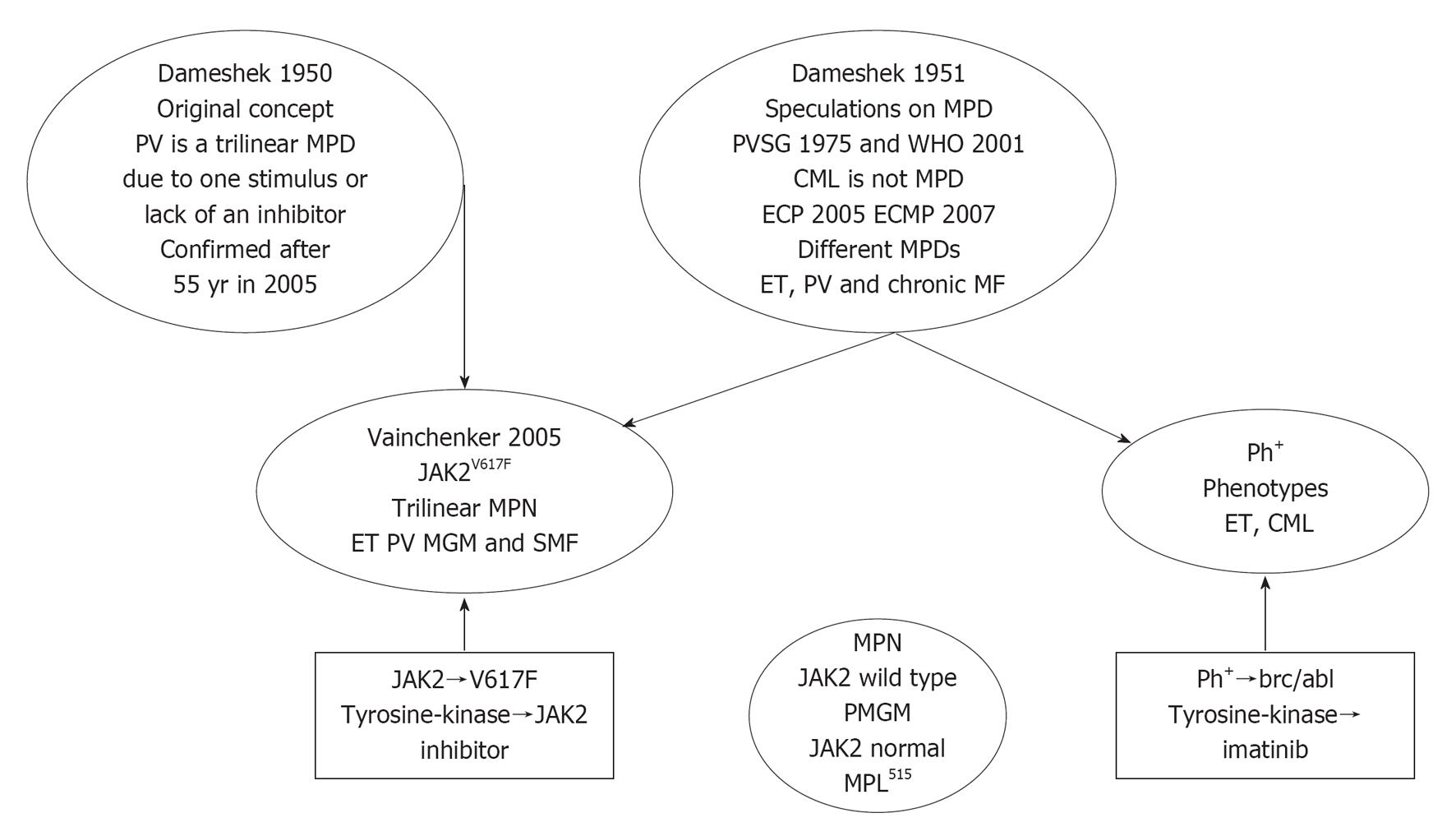
Figure 1 The concept of Dameshek in 1950 on polycythemia vera as a trilinear myeloproliferative disorder due to an unknown excessive bone marrow stimulating factor and/or a lack or a diminution in the normal inhibitory factor, which appeared to be caused by the acquired heterozygous and/or homozygous JAK2V617F mutation discovered by James et al[5].
The unifying concept of Dameshek in 1951 on lumping the chronic disorders [myeloproliferative disorder (MPDs)] essential thrombocythemia (ET), polycythemia vera (PV), agnogenic myeloid metaplasia (AMM) has been broken up by the Polycythemia Vera Study Group (PVSG) in 1975 into Ph-positive (Ph+) thrombocythemia and chronic myeloid leukemia (CML) and the Ph-negative MPDs ET, PV and myelofibrosis (MF). In 2005, PV indeed proved to be a JAK2V617F mutated trilinear MPD, whereas ET and PMF are either positive or negative for the JAK2V617F mutation. PMGM: Primary megakaryocytic and granulocytic myeloproliferation; MPN: Myeloproliferative neoplasm; WHO: World Health Organization; ECP: European Clinical and Pathologic; ECMP: European Clinical, Molecular and Pathological; MGM: Megakaryocytic, granulocytic myeloproliferation.
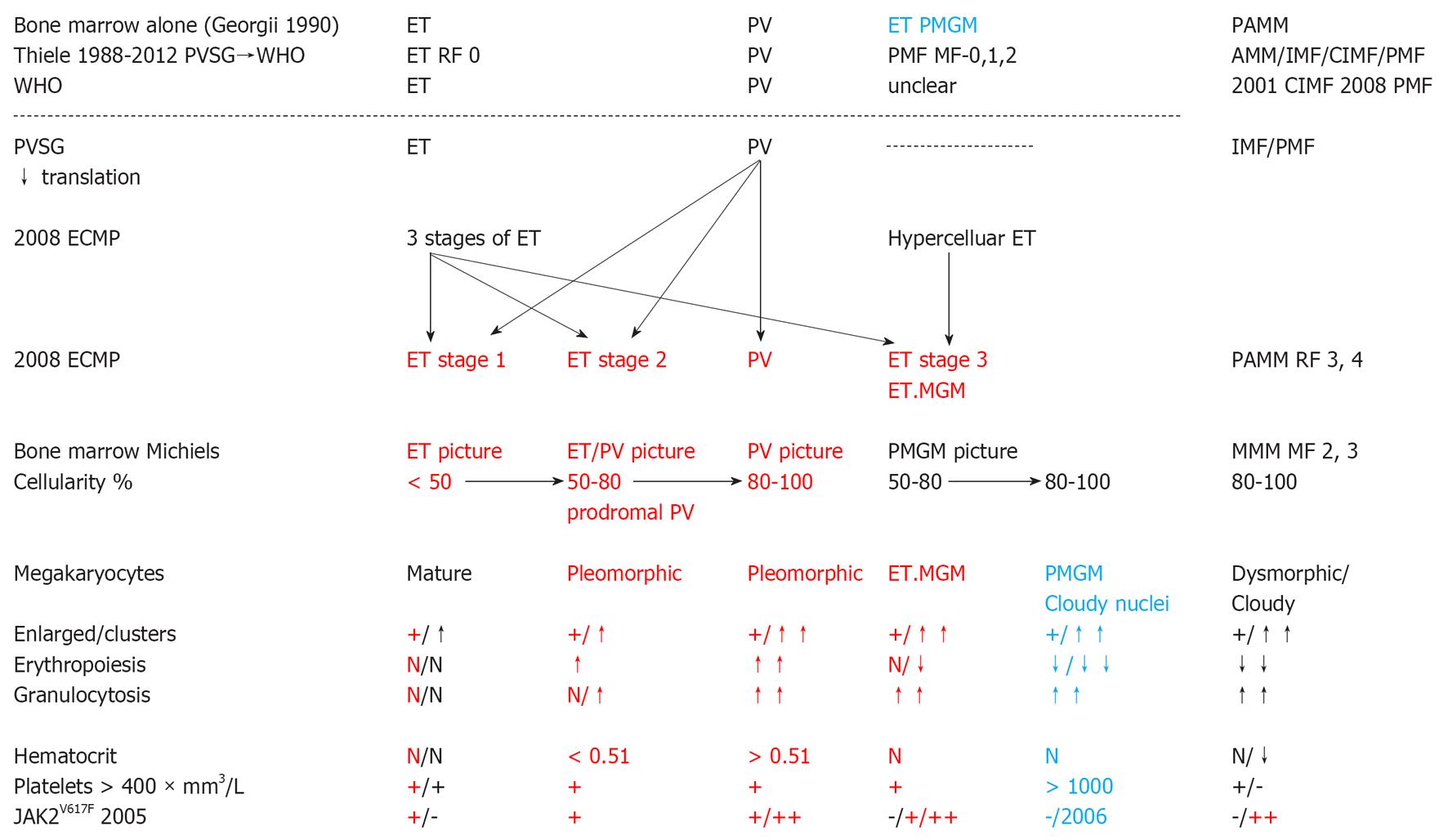
Figure 2 Bone Marrow diagnosis alone: chronic megakaryocytic granulocytic myeloproliferation by Georgii et al[24] vs chronic idiopathic myelofibrosis by Thiele et al[29,75], and comparative World Health Organization and European Clinical, Molecular and Pathological criteria for prefibrotic essential thrombocythemia, polycythemia vera and primary megakaryocytic and granulocytic myeloproliferation or chronic idiopathic myelofibrosis/primary myelofibrosis or agnogenic myeloid metaplasia.
Translation of Polycythemia Vera Study Group (PVSG) and 2008 World Health Organization (WHO) defined essential thrombocythemia (ET), polycythemia vera (PV) and chronic idiopathic myelofibrosis (CIMF), chronic megakaryocytic granulocytic myeloproliferation (CMGM) or primary megakaryocytic and granulocytic myeloproliferation (PMGM) according to European Clinical, Molecular and Pathological (ECMP) criteria subdivided in JAK2V617F mutated ET, prodromal PV, overt PV and ET.MGM (red) vs prefibrotic PMGM (blue) and 2 types of normocellular ET (JAK2V617F + left red, MPL515 + black). PMF: Primary myelofibrosis; AMM: Agnogenic myeloid metaplasia; ET.MGM: ET due to megakaryocytic, granulocytic myeloproliferation; MF: Myelofibrosis.
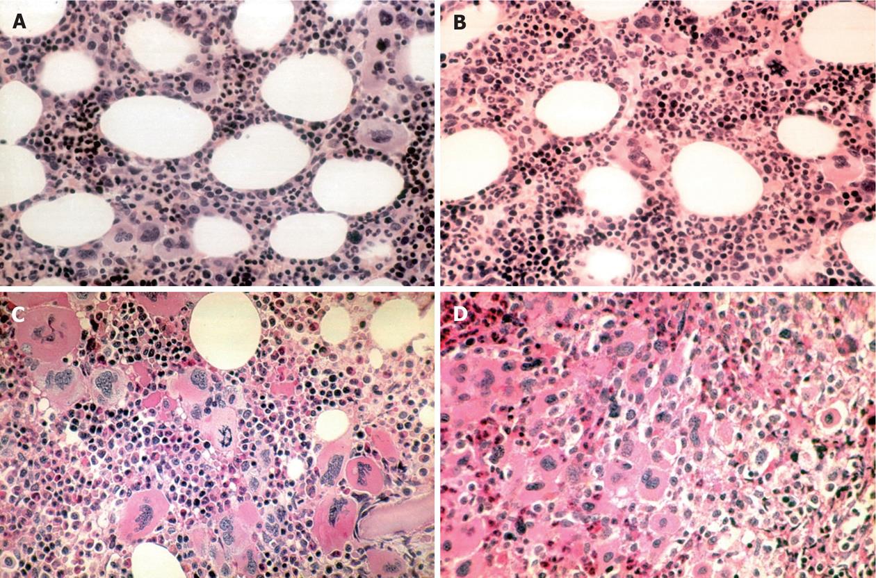
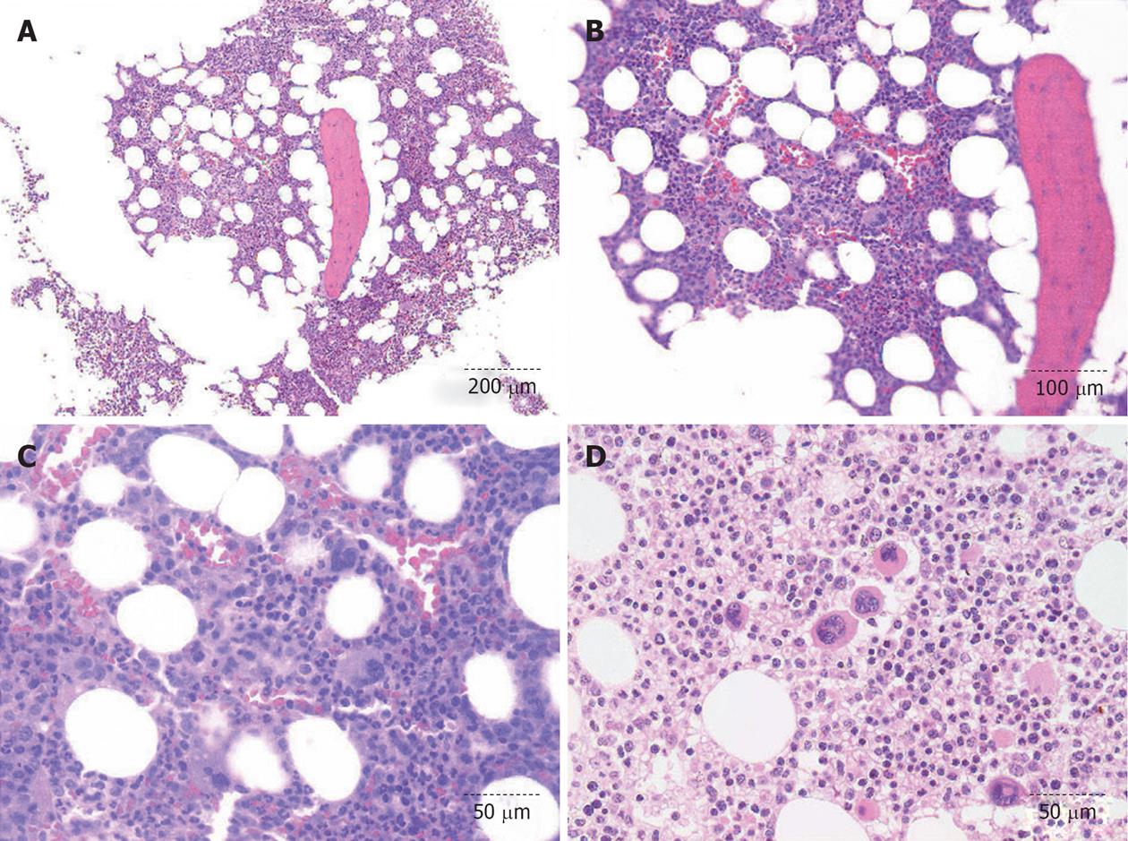
Figure 3 Bone marrow histology features in essential thrombocythemia and polycythemia vera patients.
A: Normocellular essential thrombocythemia (ET) bone marrow histology [World Health Organization (WHO)-ET] with increase of clustered pleomorphic megakaryocytes similar as in prodromal and overt polycythemia vera (PV); B: ET/PV bone marrow histology with pleomorphic megakaryocytes and increased cellularity due to increased erythropoiesis as can be seen in WHO and European Clinical, Molecular and Pathological defined prodromal PV and overt PV patients; C: PV bone marrow histology with increased cellularity due to increased erythropoiesis/granulopoiesis and increase of clustered pleomorphic megakaryocytes and no increase of reticulin fibrosis; D: Advanced PV bone marrow histology with dense clustered pleomorphic/dysmorphic megakaryocytes (not cloud-like) and increase in reticulin fibrosis grade 2.
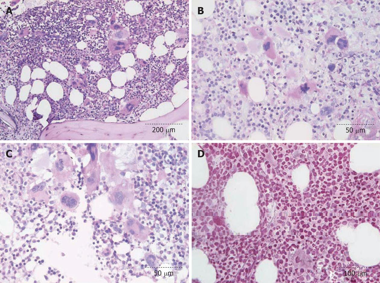
Figure 4 JAK2V617F mutated essential thrombocythemia due to megakaryocytic, granulocytic myeloproliferation with slight splenomegaly (spleen 16 cm on echogram) and a hypercellular megakaryocytic granulocytic bone marrow and clusteried pleomorphic clumpsy megakaryocytes with dysmorphic (not cloud-like) nuclei: prefibrotic essential thrombocythemia due to megakaryocytic, granulocytic myeloproliferation.
A-C: JAK2V617-positive essential thrombocythemia due to megakaryocytic, granulocytic myeloproliferation (ET.MGM) featured by hypercellular ET due to increased megakaryocytic granulocytic myeloproliferation and the presence of pleomorphic/dysmorphic megakaryocytes (not cloud-like); D: Reticulin fibrosis grade 1, myelofibrosis grade 0.
Figure 5 Forty-three-year-old female with positive polycythemia vera (platelets 405 × 109/L, low serum erythropoietin, leukocyte alkaline phosphatase score 283, hematocrit 0.
52, erythrocytes 6.1 × 10¹²/L, increased red cell mass) with a diagnostic essential thrombocythemia/polycythemia vera bone marrow picture. Such essential thrombocythemia (ET)/polycythemia vera (PV) pictures are regularly seen in prodromal PV and overt PV. A-D: JAK2V617F positive early stage PV with a ET/PV bone marrow histology with loose clusters of pleomorphic megakaryocytes.
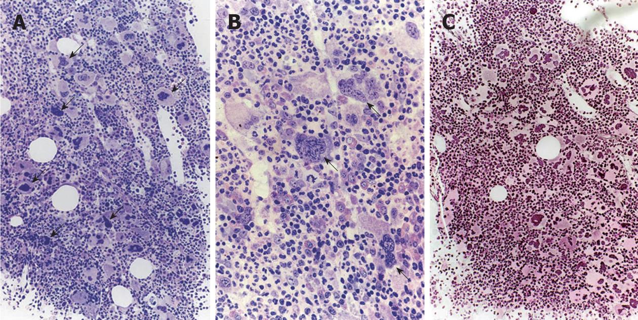
Figure 6 JAK2 wild type hypercellular essential thrombocythemia with platelet counts of 2180 × 109/L, no splenomegaly, normal lactodehydrogenase and normal white blood cell differential counts with a characteristics picture of prefibrotic primary dysmegakaryocytic granulocytic myeloproliferation.
A, B: JAK2 wild type hypercellular essential thrombocythemia with a typical primary megakaryocytic and granulocytic myeloproliferation bone marrow histology with the presence of abnormal clustering and increase in atypical giant to medium sized dysmorphic megakaryocytes containing bulky, clumsy (cloud-like) hypolobulated nuclei and definitive maturation defects; C: Reticulin fibrosis grade 0. Arrows indicate: Immature dysmorphic megakaryocytes with cloud-like nuclei.
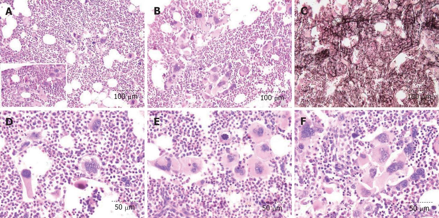
Figure 7 Thirty-seven-years old woman (asymptomatic except fatigue) with JAK2 wild type hypercellular essential thrombocythemia: platelets 1205 × 109/L, Hb 12.
5 g/dL, erythrocytes 4.9 × 10¹²/L, leukocytes 18 × 109/L, slightly increased lactodehydrogenase, no splenomegaly on palpation as the presenting features of primary megakaryocytic and granulocytic myeloproliferation (Table 5). A, B, D-F: JAK2 wild type hypercellular bone marrow histology due to primary megakaryocytic and granulocytic myeloproliferation with the presence of clustered atypical giant to medium sized dysmorphic megakaryocytes containing bulky (cloud-like) hypolobulated nuclei and definitive maturation defects; C: Reticulin fibrosis grade 2.
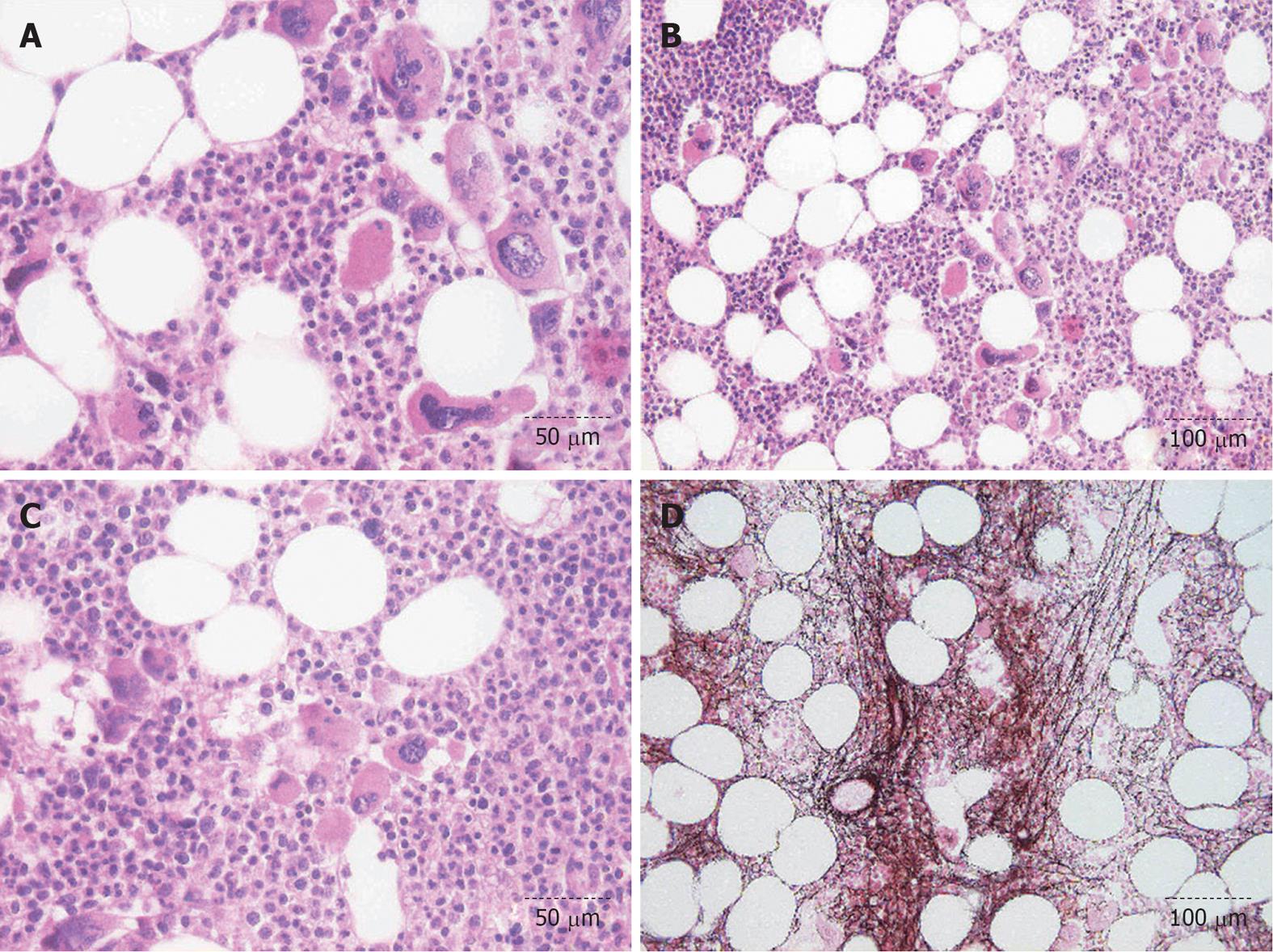
Figure 8 Chronic megakaryocytic granulocytic myelosis according to the Hannover Bone Marrow Classification at time of diagnosis in 1995, and JAK2 wild type primary megakaryocytic granulocytic myeloproliferation according to World Health Organization and European Clinical, Molecular and Pathological criteria in 2006.
A-C: Hypercellular bone marrow histology with the presence of Abnormal clustering and increase in atypical giant to medium sized dysmorphic megakaryocytes containing bulky/clumsy (cloud-like) hypolobulated nuclei and definitive maturation defects; D: Reticulin fibrosis grade 2, myelofibrosis grade 1.
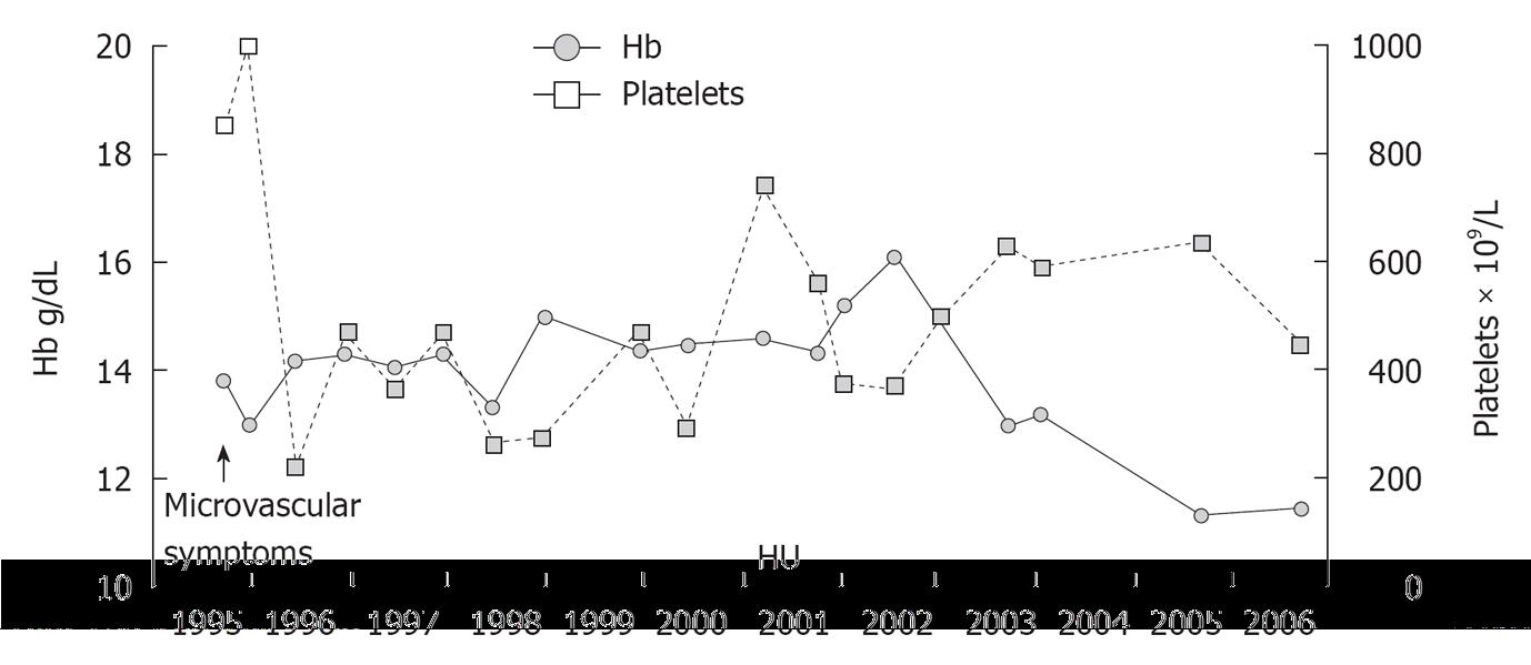
Figure 9 The case of primary megakaryocytic and granulocytic myeloproliferation in Figure 7, who presented in 1995 with microvascular circulation disturbances treated with hydroxyurea for 11 years complicated by mild anemia at platelet counts of 600 × mm3/L after 10 years of hydroxyurea (HU) for 10 years (1996-2006).
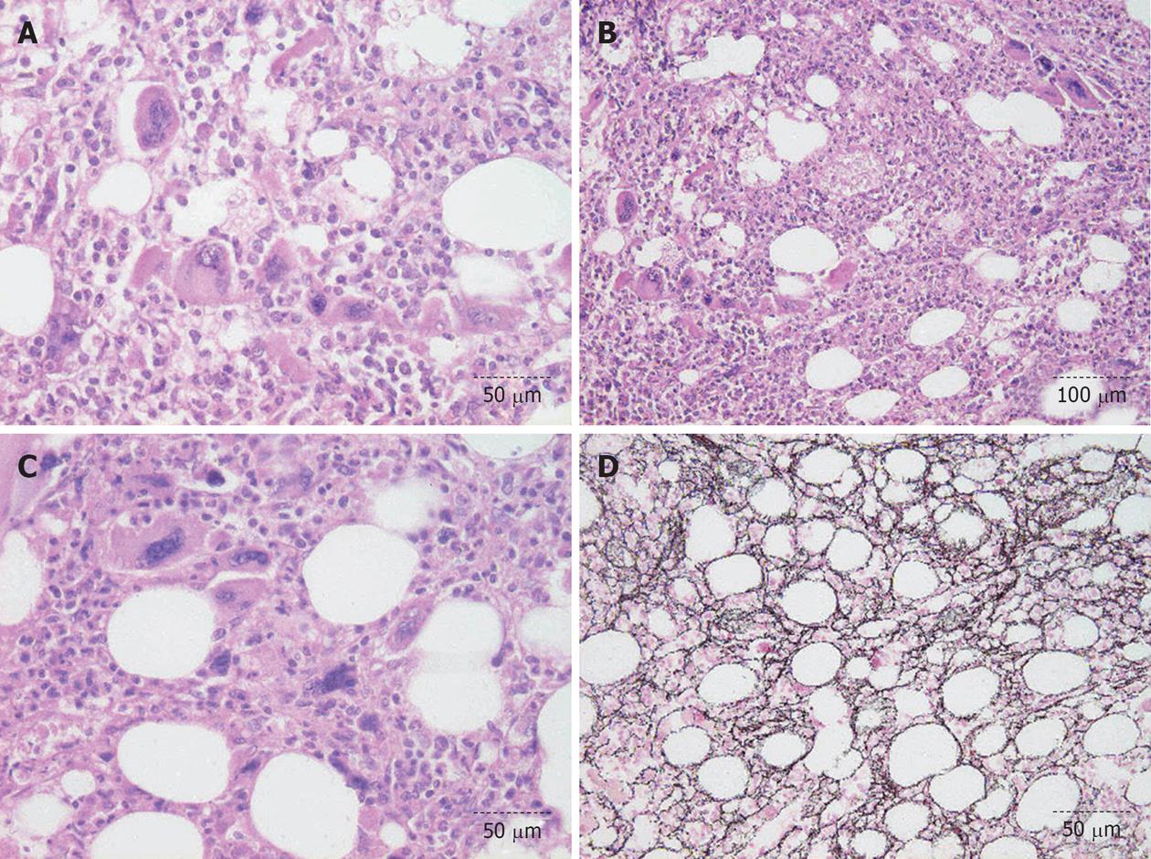
Figure 10 Essential thrombocythemia case diagnosed in 1995 as chronic megakaryocytic granulocytic myelosis, and as JAK2 wild type primary megakaryocytic granulocytic myeloproliferation in 2006 (Figure 8) was complicated by slight anemia and increased bundles of reticulin fibrosis grade 2 after 10 years of hydroxurea treatment (Figure 9).
A-C: Bone marrow histology findings in 2006 show thightly clustered immature megakaryocytes with low degree of dysmegakaryopoiesis and cloud-like nuclei. Sometimes the nuclei have an irregular contour and no real hyperchromasia; D: Increase in reticulin fibrosis with many cross-sections grade 2/myelofibrosis grade 1 (Table 5).
-
Citation: Michiels JJ, Berneman Z, Schroyens W, Lam KH, De Raeve H. PVSG and WHO
vs European Clinical, Molecular and Pathological Criteria for prefibrotic myeloproliferative neoplasms. World J Hematol 2013; 2(3): 71-88 - URL: https://www.wjgnet.com/2218-6204/full/v2/i3/71.htm
- DOI: https://dx.doi.org/10.5315/wjh.v2.i3.71


