Copyright
©2013 Baishideng.
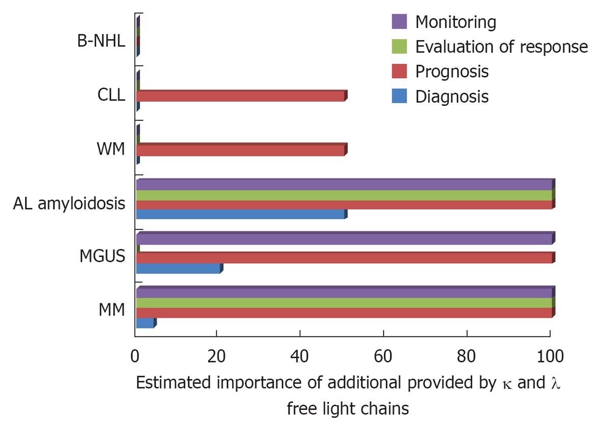
Figure 1 Estimated importance of additional information provided by free κ and λ free light chains for the diagnosis, prognosis, evaluation of response and monitoring of multiple myeloma, monoclonal gammopathy of undetermined significance, amyloid light-chain amyloidosis, chronic lymphocytic leukemia, non-Hodgkin lymphomas and Waldenstroms’ macroglobulinemia.
MM: Multiple myeloma; MGUS: Monoclonal gammopathy of undetermined significance; CLL: Chronic lymphocytic leukemia; B-NHL: B-cell non-Hodgkin lymphomas; WM: Waldenstroms’ macroglobulinemia; AL: Amyloid light-chain.
Figure 2 κ free light chain fluctuations in a light-chain κ multiple myeloma patient, during disease course.
κ-free light chain fluctuations in an LC-κ multiple myeloma patient that presented mild anaemia and bone pains and no serum or urine paraprotein by serum protein electrophoresis and immunofixation. TR: Treatment; HDT + ASCT: High dose treatment and autologous stem cell transplantation.
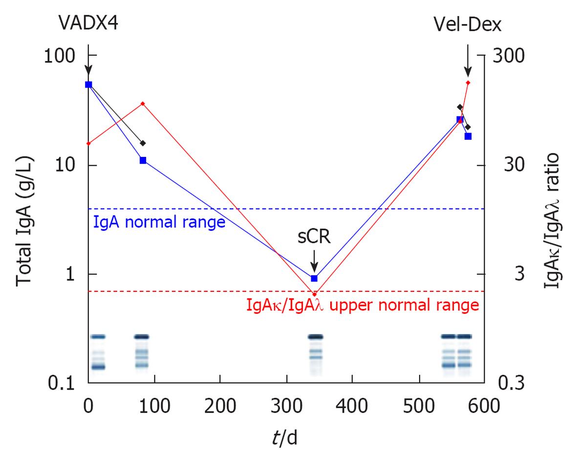
Figure 3 Heavy chain fluctuations in the course of a immunoglobulin A multiple myeloma patient.
An immunoglobulin (Ig)Aκ multiple myeloma patient achieved stringent complete remission (sCR) after second line treatment with velcade-Dexamethasone. At relapse, he was retreated with velcade-Dex. A second response by serum protein electrophoresis (SPE) was recorded after 8 d but the patient continued to clinically decline and bone marrow aspirations showed considerable plasma cell infiltration. The IgAκ/IgAλ ratio normalised at sCR, became abnormal -- relapse and increasingly abnormal during the second clinical relapse in contrast to the SPE results. Total IgA (blue line), IgA normal range (upper limit, blue dashed line), monoclonal protein by SPE densitometry (black line), IgAκ/IgAλ ratio (red line) and IgAκ/IgAλ ratio normal range (upper limit, red dashed line).
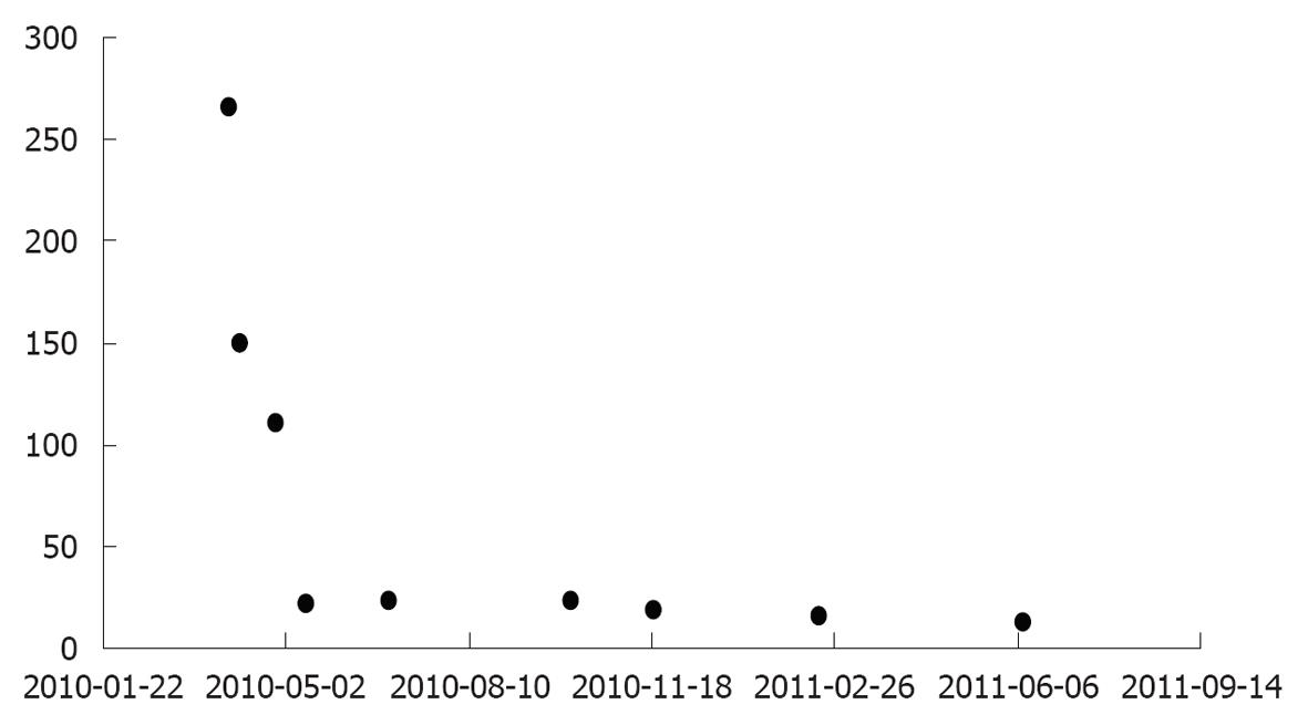
Figure 4 Lambda free light chain fluctuations in a amyloid light-chain-amyloidosis patient, in response to treatment.
Total immunoglobulin (Ig)G and IgA were decreased, immunofixation showed only κ monoclonality. λ-FLC fluctuations in response to treatment, in a LC-λ AL amyloidosis patient with bone marrow, kidney and stomach involvement FLC: Free light chain; AL: Amyloid light-chain.
- Citation: Kyrtsonis MC, Maltezas D, Koulieris E, Tzenou T, Harding SJ. Contribution of new immunoglobulin-derived biomarkers in plasma cell dyscrasias and lymphoproliferative disorders. World J Hematol 2013; 2(2): 6-12
- URL: https://www.wjgnet.com/2218-6204/full/v2/i2/6.htm
- DOI: https://dx.doi.org/10.5315/wjh.v2.i2.6


