Published online May 2, 2016. doi: 10.5314/wjd.v5.i2.109
Peer-review started: October 17, 2015
First decision: December 7, 2015
Revised: January 13, 2016
Accepted: January 27, 2016
Article in press: January 29, 2016
Published online: May 2, 2016
Processing time: 191 Days and 12.4 Hours
Ocular rosacea is an important and underdiagnosed chronic inflammatory disorder observed in children. A clinical spectrum ranging from chronic eyelid inflammation, recurrent ocular redness, photophobia and/or hordeola/chalazions and conjunctival/corneal phlyctenules evolving to neovascularization and scarring may occur. Visual impairment and consequent amblyopia are frequent and corneal perforation although rare is the most feared complication. Ocular manifestations usually precede cutaneous lesions. Although few cases of pediatric ocular rosacea (POR) have been reported in the literature, many cases must have been underdiagnosed or misdiagnosed. The delay in diagnosis is greater than one year in the large majority of cases and may lead to serious ocular sequelae. This review aims to highlight the clinical features of POR, its epidemiology, easy diagnosis and effective treatment. We also propose new diagnostic criteria, in which at least three of the five clinical criteria must be present: (1) Chronic or recurrent keratoconjunctivitis and/or red eye and/or photophobia; (2) Chronic or recurrent blepharitis and/or chalazia/hordeola; (3) Eyelid telangiectasia documented by an ophthalmologist; (4) Primary periorificial dermatitis and/or primary features of rosacea; and (5) Positive familial history of cutaneous and/or ocular rosacea.
Core tip: Ocular rosacea is a chronic inflammatory disorder with a clinical spectrum ranging from chronic eyelid inflammation, recurrent ocular redness, photophobia and/or hordeola/chalazions and conjunctival/corneal phlyctenules. Although few cases of pediatric ocular rosacea (POR) have been reported in the literature, many cases must have been underdiagnosed or misdiagnosed. This delay in diagnosis may lead to serious ocular sequelae. This review aims to highlight the clinical features of POR, its epidemiology, new diagnostic criteria, treatment and outcomes.
- Citation: Arriaga C, Domingues M, Castela G, Salgado M. Pediatric ocular rosacea, a misdiagnosed disease with high morbidity: Proposed diagnostic criteria. World J Dermatol 2016; 5(2): 109-114
- URL: https://www.wjgnet.com/2218-6190/full/v5/i2/109.htm
- DOI: https://dx.doi.org/10.5314/wjd.v5.i2.109
Rosacea is a chronic condition affecting the facial and ocular surface tissues, particularly common in the fair-skinned population[1-4]. The American National Rosacea Society developed a classification system that became the standard one. According to the patterns of signs and symptoms, four major clinical subtypes of rosacea are described: Erythematotelangiectatic, papulopustular, phymatous and ocular rosacea. In children, the phymatous type is not seen[1-4]. These subtypes may be discrete variants or may be progressive from one to another, and they can also coexist. Ocular rosacea is one of the four described types of rosacea, characterized by involvement of eyelids, conjunctiva and corneal tissue[1-4].
The prevalence of ophthalmic involvement in rosacea is probably higher than previously presumed and it varies considerably between ophthalmic and dermatological studies[2,3,5]. In most adult cases, ocular manifestations are preceded by cutaneous signs, making the diagnosis easier. However, in pediatric ocular rosacea (POR) the ocular involvement may precede dermatologic manifestations in more than half of the patients[5-8], delaying the diagnosis. The mean delay between disease onset and the diagnosis is greater than one year in most case series[6,8-11], and greater than two years in more than half of cases[6,8-10]. Before the established diagnosis, many patients are seen by multiple ophthalmologists and/or others clinicians and receive various types of therapy, including topical antibiotics, topical corticosteroids, lubricants and antiallergic drops, without success[6,8,12,13]. In fact, the management must be multidisciplinary, including dermatologists, ophthalmologists and pediatricians.
The non-recognition of POR can also be a result of the varied names adopted in the literature and the lack of overall consensus: Chronic blepharokeratoconjunctivitis[6,9-11,14-18], blepharokeratitis[10], chronic phlyctenular keratoconjunctivitis[8-10,18] phlyctenular blepharokeratoconjunctivitis[10,14,18], and meibomitis-related keratoconjunctivitis[19].
The main aim of this review is to make the children’s health care providers aware of POR, by highlighting its clinical features, epidemiology, easy diagnosis and treatment. We also propose POR criteria.
Few cases of POR have been reported in the literature. In 13 published series[5-17], a total of 259 patients were found and the number of patients in each varied between 3[16] and 51[15]. In another series, the largest ever published about POR, the sample included 615 cases, most of them described as mild, by Gupta et al[18] in 2010. In others studies, the number of cases was smaller probably because only severe cases were reported[5-17].
POR is mostly diagnosed by ophthalmologists[6,9,11,14,15], making this condition rare for other physicians and is underdiagnosed or misdiagnosed by them[5-13,15,16]. In two tertiary centers of ophthalmology, in Philadelphia[10] and in New Deli[18], chronic blepharokeratoconjunctivitis was the reason for referral in 15% and 12.3% of all children, respectively.
In the 259 patients of the 13 series[5-17], 162 are girls (62.5%) and 97 are boys (37.5%), the opposite of what was found by Gupta et al[18] (37.5% vs 62.5%).
The median age of onset in five of the 13 series (122 patients) varied between 3.2 years and 7.0 years, with extremes of 1 mo and 17 years[6,8,9,11,17]. There is a significant delay in diagnosis, often more than two years, justifying the median age at presentation in tertiary centers, between 4.6 years and 10.2 years[6,7,11,18].
A positive family history for rosacea was found in nine of the 34 patients of two series (26.5%)[5,8]. However, in most series family history wasn’t reported. Since children with rosacea are more likely to have familial rosacea[1,8], it is important to obtain this clinical data, which can help suggesting the diagnosis.
Bamford et al[20] demonstrated that having a hordeolum during childhood predisposes for rosacea in adulthood, underlying the close relationship between ocular and cutaneous inflammation. Ocular rosacea may occur without cutaneous manifestations and in individuals with any subtype of rosacea, although it is noteworthy that 50% of patients with erythematotelagiectatic and papulopustular types have eye inflammation[1,5].
Fair-skinned children of European descent are more commonly affected, although any ethnic group can be afflicted[1,4,5,8,12].
Since July of 2009, we have diagnosed and treated eight cases of POR: Three males and five females. They were referred to our tertiary Pediatric Rheumatology and Ophthalmology Units due to chronic red eye of unknown etiology, most of them after a medical peregrination and multiple ineffective topical treatments.
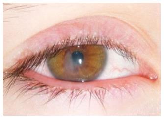
Their median age was 10 years (3-16 years) with an established diagnosis two years after the first symptoms (0-7 years); the mean previous medical consultations was eight (0-30 consultations), including at least one evaluation for an ophthalmologist in each (maximum of 13 different ophthalmologists). Two children have evolved to leukomas (Figure 1) and a decrease in visual acuity (7/10 and 8/10 respectively), what are persistent sequelae. We have already published the first two cases diagnosed at our unit[21].
The exact etiopathogenetic mechanism of rosacea and OR remains unknown. There are probably different regulatory systems involved[3-5,22]. The infection by microbial organisms may have an important role. In OR, Demodex folliculorum mites, a common inhabitant of normal human skin, possibly represents a contributing cofactor to the inflammatory reaction seen in both cutaneous and ocular disease[2-4,22,23].
Recently, bacterium Bacillus oleronius has been isolated from Demodex folliculorum mites and found to be responsible to trigger an immune system response. These seem to have a correlation with facial rosacea and OR[1,24].
Gastric coinfection with Helicobacter pylori has also been implicated, since this bacteria has the ability to produce flush-inducing toxins[3,4,7,22,23]. Staphylococcus aureus and Staphylococcus epidermidis are common organisms cultured from conjunctival or lid swabs, but their relationship with OR is questionable[7,11,12].
Recent studies focus on the role of bacterial lipases and interleukin-1 alpha and elevated concentrations of promatrix metalloproteinases in the blepharitis and corneal epitheliopathy, respectively[4]. Promatrix metalloproteinases are degrading enzymes responsible for the inferior corneal stromal thinning[4].
Rosacea induces vasodilation with increased blood flow and vessel permeability leading to erythema, telangiectasias and lymphedema of the affected tissues, especially in the eyelids[4]. The histopathological changes are unspecific, showing perifollicular infiltrates consisting of lymphohistiocytes, epithelioid and giant cells[16].
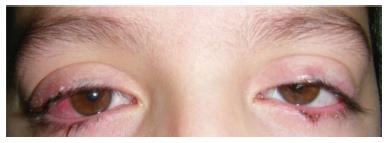
The clinical spectrum and severity of POR is variable, depending on the involvement of eyelids, conjunctiva, cornea and other ocular findings[3,4,6]. The first manifestations of POR can be chronic conjunctivitis, recurrent hordeola and/or chronic chalazia (Figure 2)[5-8,11,16,20], which are quite frequent in childhood, explaining the common delay in the diagnosis of this condition. Nevertheless, POR is often silent, painless and has unspecific clinical manifestations[2-6,12,16]. Table 1 shows the different ocular findings in POR.
| Eyelid: Telangiectasias and erythema of the lid margin, meibomian gland dysfunction, anterior blepharitis, recurrent chalazia/hordeola, madarosis (loss of eyelashes), trichiasis |
| Conjunctiva: Interpalpebral or diffuse hyperemia, papillary and/or follicular reaction, pinguecula, scarring |
| Cornea: Punctate erosions, pannus, superficial neovascularization, lipid deposition, spade-shaped infiltrate, scarring, thinning, ulceration, perforation, phlyctenula |
| Sclera: Episcleritis, scleritis |
| Insufficiency of tear film (dry eye) with abnormal Schirmer test |
| Uvea: Iritis (rare) |
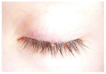
The most common manifestations are blepharitis (Figure 3), recurrent hordeola/chalazia (Figure 2), telangiectasias of the lid margin (Figure 4), dry eye, conjunctivitis and keratitis, frequently in association (blepharoconjunctivitis, blepharokeratoconjunctivitis)[2-6,8,10,15]. The typical clinical picture is a long history of hyperemic conjunctiva and intense photophobia associated with chronic blepharitis, explaining why POR is frequently called blepharoconjunctivitis[6,11,19].
Combining 12 series, including 245 patients, 185 (75.5%) had bilateral involvement, generally asymmetrical[5,6,8-17]. In the Gupta et al[18] series only 47.5% had bilateral involvement.
Eyelid involvement may precede the other features in months to years, because it is primarily an eyelid margin inflammation, such as blepharitis or meibomitis[4-6,8,11,13]. Corneal and conjunctival are secondarily involved.
The ocular symptoms include foreign body sensation, pain, burning, redness, photophobia and epiphora[3-6,8,11,19]. As a consequence of the long diagnostic delay, more than a half of the children have already corneal injuries at diagnosis, such as punctate epithelial erosions, subepithelial infiltrates, corneal phlyctenules, marginal keratitis, ulceration and corneal opacity[8,11,13]. Pediatric corneal involvement tends to be central or paracentral[6].
Depending on the severity, conjunctival and/or corneal phlyctenules may be present in 5.5%[18] up to almost 40%[8,11,15].
The primary features of pediatric facial rosacea are chronic facial flushing, non-transient erythema, papules and pustules (limited to the cheeks, chin and nasolabial areas), telangiectasias, idiopathic periorificial dermatitis and the ocular and periocular signs previously described[1,4,5,16]. Onset and severity of POR is not associated with the cutaneous signs[2,3,13].
As previously mentioned, symptoms of POR aren’t always specific and other ophthalmic disorders may present with similar findings, so the differential diagnosis includes a broad spectrum: Chronic conjunctivitis (viral, allergic, atopic), keratoconjunctivitis sicca, meibomitis, recurrent hordeola/chalazia, staphylococcal or seborrheic blepharoconjunctivitis, medication toxicity, interstitial or infectious (herpes simplex) keratitis, sterile or bacterial corneal ulcers, auto-immune diseases, sarcoidosis, among others (Table 2)[2-4,8,17].
| Chronic conjunctivitis | Medication toxicity |
| (viral, allergic, atopic) | Interstitial keratitis |
| Keratoconjunctivitis sicca | Infectious keratitis |
| Meibomitis | (herpes simplex) |
| Recurrent hordeola/chalazia | Sterile or bacterial corneal ulcers |
| Staphylococcal blepharoconjunctivitis | Auto-immune diseases |
| Seborrheic blepharoconjunctivitis | Sarcoidosis |
There are no specific clinical signs neither laboratory test nor histopathological markers for POR[2-5,12]. Chamaillard et al[5] and Hong et al[16] have proposed “dermatologic and ophthalmologic criteria for childhood rosacea”. However, in these clinical criteria four of five are cutaneous manifestations[5,16]. Cetinkaya et al[13] have also proposed the “pediatric acne rosacea diagnostic criteria” as a combination of meibomian disease, chronic blepharitis, recurrent chalazia and chronic symptoms of photophobia, ocular irritation and redness, with or without corneal vascularization, that do not respond to routine medical treatment[13]. For Léoni et al[7], the diagnostic criteria of POR requires two ophthalmologic and/or two dermatologic criteria.
Considering the above mentioned publications[5,7,13,16], in Table 3 we propose a new diagnostic criteria for POR. As in the previous proposed diagnostic criteria[5,16], ocular redness may be absent. The diagnosis of POR should be multidisciplinary, with the contribution of dermatologists, ophthalmologists and pediatricians. The presence of lid margin telangiectasia and erythema, together with meibomian gland dysfunction (chronic chalazia) and a long history of ocular irritation should suggest the diagnosis of POR[8,9,12-14], especially if there is no response to routine medical treatment[13].
| Chronic or recurrent1 keratoconjunctivitis and/or red eye and/or photophobia |
| Chronic or recurrent1 blepharitis and/or hordeola/chalazia |
| Eyelid telangiectasia documented by an ophthalmologist |
| Primary features of pediatric rosacea (facial convex areas with chronic flushing and/or erythema and/or telangiectasia, and/or papule, pustules in cheeks, chin, nose or central forehead and/or primary periorificial dermatitis) |
| Positive familial history of cutaneous and/or ocular rosacea |
The initial therapeutic approach should always include local measures, such as daily warm compresses, eyelid hygiene with neutral baby shampoo and liquefaction and removal of the thick meibomian gland secretions[1-4,8,10,11,13,15]. Prolonged topical erythromycin ointment or, more recently, azithromycin 1.5% eye drops may be useful and effective in mild cases and in association with other treatments[14]. Although very few publications support their efficacy and its administration in children is difficult, these eye drops are usually used[16]. Doan et al[14] described their experience with topical 1.5% azithromycin eye drops (monotherapy) being superior to systemic erythromycin and considered it as a first-line therapy.
Children that prove to be intolerant to prolonged topical treatment or with severe ocular involvement and/or both severe cutaneous and ocular rosacea must be treated with systemic antibiotics associated to topical care[3,5-7,12,13]. Tetracycline and doxycycline, normally used in adults, are inadvisable in children younger than 7-8 years due to their potential bone toxicity and dental staining[1,3-6,12]. Alternative safe and effective options are: Erythromycin (30-50 mg/kg per day, three times a day), clarithromycin (15 mg/kg per day divided in two doses) or azithromycin (10-12 mg/kg per day, one dose)[1,6,10,11,13,17]. Treatment with oral metronidazole is another possibility, but its frequent neurologic adverse effects, particularly peripheral neuropathy, forbids prolonged therapies[4,5,7,16]. Effective amoxicillin treatment has also been described[15].
In children older than eight years old the cyclines can be used as first systemic therapy: Minocycline, doxycycline[8,13,25]. After remission, prolonged treatment with doxycycline 40 to 100 mg once or twice daily is a good option[4,8,12,13,25].
The recurrence rate is high, especially within the first three months of treatment if systemic therapy is tapered too quickly[4,7,8,10,12]. Hence, therapeutic success is directly related to its duration, by reducing the number of recurrences. Prolonged treatments (over three months) may be required[6,8,10,13-16], with some publications recommending systemic antibiotic for at least six months[10] and others for a minimum of 12 mo[13]. Some patients will need oral antibiotics during several years, but most children may be tapered off within six months of treatment[6,10].
Intermittent treatments are necessary if shorter periods of systemic antibiotics are used[5,7,11]. Since long-term use of oral antibiotics may be problematic, it has been suggested that after six to twelve months of treatment oral therapy should be tapered slowly[4,8]. Some authors suggest that low maintenance dosages can be taken indefinitely[4], but this is questioned by others given its subtherapeutic dosages[16].
Topical (ocular) corticosteroids can prove useful for short-term exacerbations of eyelid disease and the management of inflammatory keratitis and episcleritis since they constrain eyelid and ocular inflammation[3,4,8,11-15]. However, its long-term use should be avoided due to their well-known potential side effects, such as increased intra-ocular pressure, glaucoma and cataracts. They should be discontinued as soon as possible. Furthermore, their discontinuation can frequently lead to rosacea exacerbations (topical steroid dependency)[1-4,7,13-15,17]. If indicated, topical corticosteroids must only be used during the initial weeks and the drops tapered by one drop per week[7,11,13].
Cyclosporine A 0.5% to 2% eye drops (four-six times per day) is an interesting approach for children with steroid-dependent disease and in phlyctenular blepharokeratoconjunctivitis[14,26]. Our experience shows that topic cyclosporine isn’t well tolerated by children, probably due to the lack of a suitable preparation in Europe.
It was described the efficacy of ivermectin to the treatment of refractory cases of cutaneous ocular rosacea, as an antiparasitic drug effective against mites Demodex[27]. The treatment consist in an oral single-dose, and despite being proscribed to children under five years and under 7 kg, it has been used in pediatric age[27]. This drug has primarily been reported in the treatment of immunosuppressed patients, but there are reports of its success in immunocompetent patients[27,28].
Surgical care is needed in specific cases, like corneal perforation[2,15,25]. Other options under investigation are laser and intense pulsed light therapy. Dietary intake of omega-3 has recently proven to be effective as an anti-inflammatory and in clearing meibomian gland secretions[2,6]. Flaxseed oil (∝-linoleic acid) 2.5 mL once a day for up to 12 mo, with gradual reduction to an alternate day administration, can be an option in children intolerant or non-compliant with the use of long-term systemic antibiotics[7].
POR can wax and wane with a recurrence rate of 40%[10]. Affected children suffer from chronic conjunctivitis, corneal pannus, corneal neovascularization, generalized keratitis and meibomian gland disease. Chronic symptoms and frequent exacerbations may lead to tissue hypertrophy, extensive neovascularization, scarring, corneal opacification, corneal perforation and complications from secondary infections[1,4-8,11-13,15,18]. Some patients may develop raised intraocular pressure and cataract, possibly with relation to chronic topical steroid therapy[15].
The duration of the disease and the corneal involvement are the determining factors of severity[4,6,13]. Furthermore, a prolonged therapy regimen is required to minimize corneal scarring and visual loss. Gradual tapering is recommended to avoid relapses[4,6,13]. Thus, POR can be a source of significant visual morbidity in children[4,6,8,15,18].
In comparison to adults, children seem to be more susceptible to corneal damage imposed by the inflammatory and immune response to periocular bacteria. This may compromise vision development, which combined with the position of the opacities in the cornea may be complicated by secondary amblyopia[1,4,6,8,10,12].
OR is a subtype of rosacea, which is a chronic inflammatory disease; POR is frequently under and misdiagnosed, so it is probably more common than we previously thought; POR may be associated with high morbidity, development of sequelae and it is a possible cause of loss of vision; The diagnosis is facilitated by the proposed POR criteria; An ophthalmologist observation is mandatory for the diagnosis, but it should be suggested by pediatricians or dermatologists; Treatment requires a minimum of three months’ antibiotic therapy and a subsequent gradual tapering.
We would like to acknowledge with appreciation Leonor Castendo Ramos, dermatologist of the University Hospital of Coimbra.
P- Reviewer: Manolache L, Sergi C S- Editor: Ji FF L- Editor: A E- Editor: Lu YJ
| 1. | Powell FC, Raghallaigh SN. Rosacea and Related Disorders. Dermatology. 3rd ed. USA: Elsevier Saunders 2012; 561-569. |
| 2. | Vieira AC, Mannis MJ. Ocular rosacea: common and commonly missed. J Am Acad Dermatol. 2013;69:S36-S41. [RCA] [PubMed] [DOI] [Full Text] [Cited by in Crossref: 40] [Cited by in RCA: 44] [Article Influence: 4.0] [Reference Citation Analysis (0)] |
| 3. | Vieira AC, Höfling-Lima AL, Mannis MJ. Ocular rosacea--a review. Arq Bras Oftalmol. 2012;75:363-369. [RCA] [PubMed] [DOI] [Full Text] [Cited by in Crossref: 51] [Cited by in RCA: 60] [Article Influence: 5.0] [Reference Citation Analysis (1)] |
| 4. | Oltz M, Check J. Rosacea and its ocular manifestations. Optometry. 2011;82:92-103. [RCA] [PubMed] [DOI] [Full Text] [Cited by in Crossref: 41] [Cited by in RCA: 37] [Article Influence: 2.6] [Reference Citation Analysis (0)] |
| 5. | Chamaillard M, Mortemousque B, Boralevi F, Marques da Costa C, Aitali F, Taïeb A, Léauté-Labrèze C. Cutaneous and ocular signs of childhood rosacea. Arch Dermatol. 2008;144:167-171. [RCA] [PubMed] [DOI] [Full Text] [Cited by in Crossref: 81] [Cited by in RCA: 65] [Article Influence: 3.8] [Reference Citation Analysis (0)] |
| 6. | Jones SM, Weinstein JM, Cumberland P, Klein N, Nischal KK. Visual outcome and corneal changes in children with chronic blepharokeratoconjunctivitis. Ophthalmology. 2007;114:2271-2280. [RCA] [PubMed] [DOI] [Full Text] [Cited by in Crossref: 71] [Cited by in RCA: 55] [Article Influence: 3.1] [Reference Citation Analysis (0)] |
| 7. | Léoni S, Mesplié N, Aitali F, Chamaillard M, Boralevi F, Marques da Costa C, Taïeb A, Léauté-Labrèze C, Colin J, Mortemousque B. [Metronidazole: alternative treatment for ocular and cutaneous rosacea in the pediatric population]. J Fr Ophtalmol. 2011;34:703-710. [RCA] [PubMed] [DOI] [Full Text] [Cited by in Crossref: 7] [Cited by in RCA: 10] [Article Influence: 0.7] [Reference Citation Analysis (0)] |
| 8. | Donaldson KE, Karp CL, Dunbar MT. Evaluation and treatment of children with ocular rosacea. Cornea. 2007;26:42-46. [RCA] [PubMed] [DOI] [Full Text] [Cited by in Crossref: 38] [Cited by in RCA: 45] [Article Influence: 2.5] [Reference Citation Analysis (1)] |
| 9. | Doan S, Gabison EE, Nghiem-Buffet S, Abitbol O, Gatinel D, Hoang-Xuan T. Long-term visual outcome of childhood blepharokeratoconjunctivitis. Am J Ophthalmol. 2007;143:528-529. [RCA] [PubMed] [DOI] [Full Text] [Cited by in Crossref: 27] [Cited by in RCA: 28] [Article Influence: 1.6] [Reference Citation Analysis (0)] |
| 10. | Hammersmith KM, Cohen EJ, Blake TD, Laibson PR, Rapuano CJ. Blepharokeratoconjunctivitis in children. Arch Ophthalmol. 2005;123:1667-1670. [RCA] [PubMed] [DOI] [Full Text] [Cited by in Crossref: 54] [Cited by in RCA: 55] [Article Influence: 2.9] [Reference Citation Analysis (0)] |
| 11. | Viswalingam M, Rauz S, Morlet N, Dart JK. Blepharokeratoconjunctivitis in children: diagnosis and treatment. Br J Ophthalmol. 2005;89:400-403. [RCA] [PubMed] [DOI] [Full Text] [Cited by in Crossref: 73] [Cited by in RCA: 73] [Article Influence: 3.7] [Reference Citation Analysis (0)] |
| 12. | Nazir SA, Murphy S, Siatkowski RM, Chodosh J, Siatkowski RL. Ocular rosacea in childhood. Am J Ophthalmol. 2004;137:138-144. [RCA] [PubMed] [DOI] [Full Text] [Cited by in Crossref: 59] [Cited by in RCA: 64] [Article Influence: 3.0] [Reference Citation Analysis (0)] |
| 13. | Cetinkaya A, Akova YA. Pediatric ocular acne rosacea: long-term treatment with systemic antibiotics. Am J Ophthalmol. 2006;142:816-821. [RCA] [PubMed] [DOI] [Full Text] [Cited by in Crossref: 41] [Cited by in RCA: 45] [Article Influence: 2.4] [Reference Citation Analysis (1)] |
| 14. | Doan S, Gabison E, Chiambaretta F, Touati M, Cochereau I. Efficacy of azithromycin 1.5% eye drops in childhood ocular rosacea with phlyctenular blepharokeratoconjunctivitis. J Ophthalmic Inflamm Infect. 2013;3:38. [RCA] [PubMed] [DOI] [Full Text] [Full Text (PDF)] [Cited by in Crossref: 27] [Cited by in RCA: 36] [Article Influence: 3.0] [Reference Citation Analysis (0)] |
| 15. | Teo L, Mehta JS, Htoon HM, Tan DT. Severity of pediatric blepharokeratoconjunctivitis in Asian eyes. Am J Ophthalmol. 2012;153:564-570.e1. [RCA] [PubMed] [DOI] [Full Text] [Cited by in Crossref: 24] [Cited by in RCA: 31] [Article Influence: 2.4] [Reference Citation Analysis (0)] |
| 16. | Hong E, Fischer G. Childhood ocular rosacea: considerations for diagnosis and treatment. Australas J Dermatol. 2009;50:272-275. [RCA] [PubMed] [DOI] [Full Text] [Cited by in Crossref: 14] [Cited by in RCA: 17] [Article Influence: 1.1] [Reference Citation Analysis (0)] |
| 17. | Farpour B, McClellan KA. Diagnosis and management of chronic blepharokeratoconjunctivitis in children. J Pediatr Ophthalmol Strabismus. 2001;38:207-212. [PubMed] |
| 18. | Gupta N, Dhawan A, Beri S, D’souza P. Clinical spectrum of pediatric blepharokeratoconjunctivitis. J AAPOS. 2010;14:527-529. [RCA] [PubMed] [DOI] [Full Text] [Cited by in Crossref: 28] [Cited by in RCA: 36] [Article Influence: 2.4] [Reference Citation Analysis (0)] |
| 19. | Suzuki T. Meibomitis-related keratoconjunctivitis: implications and clinical significance of meibomian gland inflammation. Cornea. 2012;31 Suppl 1:S41-S44. [RCA] [PubMed] [DOI] [Full Text] [Cited by in Crossref: 29] [Cited by in RCA: 47] [Article Influence: 3.9] [Reference Citation Analysis (0)] |
| 20. | Bamford JT, Gessert CE, Renier CM, Jackson MM, Laabs SB, Dahl MV, Rogers RS. Childhood stye and adult rosacea. J Am Acad Dermatol. 2006;55:951-955. [RCA] [PubMed] [DOI] [Full Text] [Cited by in Crossref: 33] [Cited by in RCA: 27] [Article Influence: 1.4] [Reference Citation Analysis (0)] |
| 21. | Miguel AI, Salgado MB, Lisboa MS, Henriques F, Paiva MC, Castela GP. Pediatric ocular rosacea: 2 cases. Eur J Ophthalmol. 2012;22:664-666. [RCA] [PubMed] [DOI] [Full Text] [Cited by in Crossref: 8] [Cited by in RCA: 11] [Article Influence: 0.8] [Reference Citation Analysis (0)] |
| 22. | Steinhoff M, Schauber J, Leyden JJ. New insights into rosacea pathophysiology: a review of recent findings. J Am Acad Dermatol. 2013;69:S15-S26. [RCA] [PubMed] [DOI] [Full Text] [Cited by in Crossref: 206] [Cited by in RCA: 228] [Article Influence: 20.7] [Reference Citation Analysis (0)] |
| 23. | Crawford GH, Pelle MT, James WD. Rosacea: I. Etiology, pathogenesis, and subtype classification. J Am Acad Dermatol. 2004;51:327-341; quiz 342-344. [RCA] [PubMed] [DOI] [Full Text] [Cited by in Crossref: 328] [Cited by in RCA: 320] [Article Influence: 16.0] [Reference Citation Analysis (0)] |
| 24. | Li J, O’Reilly N, Sheha H, Katz R, Raju VK, Kavanagh K, Tseng SC. Correlation between ocular Demodex infestation and serum immunoreactivity to Bacillus proteins in patients with Facial rosacea. Ophthalmology. 2010;117:870-877.e1. [RCA] [PubMed] [DOI] [Full Text] [Cited by in Crossref: 83] [Cited by in RCA: 96] [Article Influence: 6.4] [Reference Citation Analysis (0)] |
| 25. | Potz-Biedermann C, Mehra T, Deuter C, Zierhut M, Schaller M. Ophthalmic Rosacea: Case Report in a Child and Treatment Recommendations. Pediatr Dermatol. 2015;32:522-525. [RCA] [PubMed] [DOI] [Full Text] [Cited by in Crossref: 7] [Cited by in RCA: 8] [Article Influence: 0.8] [Reference Citation Analysis (0)] |
| 26. | Kharod-Dholakia B. Ocular rosacea treatment & management. Medscape - drugs and diseases, last updated in. USA: Elsevier Saunders 2014; Available from: http://emedicine.medscape.com/article/1197341-treatment#a1127. |
| 27. | Brown M, Hernández-Martín A, Clement A, Colmenero I, Torrelo A. Severe demodexfolliculorum-associated oculocutaneous rosacea in a girl successfully treated with ivermectin. JAMA Dermatol. 2014;150:61-63. [RCA] [PubMed] [DOI] [Full Text] [Cited by in Crossref: 30] [Cited by in RCA: 34] [Article Influence: 3.1] [Reference Citation Analysis (0)] |
| 28. | Patrizi A, Neri I, Chieregato C, Misciali M. Demodicidosis in immunocompetent young children: report of eight cases. Dermatology. 1997;195:239-242. [RCA] [PubMed] [DOI] [Full Text] [Cited by in Crossref: 45] [Cited by in RCA: 46] [Article Influence: 1.7] [Reference Citation Analysis (0)] |


