Copyright
©The Author(s) 2015.
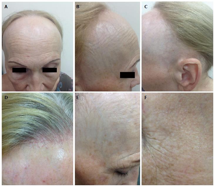
Figure 1 Clinical presentation of frontal fibrosing alopecia.
A: Fronto-temporal hairline recession in a band-like distribution. The pale, smooth, and atrophic skin of the alopetic area contrasts with the sun-damaged and pigmented skin of the forehead; B: Lateral view: Increased visibility of veins on the forehead; C: Retroauricular area involvement; D: Absence of vellus hairs, perifollicular erythema and hyperkeratosis over the frontal hairline; E: Eyebrow loss and isolated hairs in the middle of the alopetic band (“lonely hair sign”); F: Facial papules over the temporal area indicating vellus hair involvement.
- Citation: Lyakhovitsky A, Barzilai A, Amichai B. Frontal fibrosing alopecia update. World J Dermatol 2015; 4(1): 33-43
- URL: https://www.wjgnet.com/2218-6190/full/v4/i1/33.htm
- DOI: https://dx.doi.org/10.5314/wjd.v4.i1.33


