Copyright
©The Author(s) 2020.
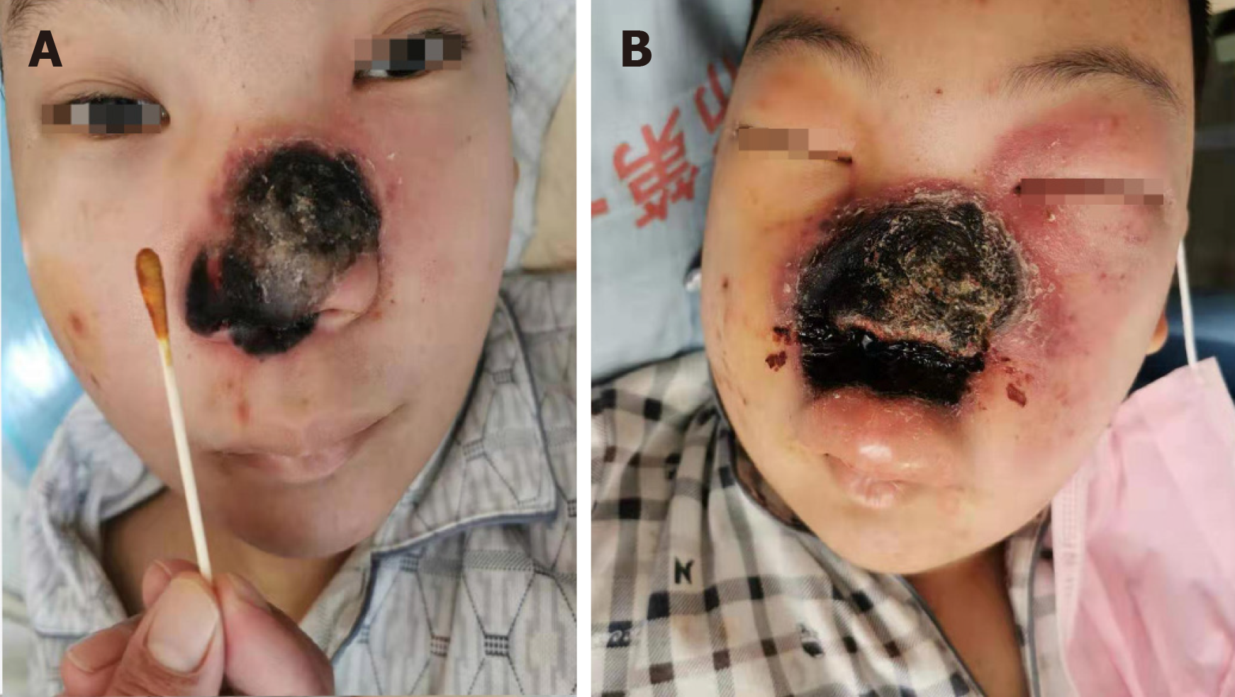
Figure 1 Before treatment with amphotericin B and one week after treatment with amphotericin B.
A: Before treatment with Amphotericin B; B: One week after treatment with amphotericin B.
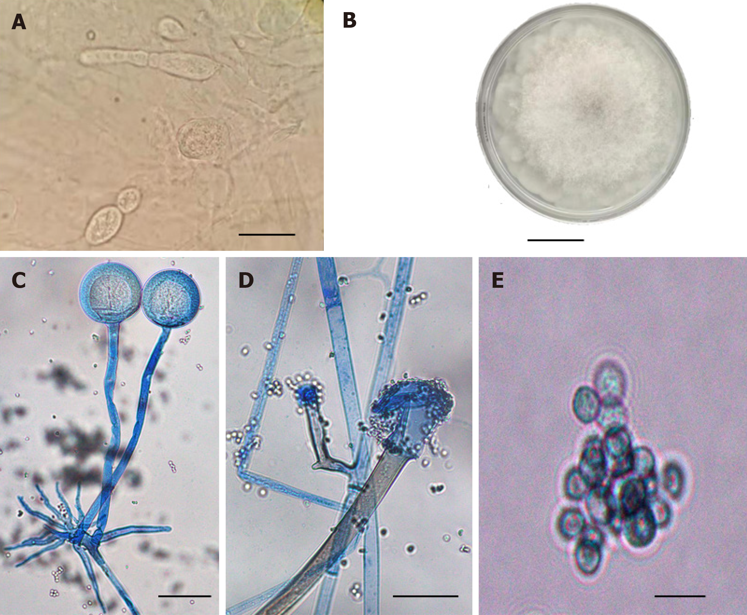
Figure 2 Pathological findings.
A: The patient's secretion was cultured at 33 °C for 3 d, and the morphology of fungal colony was observed; B: Typical sporangium and junction structure can be seen by direct microscopic examination of the patient's secretion; C: Typical spherical sporangium and developed rhizoid and sporophyte can be found by slide culture and lactophenol cotton blue staining; D: The shape of the sporangium after spores are released; E: Spore morphology of pathogenic fungi (regular round spore, with the range of diameter from 4.7 μm to 5.0 μm). All the scale bars represent 10 μm.
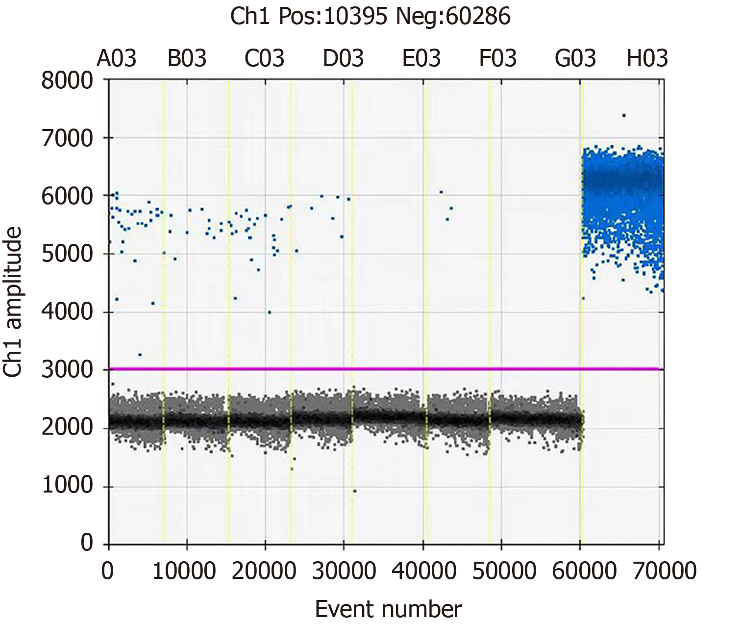
Figure 3 The patient’s blood sample was collected and detected for fungal DNA by droplet digital polymerase chain reaction.
E03, G03, and H03 represent the patient’s blood sample, negative control, and positive control, respectively. The results show that there are a large number of negative events in the patient's sample. No blood infection was detected in the patients with positive events.
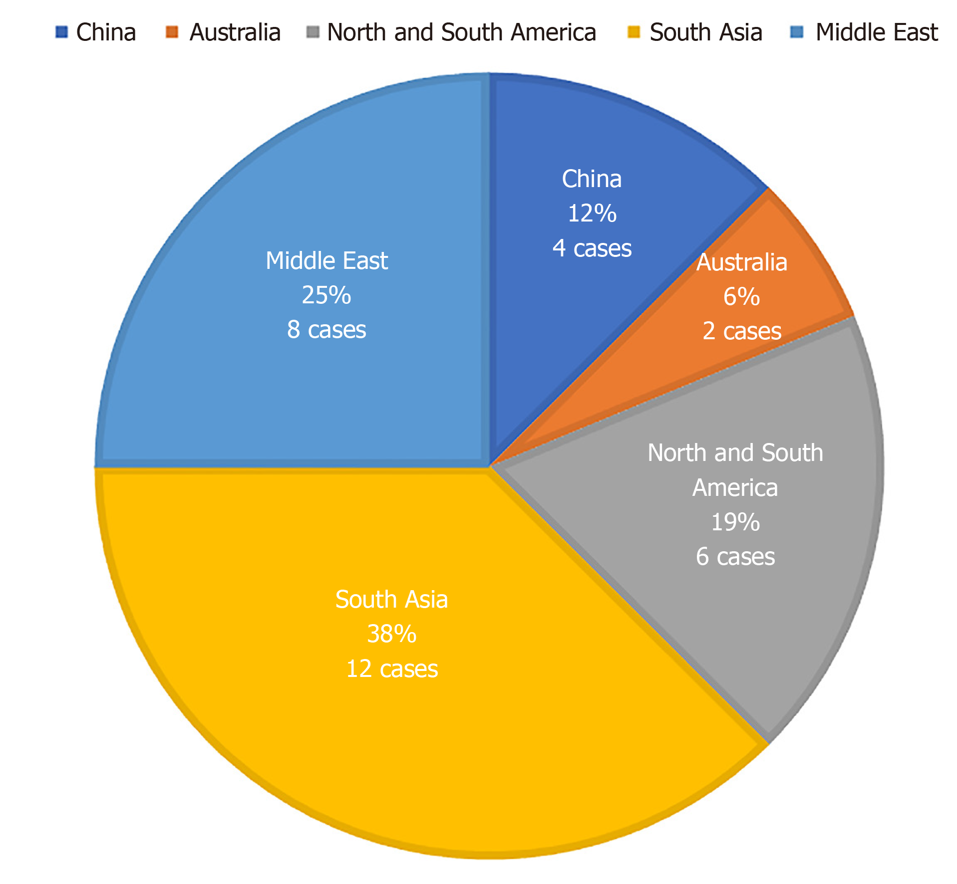
Figure 4 Distribution of rhinocerebral mucormycosis caused by Rhizopus oryzae.
- Citation: Feng YH, Guo WW, Wang YR, Shi WX, Liu C, Li DM, Qiu Y, Shi DM. Rhinocerebral mucormycosis caused by Rhizopus oryzae in a patient with acute myeloid leukemia: A case report. World J Dermatol 2020; 8(1): 1-9
- URL: https://www.wjgnet.com/2218-6190/full/v8/i1/1.htm
- DOI: https://dx.doi.org/10.5314/wjd.v8.i1.1


