Copyright
©The Author(s) 2015.
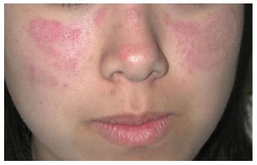
Figure 1 Acute cutaneous lupus erythematosus.
The facial erythema on the malar cheeks appears as erythematous plaques with scale. There is distinct sparing of the nasolabial folds.
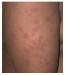
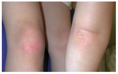
Figure 2 Acute cutaneous lupus erythematosus.
Pink edematous papules on the extremities.
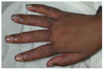
Figure 3 Acute cutaneous lupus erythematosus on the dorsal hands.
Note the erythematous edematous papules coalescing into plaques on the dorsal fingers with sparing of the phalangeal joints. Note the dilated cutaneous vessels under the proximal nail fold particularly of the index finger.
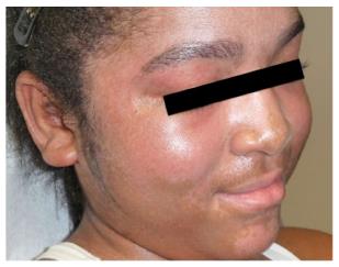
Figure 4 Discoid lupus erythematosus.
These lesions will typically favor the head and neck. A: Earlier lesions can present as erythematous papules and annular plaques with scaling; B: This patient had more chronic lesions involving her cheek, nose, chin and conchal bowl with significant dyspigmentation; C: Widespread symmetric involvement can be seen in generalized discoid lupus.
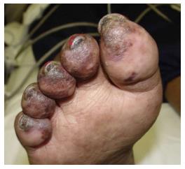
Figure 5 Chilblain lupus erythematosus.
Violaceous plaques with overlying scale on the distal toes.
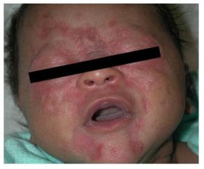
Figure 6 Neonatal lupus.
Annular lesions with central atrophy particularly concentrated around the eyes.
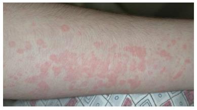
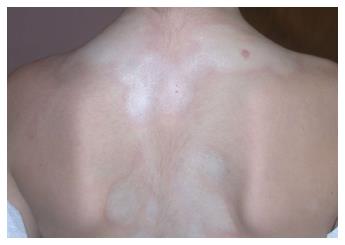
Figure 7 Juvenile idiopathic arthritis.
Erythematous papules coalescing into plaques with surrounding pallor, typically evanescent in nature.
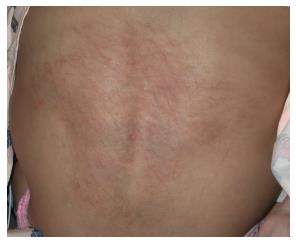
Figure 8 Juvenile idiopathic arthritis presenting as flagellate erythema.
Erythematous and hyperpigmented persistent plaques in a flagellate pattern.
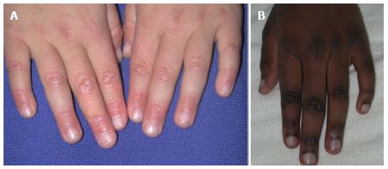
Figure 9 Dermatomyositis.
Gottron’s papules with (A) pink to (B) violaceous flat-topped papules overlying the dorsal joints of the fingers with sparing of the skin in between the joints, a finding occasionally mistaken for flat warts. Also note the erythema noted around the proximal nail folds.
Figure 10 Dermatomyositis.
Lichenoid papules and plaques over bony prominences of the knees and elbows.
Figure 11 Dermatomyositis.
Heliotrope sign with prominent capillary vasculature around the eyelids and characteristic pink-purple patches involving the cheeks, chin and temples.
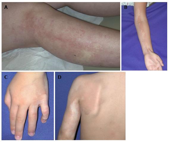
Figure 12 Linear morphea overlying joints.
Early indurated plaques with lilac-colored border (A); (B) Advanced sclerotic and hypopigmented plaques on the left arm (C) with flexion contraction of the left fifth finger; (D) residual hyperpigmentation on the left shoulder.
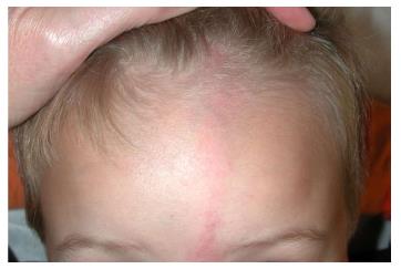
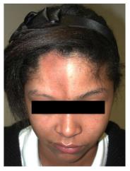
Figure 13 En coup de sabre morphea.
Early presentation with an erythematous patch in paramedian forehead location extending into the scalp with hair loss.
Figure 14 Parry-Romberg syndrome.
Note the resulting asymmetry of the right lower face due to loss of subcutaneous fat.
Figure 15 Circumscribed morphea.
Inflamed lilac border around indurated sclerotic plaques.
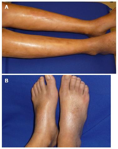
Figure 16 Pansclerotic morphea.
A: Sclerosis of the skin bound down to underlying skin making the skin appear wooden with an uneven, taut shiny texture to the skin; B: Note the sparing of the toes which differentiates pansclerotic morphea from systemic sclerosis.
- Citation: Yun D, Stein SL. Review of the cutaneous manifestations of autoimmune connective tissue diseases in pediatric patients. World J Dermatol 2015; 4(2): 80-94
- URL: https://www.wjgnet.com/2218-6190/full/v4/i2/80.htm
- DOI: https://dx.doi.org/10.5314/wjd.v4.i2.80


