Copyright
©The Author(s) 2015.
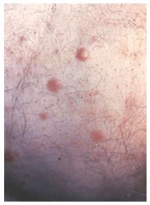
Figure 1 Primary cutaneous B-cell lymphoma.
Red-brown nodules scattered on the trunk.
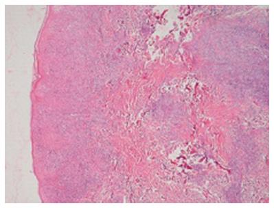
Figure 2 Widespread lymphoid infiltration with clusters of mononuclear cells involving the dermis (HE, × 10).
Figure 3 Immunophenotype of lymphoid cells: Immunohistochemical staining shows expression of CD20 (A), CD10 (B), and Bcl-6 (C) (Original magnifications × 20).
- Citation: Yilmaz F, Soyer N, Vural F. Primary cutaneous B cell lymphoma: Clinical features, diagnosis and treatment. World J Dermatol 2015; 4(1): 50-56
- URL: https://www.wjgnet.com/2218-6190/full/v4/i1/50.htm
- DOI: https://dx.doi.org/10.5314/wjd.v4.i1.50


