Copyright
©The Author(s) 2015.
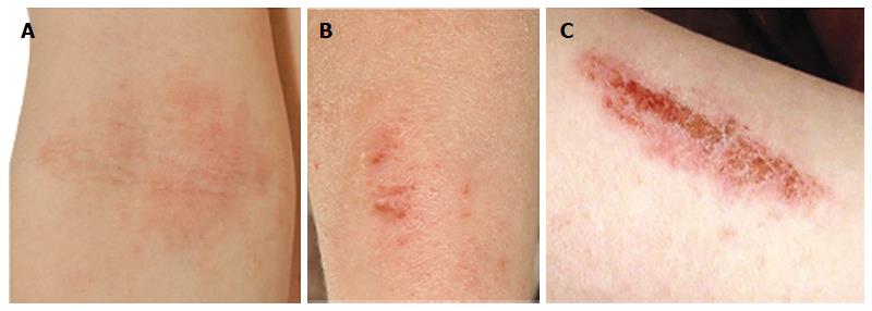
Figure 1 Clinical features of atopic dermatitis.
A: Flexural eczema; B: Eczema and ichthyosis; C: Infected excoriation. Clinical photographs (University of Dundee Computing and Media Services, Ninewells Hospital and Medical School) reproduced with patient and/or parental consent.
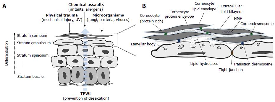
Figure 2 Outside-inside and inside-outside barrier functions of the epidermis.
A: The epidermis forms a barrier against multiple external threats and prevents excessive transepidermal water loss (TEWL, indicated by the dashed blue arrow) from the interior of the body; B: Components of the stratum corneum (SC) and stratum granulosum (SG) barriers. During terminal differentiation of SG keratinocytes into corneocytes, lamellar bodies (LBs) fuse with the plasma membrane and their contents is extruded at the SG-SC interface. LB-derived lipids are processed and arranged into continuous bilayers parallel to the corneocyte surface using the covalently bound corneocyte lipid envelope (pale blue) as a scaffold. NMF: Natural moisturising factor.
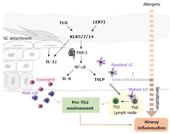
Figure 3 Hypothesised pathways from skin barrier impairment to inflammation and the atopic march.
Deficiencies in FLG and LEKTI promote enhanced activity of KLKs, which induce over-expression of TSLP and IL-6 through the PAR-2-NF-kB pathway. KLK hyperactivity also degrades transition desmosomes, inducing the release of pro-inflammatory cytokines from mechanically stressed keratinocytes. The pro-inflammatory environment triggers eosinophil and mast cell recruitment and activation. TSLP activates LCs which promote the differentiation of naïve (Th0) T cells into Th2 cells in lymph nodes. Allergen ingress (dashed red arrow) promotes allergic sensitization and may, with TSLP, stimulate downstream airway inflammation. FLG: Filaggrin; LEKTI: Lympho-epithelial Kazal-type-related inhibitor; KLKs: Kallikrein proteases; TSLP: Thymic stromal lymphopoietin; IL-6: Interleukin-6; IL-1β: Interleukin-1β; LCs: Langerhans cells; SC: Stratum corneum; PAR-2: Protease-activated receptor-2; NF-kB: Nuclear factor kappa-light-chain-enhancer of activated B cells.
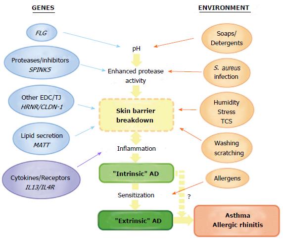
Figure 4 The combination of genetic and environmental factors in the natural history of atopic disease.
Note that some allergens (e.g., house dust mite allergens) can also contribute to total protease activity in the skin. Stress: Psychological stress; TCS: topical corticosteroids; S. aureus: Staphylococcus aureus.
- Citation: Gillespie RM, Brown SJ. From the outside-in: Epidermal targeting as a paradigm for atopic disease therapy. World J Dermatol 2015; 4(1): 16-32
- URL: https://www.wjgnet.com/2218-6190/full/v4/i1/16.htm
- DOI: https://dx.doi.org/10.5314/wjd.v4.i1.16


