Copyright
©The Author(s) 2023.
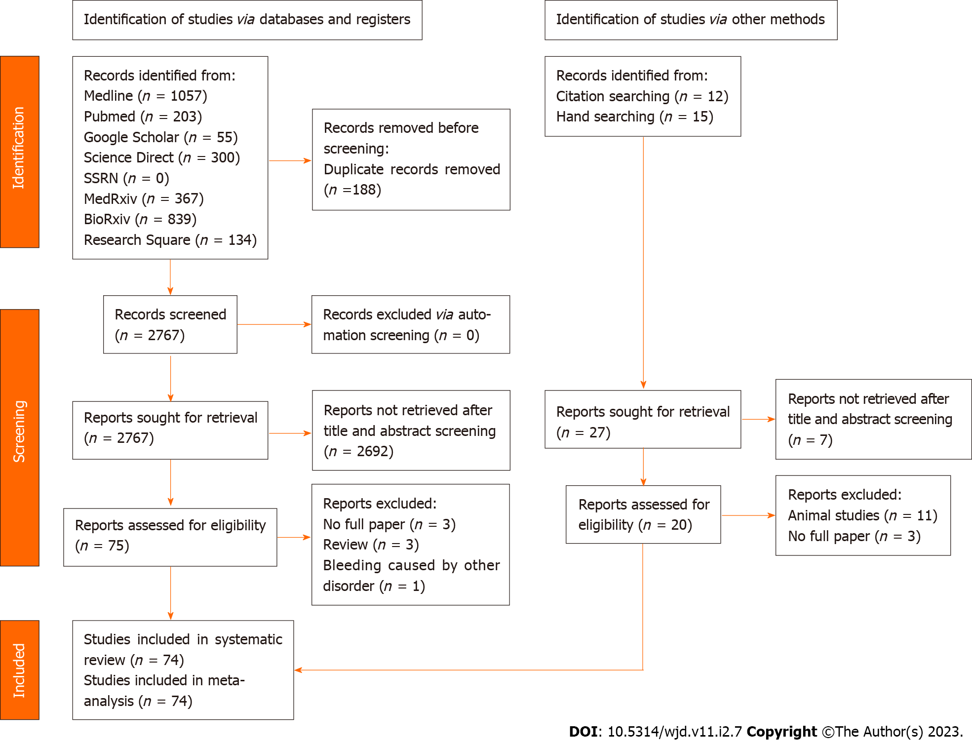
Figure 1 PRISMA flowchart for selection of included studies.
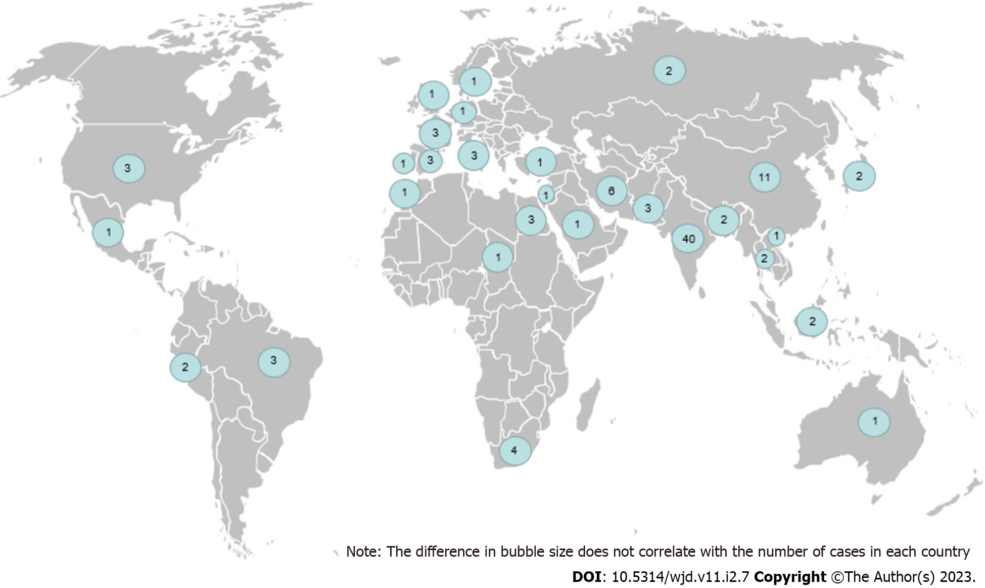
Figure 2 Worldwide distribution of 106 hematidrosis cases from 74 articles.
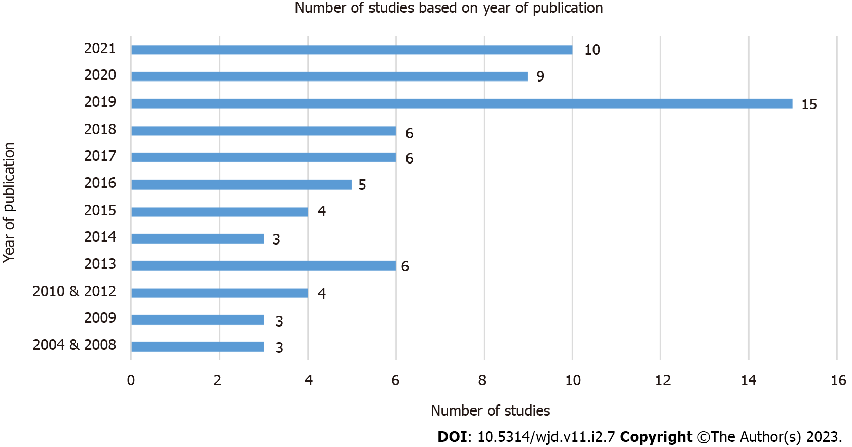
Figure 3 The number of cases is based on the year of publication.
Note that no studies were found to be published between 2005-2007 and 2011. There is one study published in 2004 and two studies in 2010.
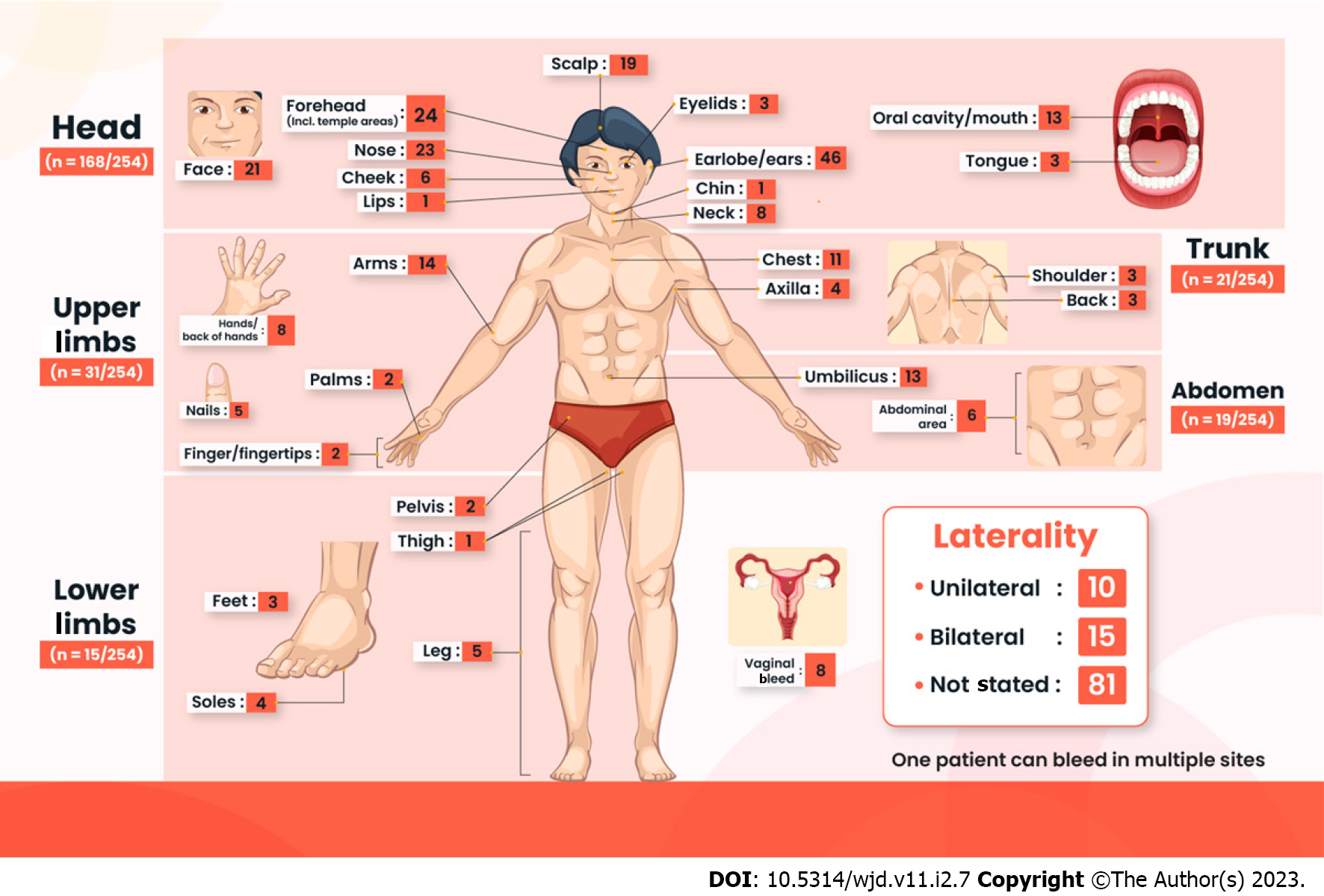
Figure 4 Characteristics of bleeding sites.
The head region is the most commonly affected (n = 168/254), especially around the ears or earlobes (n = 46), forehead (n = 24), and nose (n = 23). The next most common site is in the upper limbs (n = 31/254), with the arms being the most common site of bleeding in this region (n = 14). Although most cases do not state the laterality of bleeding (n = 81), more cases are bilateral (n = 15), as compared to being unilateral bleeding (n = 10).
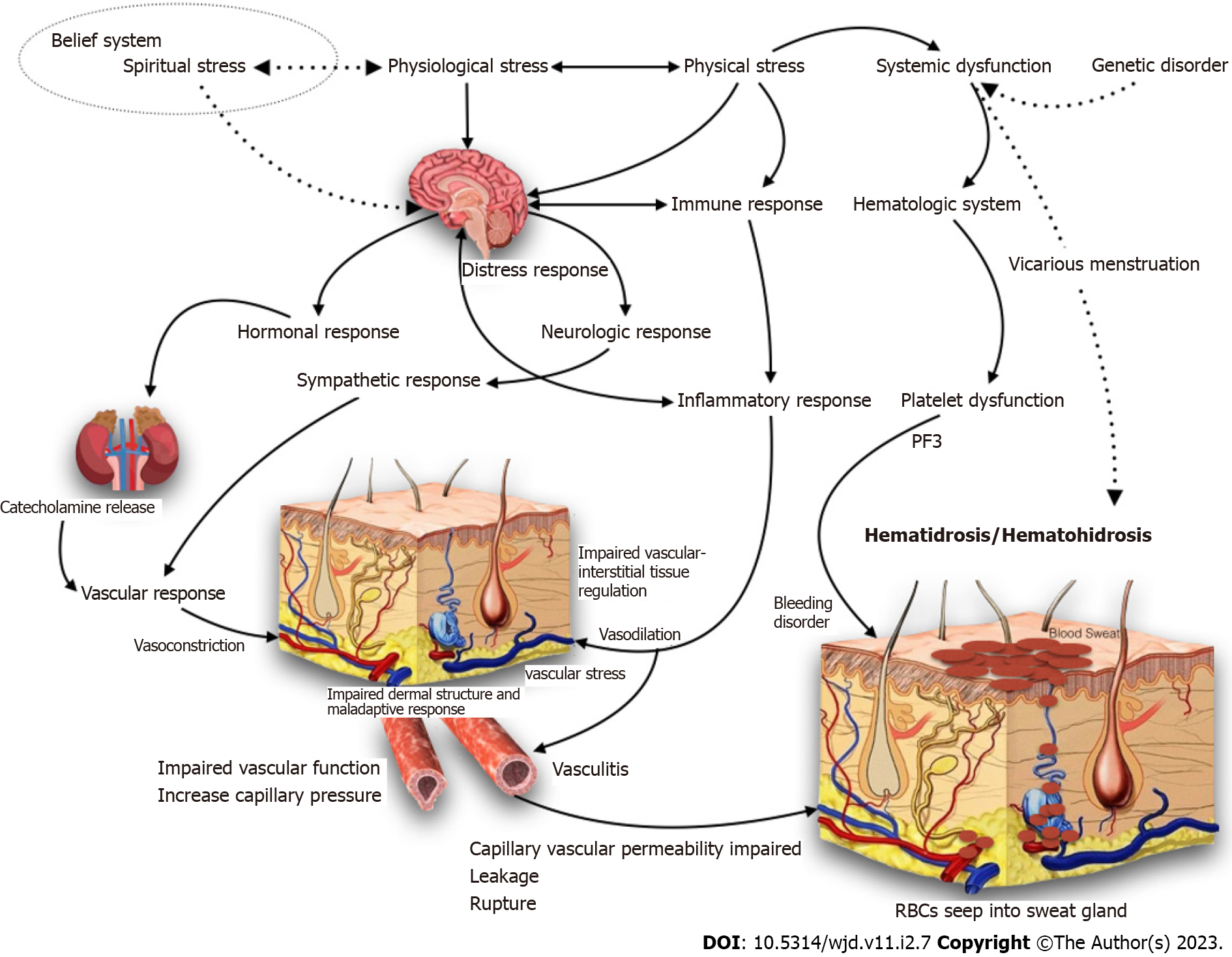
Figure 5 Postulated pathophysiology of hematidrosis.
There are several hypotheses in hematidrosis pathophysiology. The vasculitis in the dermal vessels, exacerbated by sympathetic activation related to extreme stress and anxiety, leads to periglandular vessel constriction, and subsequent expansion, allowing the blood content to seep into the sweat ducts. Another theory states that multiple blood vessels around the sweat glands are arranged in a net-like form. It is believed that under tremendous stress, the vessels contract. Once anxiety passes, the blood vessels dilate to the point of rupture. The blood, at this point, goes into the sweat glands, which push the blood to the surface and manifests as droplets of blood mixed with sweat. RBCs: Red blood cells.
- Citation: Octavius GS, Meliani F, Heriyanto RS, Yanto TA. Systematic review of hematidrosis: Time for clinicians to recognize this entity. World J Dermatol 2023; 11(2): 7-29
- URL: https://www.wjgnet.com/2218-6190/full/v11/i2/7.htm
- DOI: https://dx.doi.org/10.5314/wjd.v11.i2.7


