Copyright
©The Author(s) 2015.
World J Anesthesiol. Jul 27, 2015; 4(2): 30-38
Published online Jul 27, 2015. doi: 10.5313/wja.v4.i2.30
Published online Jul 27, 2015. doi: 10.5313/wja.v4.i2.30
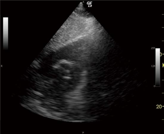
Figure 1 Subcostal right ventricular inflow outflow view confirming absence of the pulmonary artery catheter from the main pulmonary artery and right pulmonary artery prior to pulmonary artery catheter insertion.
Cardiac chamber identification labels have been omitted for clarity.
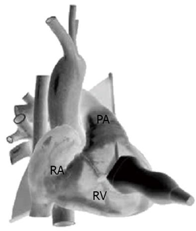
Figure 2 Parasternal right ventricular inflow outflow view, anterior projection.
Schematic diagram demonstrating transthoracic echocardiogram probe position and alignment of scanning plane. Reproduced in part with permission from Toronto General Hospital, Perioperative Interactive Education Virtual TTE (http://pie.med.utoronto.ca/TTE). TTE: Transthoracic echocardiogram; RA: Right atrium; RV: Right ventricle; PA: Pulmonary artery.
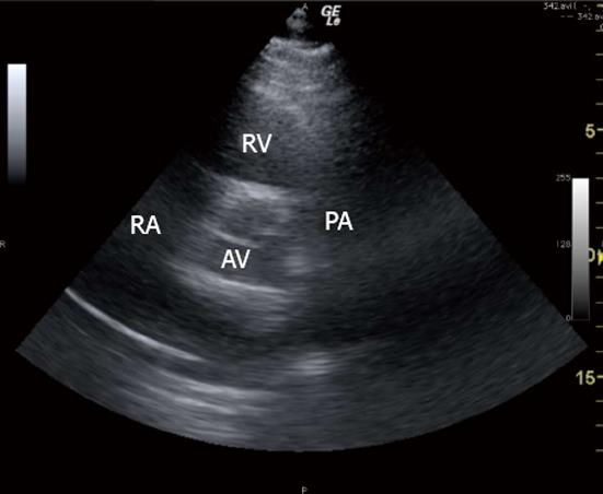
Figure 3 Parasternal right ventricular inflow outflow view, sonogram.
RA: Right atrium; RV: Right ventricle; PA: Pulmonary artery; AV: Aortic valve.
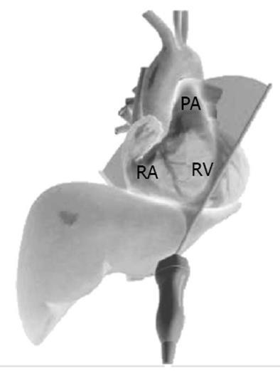
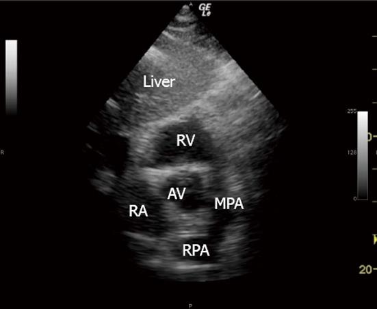
Figure 4 Subcostal right ventricular inflow outflow view, antero-inferior projection.
Schematic diagram demonstrating TTE probe position and alignment of scanning plane. Reproduced in part with permission from Toronto General Hospital, Perioperative Interactive Education Virtual TTE (http://pie.med.utoronto.ca/TTE). TTE: Transthoracic echocardiogram; RA: Right atrium; RV: Right ventricle; PA: Pulmonary artery.
Figure 5 Subcostal right ventricular inflow outflow view, sonogram.
RA: Right atrium; RV: Right ventricle; MPA: Main pulmonary artery; RPA: Right pulmonary artery; AV: Aortic valve.
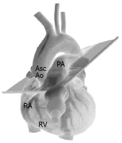
Figure 6 Parasternal ascending aorta short axis view, left anterior oblique projection.
Schematic diagram demonstrating TTE probe position and alignment of scanning plane. Reproduced in part with permission from Toronto General Hospital, Perioperative Interactive Education Virtual TTE (http://pie.med.utoronto.ca/TTE). TTE: Transthoracic echocardiogram; RA: Right atrium; RV: Right ventricle; PA: Pulmonary artery; AscAo: Ascending aorta.
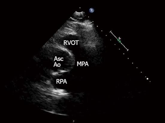
Figure 7 Parasternal ascending aorta short axis view, sonogram.
RVOT: Right ventricular outflow tract; AscAo: Ascending aorta; MPA: Main pulmonary artery; RPA: Right pulmonary.
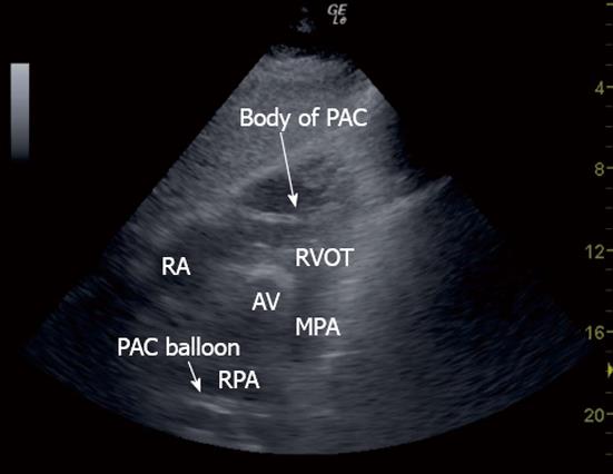
Figure 8 Subcostal right ventricular inflow outflow view showing the body of a pulmonary artery catheter in the right ventricle.
RA: Right ventricle; RVOT: Right ventricular outflow tract; AV: Aortic valve; MPA: Main pulmonary artery; RPA: Right pulmonary artery; PAC: Pulmonary artery catheter.
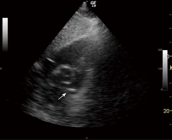
Figure 9 Subcostal right ventricular inflow outflow view confirming presence of the pulmonary artery catheter balloon in the main pulmonary artery after pulmonary artery catheter insertion (arrow).
Cardiac chamber identification labels have been omitted for clarity. Movie clip 1: Subcostal right ventricular inflow outflow view showing appearance of pulmonary artery catheter balloon in the main pulmonary artery as it is appropriately placed.
- Citation: Tan CO, Weinberg L, Story DA, McNicol L. Transthoracic echocardiography assists appropriate pulmonary artery catheter placement: An observational study. World J Anesthesiol 2015; 4(2): 30-38
- URL: https://www.wjgnet.com/2218-6182/full/v4/i2/30.htm
- DOI: https://dx.doi.org/10.5313/wja.v4.i2.30


