Copyright
©2014 Baishideng Publishing Group Co.
World J Anesthesiol. Mar 27, 2014; 3(1): 12-17
Published online Mar 27, 2014. doi: 10.5313/wja.v3.i1.12
Published online Mar 27, 2014. doi: 10.5313/wja.v3.i1.12
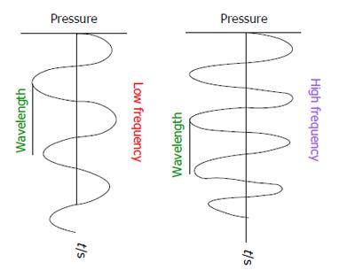
Figure 1 Ultrasound waveform.
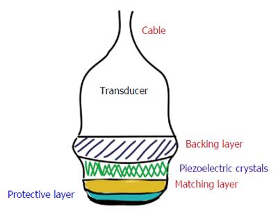
Figure 2 Typical transducer.
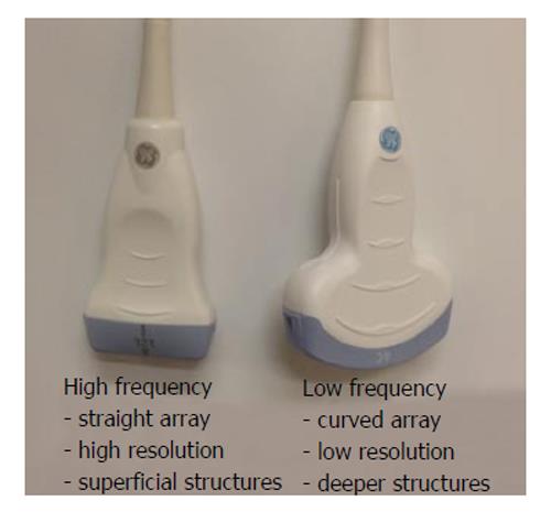
Figure 3 Commonly used transducers.
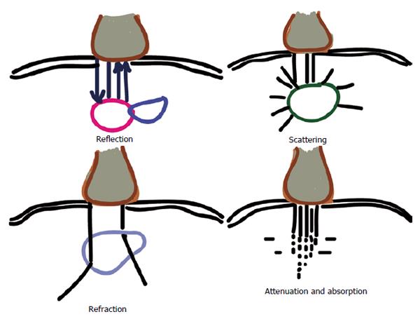
Figure 4 Ultrasound and tissue interactions.
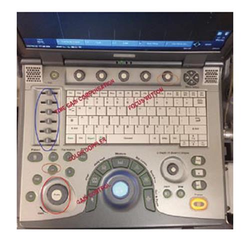
Figure 5 Necessary operating controls to obtain an optimal image.
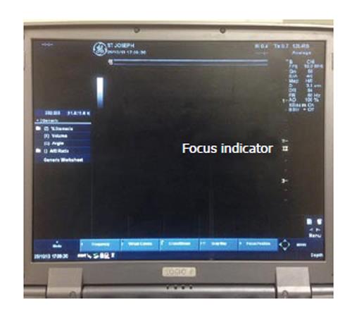
Figure 6 Focus Indicator shown on screen.
- Citation: Shanthanna H. Review of essential understanding of ultrasound physics and equipment operation. World J Anesthesiol 2014; 3(1): 12-17
- URL: https://www.wjgnet.com/2218-6182/full/v3/i1/12.htm
- DOI: https://dx.doi.org/10.5313/wja.v3.i1.12


