Copyright
©The Author(s) 2021.
World J Anesthesiol. Dec 2, 2021; 10(2): 7-15
Published online Dec 2, 2021. doi: 10.5313/wja.v10.i2.7
Published online Dec 2, 2021. doi: 10.5313/wja.v10.i2.7
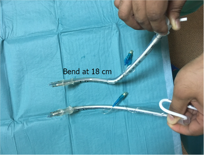
Figure 1 Regular tube (lower) and pre-formed tube (upper) with one bend at 9 cm and 30° from the horizontal plane (natural curve) and a second bend at 18 cm and at 30° right to the vertical plane.
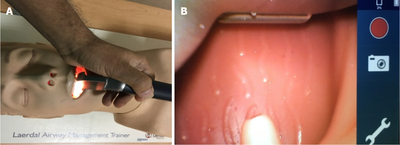
Figure 2 Passing the blade to the oropharynx.
A: Direct eye vision; B: Video monitoring screen.
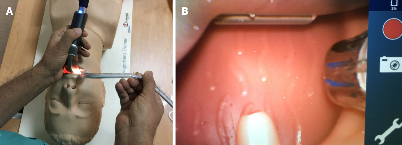
Figure 3 Passing the tube through the right angle of the mouth to pass the tonsillar pillars.
A: Under direct eye vision; B: Then, by looking at the video monitoring screen for further advancement.
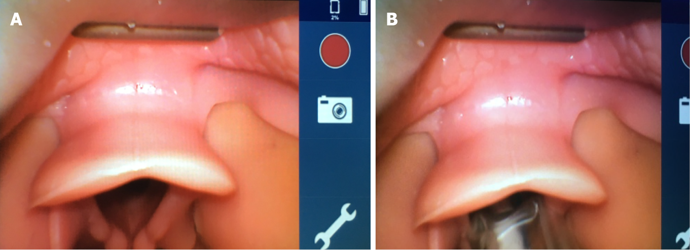
Figure 4 Advancing the blade to see the glottis (A), then advancing the tube with slow counterclockwise rotation to pass through the glottis (B), and finally removing the stylet under video monitoring screen guidance.
- Citation: Shorrab AA, Helal MA. Pre-formed endotracheal tube and stepwise insertion for more successful intubation with video laryngoscopy. World J Anesthesiol 2021; 10(2): 7-15
- URL: https://www.wjgnet.com/2218-6182/full/v10/i2/7.htm
- DOI: https://dx.doi.org/10.5313/wja.v10.i2.7


