Published online Aug 18, 2017. doi: 10.5312/wjo.v8.i8.651
Peer-review started: December 3, 2016
First decision: December 19, 2016
Revised: May 24, 2017
Accepted: June 6, 2017
Article in press: June 7, 2017
Published online: August 18, 2017
Processing time: 255 Days and 13.1 Hours
A 67-year-old female patient developed an esophagocutaneous fistula 4 mo after C4 and C5 partial corpectomy. Plain radiograph and computed tomography (CT) scan of cervical spine showed inferior screws pullout with plate migration that caused the esophageal perforation. Management included removal of anterior hardware, revision C4-5 corpectomy, iliac crest strut autograft and halo orthosis immobilization. The fistula was treated using antibiotics and a 10-french gauge rubber tube for daily irrigation and Penrose drain. At 3 mo, the esophagocutaneous fistula healed and the patient resumed oral feeding. Six months follow-up CT scan showed sound fusion with graft incorporation. At two-year follow-up, patient denied any neck pain or dysphagia. This case report presents a successful outcome of a conservative open wound management without attempted repair. The importance of this case report is to highlight this treatment method that may be considered in such a rare complication particularly if surgical repair failed.
Core tip: Esophageal perforation and subsequent fistulization is a known complication following anterior cervical spine surgery. As part of the treatment of this complication, hardware removal is commonly required. The majority of the literature advises against conservative treatment of esophageal injury due to the associated morbidity and mortality.
- Citation: Elgafy H, Khan M, Azurdia J, Peters N. Open wound management of esophagocutaneous fistula in unstable cervical spine after corpectomy and multilevel laminectomy: A case report and review of the literature. World J Orthop 2017; 8(8): 651-655
- URL: https://www.wjgnet.com/2218-5836/full/v8/i8/651.htm
- DOI: https://dx.doi.org/10.5312/wjo.v8.i8.651
Anterior cervical spine discectomy and corpectomy are reliable with good outcomes for the treatment of neck pain with radiculopathy or myelopathy. The incidence of esophageal perforation in anterior cervical spine surgery is 0.2% to 0.4%. High mortality rates up to 20% have been reported with injury even when the patient is treated within the first 24 h. This increases to 50% when treatment is further delayed. In rare circumstances with delayed diagnosis, esophagocutaneous fistulous tract may form and presents with discharge of food particles from the surgical wound. As with most infections involving orthopedic implants, management involves hardware removal, debridement of soft tissues and culture specific antibiotic[1-4]. The objective of this case report is to present a successful open wound management without attempted repair of a patient with an esophagocutaneous fistula.
A 67-year-old female patient was hospitalized at the authors’ institution for left distal femur fracture that was treated with open reduction and internal fixation. During her postoperative stay, it was noted that food particles were draining from an anterior cervical wound. Patient had a history of two previous cervical spine surgeries, both performed at other institutions. The first was a C4-6 posterior laminectomy without fusion, performed eight years prior to this hospitalization. The second surgery was performed 4 mo prior to her admission to the authors’ institution. It consisted of C4 and C5 partial corpectomy with insertion of a polyetheretherketone (PEEK) cage and C3-6 anterior cervical instrumentation.
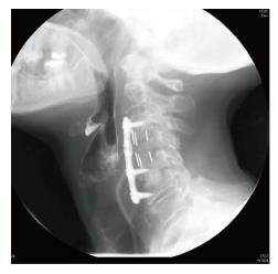
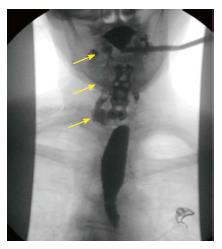
The spine service was consulted and plain radiograph demonstrated inferior screws pullout with plate migration (Figure 1). Computed tomography (CT) scan showed subcutaneous air tracking along the neck soft tissues. General surgery and otolaryngology were consulted and an esophagram (Figure 2) revealed ingested oral contrast tracking along the right subcutaneous tissues of the neck confirming perforation of the esophagus at the level of the inferior screws with fistulization through the anterior surgical wound. Blood work showed normal white cell count 8000 (normal 4500-10000), decreased prealbumin 6.1 mg/dL (normal 17-34) and serum iron level 15 mg/dL (normal 50-212) that confirmed malnutrition.
The patient’s oral intake was suspended and a nasogastric tube placed to facilitate feeding. The patient was taken to the operating room and underwent removal of the anterior hardware, drainage of cervical abscess, revision C4-5 corpectomy, C3-C6 fusion using tricortical iliac crest strut autograft and halo vest immobilization. The wound was left open and managed by the general surgery and otolaryngology services. One week after the revision cervical fusion, the patient was taken to the operating room by general surgery for irrigation and debridement, insertion of a 10 French gauge rubber tube for irrigation and Penrose drain. The wound was irrigated via the rubber tube two times daily with a dilute hydrogen peroxide solution. The patient was placed on ceftriaxone and flagyl for 6 wk as cultures grew polymicrobial mouth flora.
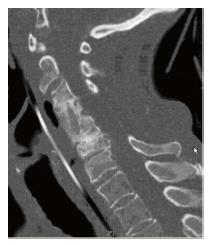
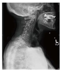
The halo vest removed at 3 mo. The fistulous tract healed at 3 mo and patient resumed oral feeding. Six months follow-up CT scan showed graft incorporation (Figure 3). At two years follow up, patient denied any neck pain or dysphagia and plain radiograph showed maintenance of the cervical spine alignment (Figure 4).
The incidence of esophageal perforation after anterior cervical spine surgery is 0.2% to 0.4% and may present intraoperatively or in the postoperative period[1-5]. Graft dislodgment, prominent hardware or migration can result in chronic pressure on the esophagus, which leads to ischemic tissue breakdown[4,6,7]. It has been reported that 50% of esophageal fistulas occur at C5-6 level instrumentation. At this anatomic landmark, known as Lannier’s triangle, the pharynx transitions to the esophagus and the posterior esophageal mucosa is extremely thin and covered only by fascia[8-11].
Patients with delayed esophageal injury commonly present with surgical wound infection, odynophagia (pain on swallowing) and dysphagia[1,4,12,13]. When esophageal injury is suspected, contrast swallow studies may reveal extravasation of the contrast material and CT scan may demonstrate subcutaneous air. The patient in the current report had loose hardware, prior corpectomy and presented with food particles draining from an anterior cervical wound, which is pathognomonic for esophageal fistula.
Treatment strategies for esophageal perforation and fistula are debated (Table 1). The majority of publications recommended surgical repair of esophageal injury due to the associated morbidity and mortality[1,3,4,7,9,10,14-19]. However, some have reported successful conservative management[20,21]. The key aspects of the treatment strategy include: Anterior hardware removal, posterior fusion for patients in whom primary fusion has not yet occurred, primary closure of the esophageal perforation, and intravenous antibiotics. The patient in the current report was treated with anterior hardware removal and revision interbody fusion with iliac crest tricortical autograft. Patient’s prior multilevel laminectomy rendered the cervical spine unstable after anterior hardware removal. In the setting of esophageal perforation and active infection re-instrumentation of the anterior cervical spine was not possible. Commonly a posterior cervical instrumentation and fusion would be the approach considered. The patient presented in the current study had an increased risk of postoperative posterior cervical spine surgical wound infection related to the existing anterior wound infection and malnutrition. Furthermore, the previous multilevel wide posterior laminectomy would have made the posterior cervical approach challenging with increased risk of dural tear and spinal cord injury. Given those risks associated with a posterior approach in this patient, the authors opted to use a halo vest immobilization postoperatively for cervical stabilization in place of posterior instrumentation. The esophageal perforation and fistulous tract in this patient successfully resolved without attempted repair by two times daily wound irrigation through a rubber tubing and Penrose drain.
| Ref. | No of patients with perforation | Time of diagnosis | Management | Outcome |
| Zhong et al[1] | 6 | Early postoperative | Wound debrided in 3 patients, implant removed and primary suture of perforation in 2 patients | 5 healed 1 died due to pneumonia |
| Ardon et al[3] | 4 | Early postoperative in 3 patients | Hardware removed with primary suture of the perforation in 2 patients and in one of these an additional sternocleidomastoid myoplasty was done | 3 healed 1 patient died due to systemic complication, indirectly related to the perforation |
| Yin et al[4] | 1 | 3 yr after surgery | Emergency tracheostomy, hardware removal, abscess drainage and infected tissue debridement | Healed |
| Jamjoom et al[7] | 1 | Early postoperative | No definite perforation detected at reoperation, pharyngocutaneous fistula formed subsequently No attempted repair Open drainage in association with broad spectrum antibiotics, continuous nasopharyngeal suctioning, stopping of oral intake and gastrostomy feeding | Fistula recurred twice soon after resumption of oral feeding |
| Orlando et al[9] | 5 | 2 during surgery 2 early postoperative 6 mo postoperative in 1 | Hardware removal in 2 Hardware retained in 1 No hardware inserted in 2 Esophagus repaired in 4 | All healed |
| Sun et al[10] | 5 | 1 during surgery 4 early postoperative | Hardware removal in 2 Esophagus repaired in 4 reinforcement with a sternocleidomastoid muscle flap in 1 patient | All healed |
| Balmaseda et al[20] | 1 | Early postoperative | Hardware retained No repair | Healed |
| Ji et al[21] | 1 | Early postoperative | Hardware retained repaired and reinforced with sternocleidomastoid flap Recurrent esophageal leakage 2 d after the repair Wound reopened and a continuous irrigation and drainage system used | Healed |
In conclusion, the current report shows that this complication can be successfully treated with open wound management. This highlights the value of wound management for such a rare complication that could be considered after failed surgical repair of esophageal injury.
A 67-year-old female patient presented with food particles draining from an anterior cervical wound. Patient had a history of two previous cervical spine surgeries; the first was a C4-6 posterior laminectomy without fusion, performed eight years prior current presentation. The second surgery was performed 4 mo prior to her admission to the authors’ institution. It consisted of C4 and C5 partial corpectomy with insertion of a PEEK cage and C3-6 anterior cervical instrumentation.
Esophagus perforation with fistulization through the anterior surgical wound.
Blood work showed normal white cell count 8000 (normal 4500-10000), decreased prealbumin 6.1 mg/dL (normal 17-34) and serum iron level 15 mcg/dL (normal 50-212) that confirmed malnutrition.
Plain radiograph demonstrated inferior screws pullout with plate migration. Computed tomography (CT) scan showed subcutaneous air tracking along the neck soft tissues. Esophagram revealed ingested oral contrast tracking along the right subcutaneous tissues of the neck.
Management included suspended oral intake, a nasogastric tube feeding, removal of anterior hardware, revision C4-5 corpectomy, iliac crest strut autograft and halo orthosis immobilization. The wound left opened and a 10-french gauge rubber tube was placed for daily irrigation. The patient was placed on ceftriaxone and flagyl for 6 wk.
The majority of publications recommended surgical repair of esophageal injury due to the associated morbidity and mortality. However, some have reported successful conservative management.
Fifty percent of esophageal fistulas occur at C5-6 level instrumentation. At this anatomic landmark, known as Lannier’s triangle, the pharynx transitions to the esophagus and the posterior esophageal mucosa is extremely thin and covered only by fascia.
When esophageal injury is suspected, contrast swallow studies may reveal extravasation of the contrast material and CT scan may demonstrate subcutaneous air. The current report shows that this complication can be successfully treated with open wound management.
Text well written and easily comprehensible with clear figures.
Manuscript source: Invited manuscript
Specialty type: Orthopedics
Country of origin: United States
Peer-review report classification
Grade A (Excellent): A
Grade B (Very good): 0
Grade C (Good): C, C
Grade D (Fair): 0
Grade E (Poor): 0
P- Reviewer: Lakhdar F, Leonardi M, Teli MGA S- Editor: Kong JX L- Editor: A E- Editor: Lu YJ
| 1. | Zhong ZM, Jiang JM, Qu DB, Wang J, Li XP, Lu KW, Xu B, Chen JT. Esophageal perforation related to anterior cervical spinal surgery. J Clin Neurosci. 2013;20:1402-1405. [RCA] [PubMed] [DOI] [Full Text] [Cited by in Crossref: 31] [Cited by in RCA: 36] [Article Influence: 3.0] [Reference Citation Analysis (0)] |
| 2. | Memtsoudis SG, Hughes A, Ma Y, Chiu YL, Sama AA, Girardi FP. Increased in-hospital complications after primary posterior versus primary anterior cervical fusion. Clin Orthop Relat Res. 2011;469:649-657. [RCA] [PubMed] [DOI] [Full Text] [Cited by in Crossref: 71] [Cited by in RCA: 82] [Article Influence: 5.9] [Reference Citation Analysis (0)] |
| 3. | Ardon H, Van Calenbergh F, Van Raemdonck D, Nafteux P, Depreitere B, van Loon J, Goffin J. Oesophageal perforation after anterior cervical surgery: management in four patients. Acta Neurochir (Wien). 2009;151:297-302; discussion 302. [RCA] [PubMed] [DOI] [Full Text] [Cited by in Crossref: 32] [Cited by in RCA: 34] [Article Influence: 2.1] [Reference Citation Analysis (0)] |
| 4. | Yin DH, Yang XM, Huang Q, Yang M, Tang QL, Wang SH, Wang S, Liu JJ, Yang T, Li SS. Pharyngoesophageal perforation 3 years after anterior cervical spine surgery: a rare case report and literature review. Eur Arch Otorhinolaryngol. 2015;272:2077-2082. [RCA] [PubMed] [DOI] [Full Text] [Cited by in Crossref: 10] [Cited by in RCA: 9] [Article Influence: 0.9] [Reference Citation Analysis (0)] |
| 5. | Fuji T, Kuratsu S, Shirasaki N, Harada T, Tatsumi Y, Satani M, Kubo M, Hamada H. Esophagocutaneous fistula after anterior cervical spine surgery and successful treatment using a sternocleidomastoid muscle flap. A case report. Clin Orthop Relat Res. 1991;8-13. [PubMed] |
| 6. | Kim SJ, Ju CI, Kim DM, Kim SW. Delayed esophageal perforation after cervical spine plating. Korean J Spine. 2013;10:174-176. [RCA] [PubMed] [DOI] [Full Text] [Full Text (PDF)] [Cited by in Crossref: 13] [Cited by in RCA: 15] [Article Influence: 1.3] [Reference Citation Analysis (0)] |
| 7. | Jamjoom ZA. Pharyngo-cutaneous fistula following anterior cervical fusion. Br J Neurosurg. 1997;11:69-74. [RCA] [PubMed] [DOI] [Full Text] [Cited by in Crossref: 21] [Cited by in RCA: 24] [Article Influence: 0.9] [Reference Citation Analysis (0)] |
| 8. | Patel NP, Wolcott WP, Johnson JP, Cambron H, Lewin M, McBride D, Batzdorf U. Esophageal injury associated with anterior cervical spine surgery. Surg Neurol. 2008;69:20-24; discission 24. [RCA] [PubMed] [DOI] [Full Text] [Cited by in Crossref: 58] [Cited by in RCA: 55] [Article Influence: 3.1] [Reference Citation Analysis (0)] |
| 9. | Orlando ER, Caroli E, Ferrante L. Management of the cervical esophagus and hypofarinx perforations complicating anterior cervical spine surgery. Spine (Phila Pa 1976). 2003;28:E290-E295. [RCA] [PubMed] [DOI] [Full Text] [Cited by in Crossref: 59] [Cited by in RCA: 71] [Article Influence: 3.2] [Reference Citation Analysis (0)] |
| 10. | Sun L, Song YM, Liu LM, Gong Q, Liu H, Li T, Kong QQ, Zeng JC. Causes, treatment and prevention of esophageal fistulas in anterior cervical spine surgery. Orthop Surg. 2012;4:241-246. [RCA] [PubMed] [DOI] [Full Text] [Cited by in Crossref: 13] [Cited by in RCA: 18] [Article Influence: 1.5] [Reference Citation Analysis (0)] |
| 11. | Newhouse KE, Lindsey RW, Clark CR, Lieponis J, Murphy MJ. Esophageal perforation following anterior cervical spine surgery. Spine (Phila Pa 1976). 1989;14:1051-1053. [RCA] [PubMed] [DOI] [Full Text] [Cited by in Crossref: 129] [Cited by in RCA: 114] [Article Influence: 3.2] [Reference Citation Analysis (0)] |
| 12. | Kelly MF, Spiegel J, Rizzo KA, Zwillenberg D. Delayed pharyngoesophageal perforation: a complication of anterior spine surgery. Ann Otol Rhinol Laryngol. 1991;100:201-205. [RCA] [PubMed] [DOI] [Full Text] [Cited by in Crossref: 68] [Cited by in RCA: 63] [Article Influence: 1.9] [Reference Citation Analysis (0)] |
| 13. | Zdichavsky M, Blauth M, Bosch U, Rosenthal H, Knop C, Bastian L. Late esophageal perforation complicating anterior cervical plate fixation in ankylosing spondylitis: a case report and review of the literature. Arch Orthop Trauma Surg. 2004;124:349-353. [RCA] [PubMed] [DOI] [Full Text] [Cited by in Crossref: 27] [Cited by in RCA: 25] [Article Influence: 1.2] [Reference Citation Analysis (0)] |
| 14. | Gaudinez RF, English GM, Gebhard JS, Brugman JL, Donaldson DH, Brown CW. Esophageal perforations after anterior cervical surgery. J Spinal Disord. 2000;13:77-84. [RCA] [PubMed] [DOI] [Full Text] [Cited by in Crossref: 152] [Cited by in RCA: 148] [Article Influence: 5.9] [Reference Citation Analysis (0)] |
| 15. | von Rahden BH, Stein HJ, Scherer MA. Late hypopharyngo-esophageal perforation after cervical spine surgery: proposal of a therapeutic strategy. Eur Spine J. 2005;14:880-886. [RCA] [PubMed] [DOI] [Full Text] [Cited by in Crossref: 30] [Cited by in RCA: 33] [Article Influence: 1.7] [Reference Citation Analysis (0)] |
| 16. | Sansur CA, Early S, Reibel J, Arlet V. Pharyngocutaneous fistula after anterior cervical spine surgery. Eur Spine J. 2009;18:586-591. [RCA] [PubMed] [DOI] [Full Text] [Cited by in Crossref: 9] [Cited by in RCA: 13] [Article Influence: 0.8] [Reference Citation Analysis (0)] |
| 17. | Dakwar E, Uribe JS, Padhya TA, Vale FL. Management of delayed esophageal perforations after anterior cervical spinal surgery. J Neurosurg Spine. 2009;11:320-325. [RCA] [PubMed] [DOI] [Full Text] [Cited by in Crossref: 34] [Cited by in RCA: 39] [Article Influence: 2.4] [Reference Citation Analysis (0)] |
| 18. | Hanci M, Toprak M, Sarioğlu AC, Kaynar MY, Uzan M, Işlak C. Oesophageal perforation subsequent to anterior cervical spine screw/plate fixation. Paraplegia. 1995;33:606-609. [RCA] [PubMed] [DOI] [Full Text] [Cited by in Crossref: 33] [Cited by in RCA: 32] [Article Influence: 1.1] [Reference Citation Analysis (0)] |
| 19. | Iyoob VA. Postoperative pharyngocutaneous fistula: treated by sternocleidomastoid flap repair and cricopharyngeus myotomy. Eur Spine J. 2013;22:107-112. [RCA] [PubMed] [DOI] [Full Text] [Cited by in Crossref: 7] [Cited by in RCA: 10] [Article Influence: 0.8] [Reference Citation Analysis (0)] |
| 20. | Balmaseda MT Jr, Pellioni DJ. Esophagocutaneous fistula in spinal cord injury: a complication of anterior cervical fusion. Arch Phys Med Rehabil. 1985;66:783-784. [PubMed] |
| 21. | Ji H, Liu D, You W, Zhou F, Liu Z. Success in esophageal perforation repair with open-wound management after revision cervical spine surgery: a case report. Spine (Phila Pa 1976). 2015;40:E183-E185. [RCA] [PubMed] [DOI] [Full Text] [Cited by in Crossref: 8] [Cited by in RCA: 11] [Article Influence: 1.1] [Reference Citation Analysis (0)] |


