Published online Dec 18, 2017. doi: 10.5312/wjo.v8.i12.913
Peer-review started: November 23, 2016
First decision: February 17, 2017
Revised: February 23, 2017
Accepted: October 29, 2017
Article in press: October 29, 2017
Published online: December 18, 2017
Processing time: 390 Days and 22.7 Hours
To quantitatively assess rotatory and anterior-posterior instability in vivo after anterior cruciate ligament (ACL) reconstruction using bone-patellar tendon-bone (BTB) autografts, and to clarify the influence of tunnel positions on the knee stability.
Single-bundle ACL reconstruction with BTB autograft was performed on 50 patients with a mean age of 28 years using the trans-tibial (TT) (n = 20) and trans-portal (TP) (n = 30) techniques. Femoral and tibial tunnel positions were identified from the high-resolution 3D-CT bone models two weeks after surgery. Anterolateral rotatory translation was examined using a Slocum anterolateral rotatory instability test in open magnetic resonance imaging (MRI) 1.0-1.5 years after surgery, by measuring anterior tibial translation at the medial and lateral compartments on its sagittal images. Anterior-posterior stability was evaluated with a Kneelax3 arthrometer.
A total of 40 patients (80%) were finally followed up. Femoral tunnel positions were shallower (P < 0.01) and higher (P < 0.001), and tibial tunnel positions were more posterior (P < 0.05) in the TT group compared with the TP group. Anterolateral rotatory translations in reconstructed knees were significantly correlated with the shallow femoral tunnel positions (R = 0.42, P < 0.01), and the rotatory translations were greater in the TT group (3.2 ± 1.6 mm) than in the TP group (2.0 ± 1.8 mm) (P < 0.05). Side-to-side differences of Kneelax3 arthrometer were 1.5 ± 1.3 mm in the TT, and 1.7 ± 1.6 mm in the TP group (N.S.). Lysholm scores, KOOS subscales and re-injury rate showed no difference between the two groups.
Anterolateral rotatory instability significantly correlated shallow femoral tunnel positions after ACL reconstruction using BTB autografts. Clinical outcomes, rotatory and anterior-posterior stability were overall satisfactory in both techniques, but the TT technique located femoral tunnels in shallower and higher positions, and tibial tunnels in more posterior positions than the TP technique, thus increased the anterolateral rotation. Anatomic ACL reconstruction with BTB autografts may restore knee function and stability.
Core tip: Anterolateral rotatory instability was quantitatively assessed in 40 anterior cruciate ligament-reconstructed knees with bone-patellar tendon-bone autografts using a Slocum anterolateral rotatory instability test in open magnetic resonance imaging 1-1.5 years after surgery, and correlated to tunnel positions evaluated by high resolution computed tomography scan 2 wk after surgery. Femoral tunnel positions were shallower (P < 0.01) and higher (P < 0.001), and tibial tunnel positions were more posterior (P < 0.05) in the trans-tibial (TT) group, compared with the trans-portal (TP) group. Anterolateral rotatory translations were significantly correlated with the shallow femoral tunnel positions, and they were greater in the TT group (3.2 ± 1.6 mm) than in the TP group (2.0 ± 1.8 mm) (P < 0.05).
- Citation: Tashiro Y, Okazaki K, Murakami K, Matsubara H, Osaki K, Iwamoto Y, Nakashima Y. Anterolateral rotatory instability in vivo correlates tunnel position after anterior cruciate ligament reconstruction using bone-patellar tendon-bone graft. World J Orthop 2017; 8(12): 913-921
- URL: https://www.wjgnet.com/2218-5836/full/v8/i12/913.htm
- DOI: https://dx.doi.org/10.5312/wjo.v8.i12.913
It is the goal of anterior cruciate ligament (ACL) reconstruction to restore normal knee function and kinematics, finally achieving patient’s return to sports and daily activities. Recently, anatomic ACL reconstruction which reproduces dimensions, fiber orientations and insertion sites of the native ACL has been reported to improve knee stability and clinical outcomes after surgery[1-4]. Oblique fiber orientation based on anatomical location of bone tunnels is more favorable for controlling rotation, as well as resisting anterior tibial force, compared with a vertical graft orientation[5,6]. ACL reconstruction creating femoral tunnels independently from tibial tunnels has been shown to locate femoral tunnels more closely to anatomical footprint than the trans-tibial (TT) technique[7-9]. A double-bundle technique has been one of the popular methods to perform anatomic ACL reconstruction, principally using soft tissue grafts such as hamstring tendon[10-13]. However, anatomic single-bundle technique has developed recently, showing comparable outcomes as double-bundle techniques[14-17]. Therefore, it may be possible that single-bundle ACL reconstruction with bone-patellar tendon-bone (BTB) grafts, which is based on the modern concept of ACL anatomy[18-21], could restore close to normal ACL function.
One of the great advantages of BTB autograft is its better graft-tunnel healing, as well as the stable initial fixation with bone block, compared with other soft tissue grafts[22-25]. Although several original studies have reported kinematics after ACL reconstruction with BTB grafts, they were based on cadaveric specimens measured by testing machine or robotic system[5,6,26-28], which could not reflect better graft-tunnel healing of BTB grafts. Recent in vivo studies using BTB grafts have introduced the anatomic single-bundle technique, which locates bone tunnels within the native insertion site, and have shown favorable clinical results after for ACL reconstruction, but the degree of rotatory instability was mainly assessed by manual pivot-shift test[18,29-31], not quantitatively. Only a few studies from limited research groups so far have reported quantitative results of rotatory instability after anatomic ACL reconstruction using BTB grafts[32-34]. Therefore, it would be clinically relevant to assess in vivo rotatory instability objectively after ACL reconstruction using BTB autografts.
For the surgical technique of creating femoral tunnels, we had used the TT technique until 2010, modifying the position and orientation of the graft more obliquely[12,35,36]. But this technique sometimes made it difficult for us to place femoral tunnels within the anatomical footprint[9,37-40], thus since the late 2010, we’ve shifted to the trans-portal (TP) technique, which enables femoral tunnel placement independently from tibial tunnels[8,41,42]. In addition, we have utilized open MRI to assess anterolateral rotatory instability of ACL-deficient and ACL-reconstructed knees since 2005, and have shown its usefulness in quantification[35,43-45].
The purpose of this study was to: (1) Compare the knee stability in vivo after ACL reconstruction using BTB autografts via TT and TP techniques; and (2) clarify the influence of tunnel position on the knee stability. We hypothesized that: (1) The TP technique would show less instability; and (2) tunnel positions may affect knee stability after single-bundle ACL reconstruction using BTB autografts.
From April 2009 to March 2013, single-bundle primary ACL reconstruction was performed on 52 knees with a BTB autograft. Patients with any history of significant injury to other knee ligaments, articular cartilage and bilateral ACL cases (2 knees) were excluded. Consequently, 50 patients with a mean age of 28 years (range: 17-45) were enrolled. All patients were male. TT technique was used in 20 knees from April 2009 to 2010, and TP technique was used in 30 patients from August 2010 to March 2013 (Table 1). A computed tomography (CT) scan was performed with 1-2 mm slices in order to determine tunnel positions 2 wk after surgery. Anterolateral rotatory instability in vivo was assessed quantitatively in 40 patients (80%) using open MRI an average of 1.2 years (range: 1.0-1.5 years) after surgery. All aspects of this study was approved by the institutional review board (IRB) of our university (ID: 24-108), and all subjects gave their informed consent before they were included.
| TT group | TP group | Significance | |
| n | 20 | 30 | |
| Period of surgery | Apr 2009- Dec 2010 | Aug 2010-Mar 2013 | |
| Age | 29 ± 9 | 27 ± 9 | NS |
| Height (cm) | 171.3 ± 7.1 | 171.7 ± 6.0 | NS |
| Weight (kg) | 73.8 ± 6.9 | 75.5 ± 12.2 | NS |
| Lysholm score | 65 ± 11 | 63 ± 14 | NS |
The subjects underwent arthroscopic ACL reconstruction at a median of 6 wk after the injury. An arthroscopic leg holder was utilized to hold the affected knee in 90º of flexion. A 10-mm BTB autograft was harvested. The anterolateral portal was positioned as high as the inferior pole of the patella so that it gave an excellent arthroscopic view over the tibial footprint of the ACL. The tibial tunnel was targeted in the center of the native ACL insertion site, avoiding impingement during knee extension.
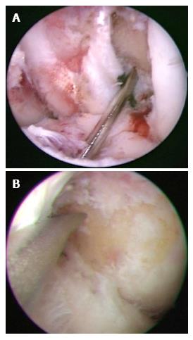
In the TT group, a femoral guide wire was inserted via the tibial tunnel, and then it was centered at the 1:30-2:00 o’clock position for the left knees (10:00-10:30 for right) (Figure 1A). The femoral tunnel was drilled trans-tibially with the knee in 90° of flexion. In the TP group, the anteromedial portal was used to allow optimal visualization of the lateral wall of the intercondylar notch, including the ACL femoral insertion site[13,41]. In addition, the accessory medial portal was established far medially, just above the anterior horn of the medial meniscus, in a position allowing direct access to the center of the ACL femoral insertion site and avoiding damage to articular cartilage during femoral drilling (Figure 1B). A guide wire was introduced through the accessory medial portal and placed at the center of femoral insertion site. The femoral tunnel was drilled using a 2.4-mm straight guide pin and rigid drills, with the knee kept in maximal flexion.
In all cases, the BTB graft was fixed to the femur using extracortical fixation (EndoButton CL BTB, Smith and Nephew Endoscopy). Tibial side was fixed with interference screws (Softsilk 1.5 Fixation Screws, Smith and Nephew Endoscopy). A notch plasty was not performed in any of our patients. All of the patients underwent a standard rehabilitation program with early weight bearing and range of motion exercise. Sports activities were permitted 9 mo after the reconstruction, if the patients had regained functional strength and stability.
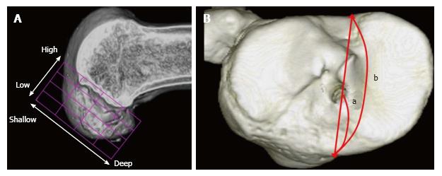
The locations of the femoral and tibial tunnel aperture centers were identified from 3D bone models generated from the high-resolution CT scan two weeks after surgery. Femoral tunnel positions were measured according to the quadrant method (Figure 2A)[46]. For the tibial side, the technique of Staubli and Rauschning was used for the measurement (Figure 2B)[47]. A commercially available medical imaging software (Real INTAGE, Cybernet Systems Co, Ltd, Tokyo, Japan) was used in these analysis.
The assessment of in vivo anterolateral rotatory instability (ALRI) was performed by applying the Slocum ALRI test[48] to stress the tibia rotating anteriorly and internally in a horizontal open MRI Scanner, as previously described[35,43-45]. The MRI system used in this study was an open MRI at 0.4 T (APERTO, Hitachi Medical Co, Tokyo, Japan). Briefly, the patient was kept in a semilateral recumbent position on the table. The hip and knee of the contra lateral side were flexed. The affected knee was placed in 10° of flexion and the medial side of the foot was rested on a pad so that the weight of the leg was borne on the heel and the knee sagged into valgus. The examiner placed his one hand on the distal femur and the other hand on the proximal tibia from the posterior side. He pushed the fibular head anteriorly with his thumb to increase the stress that makes the tibia rotate anteriorly and internally.
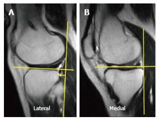
The anterior translation of the tibia with respect to the femoral condyle was measured on sagittal images scanned at each center of the medial and lateral compartments, respectively, in order to evaluate rotatory instability (Figure 3). The image plane scanned under stress was adjusted to the same sagittal plane scanned before stress, using the Interactive Scan Control (ISC) software program. The ISC program determines the image plane interactively on the basis of fluoroscopic images displayed on a user interface with an update time of 2 s, including the scan time. The MRI operator can change the image plane, oblique angle and phase encoding direction during the scan. It usually takes less than 3 min from applying stress to completing the scan, including the fine-tuning of the plane, when the ISC is used. The anterolateral rotatory translation, determined from anterolateral minus anteromedial tibial translation, was calculated to assess ALRI. Side-to-side differences of anterolateral tibial translation and anteromedial tibial translation were also analyzed, respectively. High intra- and inter-observer reproducibility (correlation coefficient = 0.98, 0.91, respectively) have been demonstrated between 2 successive examinations in our previous study, using this assessment technique[43].
The subjective knee function was assessed with the Lysholm scores and Knee injury and Osteoarthritis Outcome Score (KOOS) scales[49,50]. Anterior-posterior stability was evaluated with a Kneelax3 arthrometer (MR Systems, Haarlem, The Netherlands) at 134 N anterior force.
Femoral and tibial tunnel positions were compared between TT and TP groups using Student’s t-test. The side-to-side differences of tibial translations, anterolateral rotatory translation and clinical outcomes were also compared between the 2 groups using Student’s t-test. The relationships between tunnel positions and knee stability parameters were analyzed using Pearson’s correlations. For those statistical analyses, the StatView 5.0 software (SAS Institute Inc., Cary, NC, United States) was used with a significance level of P < 0.05. All statistical analyses of this study were reviewed by a biomedical statistician.
Femoral tunnels were located significantly shallower (P < 0.01) and higher (P < 0.001) in the TT group, compared with the TP group. Tibial tunnel positions in the TT group were significantly posterior than those of the TP group (P < 0.05) (Table 2).
| TT technique (%) | TP technique (%) | Significance | ||
| Femur | Depth | 34.0 ± 4.9 | 29.7 ± 4.9 | P < 0.01 |
| Height | 30.3 ± 5.6 | 39.3 ± 7.3 | P < 0.001 | |
| Tibia | Anterior-posterior | 47.1 ± 7.5 | 42.0 ± 4.9 | P < 0.05 |
In open MRI analysis, the anterolateral rotatory translation (= anterolateral minus anteromedial tibial translation) of the affected knees were 3.2 ± 1.6 mm in the TT group and 2.0 ± 1.8 mm in the TP group, and significantly larger in the TT group (P < 0.05). The side-to-side differences of anterolateral tibial translation were 1.4 ± 1.6 mm in the TT group and 0.9 ± 1.9 mm in the TP group (N.S.). There was no significant difference in the side-to-side difference of Kneelax3 arthrometer, Lysholm scores, KOOS and re-injury rate between the two groups (Table 3).
| TT technique | TP technique | Significance | |
| Lysholm score | 94 ± 7 | 95 ± 7 | NS |
| KOOS subscale | |||
| Symptoms | 89 ± 9 | 90 ± 12 | NS |
| Pain | 87 ± 7 | 89 ± 8 | NS |
| ADL | 92 ± 12 | 96 ± 10 | NS |
| Sport/Rec | 82 ± 14 | 84 ± 9 | NS |
| QoL | 78 ± 13 | 80 ± 11 | NS |
| Re-injury (ipsilateral) | 0 | 0 | NS |
| Kneelax3 | NS | ||
| Side-to-side diff. (mm) | 1.5 ± 1.3 | 1.7 ± 1.6 | |
| MRI analysis | |||
| Anterolateral rotatory translation | |||
| Affected side (mm) | 3.2 ± 1.6 | 2.0 ± 1.8 | P < 0.05 |
| Contra-lateral side (mm) | 2.4 ± 1.6 | 2.5 ± 2.7 | NS |
| Side-to-side diff. (mm) of | |||
| Anteromedial tibial translation | 0.6 ± 0.8 | 1.4 ± 2.3 | NS |
| Anterolateral tibial translation | 1.4 ± 1.6 | 0.9 ± 1.9 | NS |
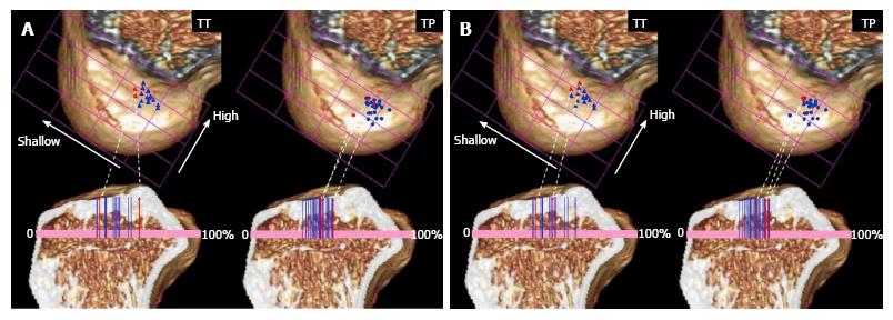
The anterolateral rotatory translation were significantly correlated with the shallow (distal and anterior in anatomy) femoral tunnel position (R = 0.42, P < 0.01), while the correlation between the side-to-side differences of Kneelax3 arthrometer and shallow femoral tunnel positions was weak and not statistically significant (R = 0.27, P = 0.14) (Table 4). Femoral and tibial tunnel positions are plotted in both groups, according to the quadrant method and Staubli’s technique, together with the relationship with stability results of MRI and Kneelax3 arthrometer (Figure 4).
| Femur | Tibia | |||
| Shallow (+)-Deep (-) | Low (+)-High (-) | Posterior (+)-Anterior (-) | ||
| Kneelax3 | Corr (R) | 0.27 | -0.02 | 0.15 |
| side-to-side differences | Significance | NS (P = 0.14) | NS | NS |
| MRI analysis | ||||
| Anterolateral | Corr (R) | 0.42 | -0.13 | 0.12 |
| rotatory translation | Significance | P < 0.01 | NS | NS |
We aimed to clarify in vivo rotatory knee stability as well as the anterior-posterior stability after ACL reconstruction using BTB autografts, and correlate knee stability to tunnel positions. The most important findings of this study were that the anterolateral rotatory translations (= anterolateral minus anteromedial tibial translation) were significantly correlated with the shallow (distal and anterior in anatomy) femoral tunnel positions. A previous in vivo study has also reported that ACL reconstruction using BTB autografts with non-anatomic tunnel position resulted in significantly increased positive pivot-shift test cases, compared with those with anatomic tunnel positions at 1-year follow-up[30]. Another robotic study using cadaveric knees has reported that anatomic ACL reconstruction with rectangular BTB grafts restored knee kinematics better than the one with oval femoral tunnels located in shallower and higher positions[6], and these were consistent with our study.
Comparison between TT and TP groups showed shallower and higher femoral tunnel positions, more posterior tibial tunnel positions and increased anterolateral rotatory translation in the TT group. Previous studies have reported that it is more difficult for TT technique to locate femoral tunnels anatomically and restore normal kinematics, compared with TP technique[7-9,37,41,42], whereas no significant difference was found in side-to-side differences of Kneelax3 measurement, anterolateral and anteromedial tibial translation in MRI, or other clinical outcomes. The reasons why these stability parameters and clinical outcomes showed no difference between the two techniques may be that the TT-techniques we used did not locate femoral tunnels in “high-noon” isometric position, but located them in oblique positions which are mostly within the femoral footprint, as shown in Figure 4, thus the two groups resulted in less than 2 mm of mean side-to-side difference of anterolateral tibial translation and Kneelax3 measurement with small differences. A recent study using modified TT technique has reported similar anatomic femoral tunnel positions and good clinical results which are comparable to TP technique[51], although TT technique still runs a risk of creating posterior tibial tunnels and resulting vertical graft orientation[52,53]. A vertical graft orientation, created by shallow femoral tunnels and posterior tibial tunnels, may result in residual rotatory knee instability[40,54].
It is well known that merits of using a BTB autograft are its stable initial fixation and good bone-graft healing[23-25]. BTB cases in our cohort also showed sufficient stability within 2 mm of mean side-to-side difference of anterior tibial translation in rotatory and anterior-posterior evaluation and excellent clinical outcomes. To our knowledge, only a few studies so far have reported quantitative assessment of rotatory instability in vivo after anatomic ACL reconstruction using BTB autografts[32-34]. Most of the previous studies about BTB grafts were in vitro kinematic study using cadaveric specimens[5,6,26-28], or in vivo study evaluated by manual testing of pivot-shift[18,29-31]. We added the quantitatively assessed evidence of rotatory instability after anatomic ACL reconstruction using BTB autografts to the current knowledge. Our results suggest that anatomical placement of BTB autografts would restore knee stability and function after ACL reconstruction.
One of the limitations of this study was that all the subjects included were male patients, thus it might have affected the results[55]. However, recent large cohort studies have reported gender is not a risk factor for knee instability or revision after ACL reconstruction[56-58]. Secondly, our sample size was relatively small. It was because we usually used hamstring grafts for female patients and for those who had habits of frequent kneeling. The size might not be enough to detect small differences of anterolateral tibial translation between the two techniques.
Anterolateral rotatory instability in vivo significantly correlated shallow (distal and anterior in anatomy) femoral tunnel positions after ACL reconstruction using BTB autografts. TT technique located femoral tunnels in shallower and higher positions, and tibial tunnels in more posterior positions than the TP technique, thus increased the anterolateral rotation in reconstructed knees. Clinical outcomes and knee stability in both techniques were overall satisfactory with less than 2 mm of side-to-side differences in rotatory and anterior-posterior instability. As for clinical relevance, anatomic reconstruction of the ACL using BTB autografts may restore knee function and stability.
Anatomic single-bundle anterior cruciate ligament (ACL) reconstruction using bone-patellar tendon-bone (BTB) autograft may restore close to normal ACL function. However, quantitative studies showing in vivo rotatory instability after anatomic ACL reconstruction using BTB graft are sparse.
In vivo anterolateral rotatory instability (ALRI) can be assessed quantitatively by applying the Slocum ALRI test in a horizontal open MRI Scanner.
This study added the quantitatively assessed evidence of rotatory instability after anatomic ACL reconstruction using BTB autografts to the current knowledge.
It was suggested that anatomical placement of BTB autografts would restore knee stability and function after ACL reconstruction.
ALRI: Anterolateral rotatory instability.
The manuscript is well-written.
We thank Dr. Brandon Marshall PhD (University of Pittsburgh) for his assistance in editing the manuscript.
Manuscript source: Invited manuscript
Specialty type: Orthopedics
Country of origin: Japan
Peer-review report classification
Grade A (Excellent): 0
Grade B (Very good): B
Grade C (Good): C, C
Grade D (Fair): 0
Grade E (Poor): 0
P- Reviewer: Cui Q, Jiao C, Luo XH S- Editor: Ji FF L- Editor: A E- Editor: Lu YJ
| 1. | Kondo E, Merican AM, Yasuda K, Amis AA. Biomechanical comparison of anatomic double-bundle, anatomic single-bundle, and nonanatomic single-bundle anterior cruciate ligament reconstructions. Am J Sports Med. 2011;39:279-288. [RCA] [PubMed] [DOI] [Full Text] [Cited by in Crossref: 164] [Cited by in RCA: 143] [Article Influence: 10.2] [Reference Citation Analysis (0)] |
| 2. | Zantop T, Diermann N, Schumacher T, Schanz S, Fu FH, Petersen W. Anatomical and nonanatomical double-bundle anterior cruciate ligament reconstruction: importance of femoral tunnel location on knee kinematics. Am J Sports Med. 2008;36:678-685. [RCA] [PubMed] [DOI] [Full Text] [Cited by in Crossref: 197] [Cited by in RCA: 193] [Article Influence: 11.4] [Reference Citation Analysis (0)] |
| 3. | Forsythe B, Kopf S, Wong AK, Martins CA, Anderst W, Tashman S, Fu FH. The location of femoral and tibial tunnels in anatomic double-bundle anterior cruciate ligament reconstruction analyzed by three-dimensional computed tomography models. J Bone Joint Surg Am. 2010;92:1418-1426. [RCA] [PubMed] [DOI] [Full Text] [Cited by in Crossref: 263] [Cited by in RCA: 256] [Article Influence: 17.1] [Reference Citation Analysis (0)] |
| 4. | Rayan F, Nanjayan SK, Quah C, Ramoutar D, Konan S, Haddad FS. Review of evolution of tunnel position in anterior cruciate ligament reconstruction. World J Orthop. 2015;6:252-262. [RCA] [PubMed] [DOI] [Full Text] [Full Text (PDF)] [Cited by in CrossRef: 75] [Cited by in RCA: 81] [Article Influence: 8.1] [Reference Citation Analysis (1)] |
| 5. | Loh JC, Fukuda Y, Tsuda E, Steadman RJ, Fu FH, Woo SL. Knee stability and graft function following anterior cruciate ligament reconstruction: Comparison between 11 o’clock and 10 o’clock femoral tunnel placement. 2002 Richard O’Connor Award paper. Arthroscopy. 2003;19:297-304. [RCA] [PubMed] [DOI] [Full Text] [Cited by in Crossref: 554] [Cited by in RCA: 502] [Article Influence: 22.8] [Reference Citation Analysis (0)] |
| 6. | Suzuki T, Shino K, Otsubo H, Suzuki D, Mae T, Fujimiya M, Yamashita T, Fujie H. Biomechanical comparison between the rectangular-tunnel and the round-tunnel anterior cruciate ligament reconstruction procedures with a bone-patellar tendon-bone graft. Arthroscopy. 2014;30:1294-1302. [RCA] [PubMed] [DOI] [Full Text] [Cited by in Crossref: 41] [Cited by in RCA: 42] [Article Influence: 3.8] [Reference Citation Analysis (0)] |
| 7. | Abebe ES, Kim JP, Utturkar GM, Taylor DC, Spritzer CE, Moorman CT, Garrett WE, DeFrate LE. The effect of femoral tunnel placement on ACL graft orientation and length during in vivo knee flexion. J Biomech. 2011;44:1914-1920. [RCA] [PubMed] [DOI] [Full Text] [Full Text (PDF)] [Cited by in Crossref: 50] [Cited by in RCA: 46] [Article Influence: 3.3] [Reference Citation Analysis (0)] |
| 8. | Bedi A, Musahl V, Steuber V, Kendoff D, Choi D, Allen AA, Pearle AD, Altchek DW. Transtibial versus anteromedial portal reaming in anterior cruciate ligament reconstruction: an anatomic and biomechanical evaluation of surgical technique. Arthroscopy. 2011;27:380-390. [RCA] [PubMed] [DOI] [Full Text] [Cited by in Crossref: 229] [Cited by in RCA: 215] [Article Influence: 15.4] [Reference Citation Analysis (0)] |
| 9. | Wang H, Fleischli JE, Zheng NN. Transtibial versus anteromedial portal technique in single-bundle anterior cruciate ligament reconstruction: outcomes of knee joint kinematics during walking. Am J Sports Med. 2013;41:1847-1856. [RCA] [PubMed] [DOI] [Full Text] [Cited by in Crossref: 70] [Cited by in RCA: 65] [Article Influence: 5.4] [Reference Citation Analysis (0)] |
| 10. | Shen W, Forsythe B, Ingham SM, Honkamp NJ, Fu FH. Application of the anatomic double-bundle reconstruction concept to revision and augmentation anterior cruciate ligament surgeries. J Bone Joint Surg Am. 2008;90 Suppl 4:20-34. [RCA] [PubMed] [DOI] [Full Text] [Cited by in Crossref: 87] [Cited by in RCA: 72] [Article Influence: 4.2] [Reference Citation Analysis (0)] |
| 11. | Shaerf DA, Pastides PS, Sarraf KM, Willis-Owen CA. Anterior cruciate ligament reconstruction best practice: A review of graft choice. World J Orthop. 2014;5:23-29. [RCA] [PubMed] [DOI] [Full Text] [Full Text (PDF)] [Cited by in CrossRef: 86] [Cited by in RCA: 91] [Article Influence: 8.3] [Reference Citation Analysis (2)] |
| 12. | Yasuda K, Kondo E, Ichiyama H, Tanabe Y, Tohyama H. Clinical evaluation of anatomic double-bundle anterior cruciate ligament reconstruction procedure using hamstring tendon grafts: comparisons among 3 different procedures. Arthroscopy. 2006;22:240-251. [RCA] [PubMed] [DOI] [Full Text] [Cited by in Crossref: 429] [Cited by in RCA: 377] [Article Influence: 19.8] [Reference Citation Analysis (0)] |
| 13. | van Eck CF, Lesniak BP, Schreiber VM, Fu FH. Anatomic single- and double-bundle anterior cruciate ligament reconstruction flowchart. Arthroscopy. 2010;26:258-268. [RCA] [PubMed] [DOI] [Full Text] [Cited by in Crossref: 240] [Cited by in RCA: 217] [Article Influence: 14.5] [Reference Citation Analysis (0)] |
| 14. | Porter MD, Shadbolt B. “Anatomic” single-bundle anterior cruciate ligament reconstruction reduces both anterior translation and internal rotation during the pivot shift. Am J Sports Med. 2014;42:2948-2954. [RCA] [PubMed] [DOI] [Full Text] [Cited by in Crossref: 18] [Cited by in RCA: 21] [Article Influence: 1.9] [Reference Citation Analysis (0)] |
| 15. | Hussein M, van Eck CF, Cretnik A, Dinevski D, Fu FH. Individualized anterior cruciate ligament surgery: a prospective study comparing anatomic single- and double-bundle reconstruction. Am J Sports Med. 2012;40:1781-1788. [RCA] [PubMed] [DOI] [Full Text] [Cited by in Crossref: 116] [Cited by in RCA: 104] [Article Influence: 8.0] [Reference Citation Analysis (0)] |
| 16. | Claes S, Neven E, Callewaert B, Desloovere K, Bellemans J. Tibial rotation in single- and double-bundle ACL reconstruction: a kinematic 3-D in vivo analysis. Knee Surg Sports Traumatol Arthrosc. 2011;19 Suppl 1:S115-S121. [RCA] [PubMed] [DOI] [Full Text] [Cited by in Crossref: 25] [Cited by in RCA: 25] [Article Influence: 1.8] [Reference Citation Analysis (0)] |
| 17. | Tsuda E, Ishibashi Y, Fukuda A, Tsukada H, Toh S. Comparable results between lateralized single- and double-bundle ACL reconstructions. Clin Orthop Relat Res. 2009;467:1042-1055. [RCA] [PubMed] [DOI] [Full Text] [Cited by in Crossref: 51] [Cited by in RCA: 44] [Article Influence: 2.8] [Reference Citation Analysis (0)] |
| 18. | Sasaki S, Tsuda E, Hiraga Y, Yamamoto Y, Maeda S, Sasaki E, Ishibashi Y. Prospective Randomized Study of Objective and Subjective Clinical Results Between Double-Bundle and Single-Bundle Anterior Cruciate Ligament Reconstruction. Am J Sports Med. 2016;44:855-864. [RCA] [PubMed] [DOI] [Full Text] [Cited by in Crossref: 38] [Cited by in RCA: 49] [Article Influence: 5.4] [Reference Citation Analysis (0)] |
| 19. | Shino K, Mae T, Tachibana Y. Anatomic ACL reconstruction: rectangular tunnel/bone-patellar tendon-bone or triple-bundle/semitendinosus tendon grafting. J Orthop Sci. 2015;20:457-468. [RCA] [PubMed] [DOI] [Full Text] [Full Text (PDF)] [Cited by in Crossref: 68] [Cited by in RCA: 66] [Article Influence: 6.6] [Reference Citation Analysis (0)] |
| 20. | Domnick C, Raschke MJ, Herbort M. Biomechanics of the anterior cruciate ligament: Physiology, rupture and reconstruction techniques. World J Orthop. 2016;7:82-93. [RCA] [PubMed] [DOI] [Full Text] [Full Text (PDF)] [Cited by in CrossRef: 32] [Cited by in RCA: 63] [Article Influence: 7.0] [Reference Citation Analysis (1)] |
| 21. | Iliopoulos E, Galanis N, Zafeiridis A, Iosifidis M, Papadopoulos P, Potoupnis M, Geladas N, Vrabas IS, Kirkos J. Anatomic single-bundle anterior cruciate ligament reconstruction improves walking economy: hamstrings tendon versus patellar tendon grafts. Knee Surg Sports Traumatol Arthrosc. 2017;25:3155-3162. [RCA] [PubMed] [DOI] [Full Text] [Cited by in Crossref: 5] [Cited by in RCA: 7] [Article Influence: 0.9] [Reference Citation Analysis (0)] |
| 22. | Suzuki T, Shino K, Nakagawa S, Nakata K, Iwahashi T, Kinugasa K, Otsubo H, Yamashita T. Early integration of a bone plug in the femoral tunnel in rectangular tunnel ACL reconstruction with a bone-patellar tendon-bone graft: a prospective computed tomography analysis. Knee Surg Sports Traumatol Arthrosc. 2011;19 Suppl 1:S29-S35. [RCA] [PubMed] [DOI] [Full Text] [Cited by in Crossref: 38] [Cited by in RCA: 34] [Article Influence: 2.4] [Reference Citation Analysis (0)] |
| 23. | Petersen W, Laprell H. Insertion of autologous tendon grafts to the bone: a histological and immunohistochemical study of hamstring and patellar tendon grafts. Knee Surg Sports Traumatol Arthrosc. 2000;8:26-31. [RCA] [PubMed] [DOI] [Full Text] [Cited by in Crossref: 94] [Cited by in RCA: 81] [Article Influence: 3.2] [Reference Citation Analysis (0)] |
| 24. | Yoshiya S, Nagano M, Kurosaka M, Muratsu H, Mizuno K. Graft healing in the bone tunnel in anterior cruciate ligament reconstruction. Clin Orthop Relat Res. 2000;376:278-286. [PubMed] |
| 25. | Park MJ, Lee MC, Seong SC. A comparative study of the healing of tendon autograft and tendon-bone autograft using patellar tendon in rabbits. Int Orthop. 2001;25:35-39. [RCA] [PubMed] [DOI] [Full Text] [Cited by in Crossref: 76] [Cited by in RCA: 80] [Article Influence: 3.3] [Reference Citation Analysis (0)] |
| 26. | Driscoll MD, Isabell GP, Conditt MA, Ismaily SK, Jupiter DC, Noble PC, Lowe WR. Comparison of 2 femoral tunnel locations in anatomic single-bundle anterior cruciate ligament reconstruction: a biomechanical study. Arthroscopy. 2012;28:1481-1489. [RCA] [PubMed] [DOI] [Full Text] [Cited by in Crossref: 64] [Cited by in RCA: 64] [Article Influence: 4.9] [Reference Citation Analysis (0)] |
| 27. | Herbort M, Tecklenburg K, Zantop T, Raschke MJ, Hoser C, Schulze M, Petersen W, Fink C. Single-bundle anterior cruciate ligament reconstruction: a biomechanical cadaveric study of a rectangular quadriceps and bone--patellar tendon--bone graft configuration versus a round hamstring graft. Arthroscopy. 2013;29:1981-1990. [RCA] [PubMed] [DOI] [Full Text] [Cited by in Crossref: 44] [Cited by in RCA: 45] [Article Influence: 3.8] [Reference Citation Analysis (0)] |
| 28. | Scopp JM, Jasper LE, Belkoff SM, Moorman CT. The effect of oblique femoral tunnel placement on rotational constraint of the knee reconstructed using patellar tendon autografts. Arthroscopy. 2004;20:294-299. [RCA] [PubMed] [DOI] [Full Text] [Cited by in Crossref: 235] [Cited by in RCA: 204] [Article Influence: 9.7] [Reference Citation Analysis (0)] |
| 29. | Alentorn-Geli E, Samitier G, Alvarez P, Steinbacher G, Cugat R. Anteromedial portal versus transtibial drilling techniques in ACL reconstruction: a blinded cross-sectional study at two- to five-year follow-up. Int Orthop. 2010;34:747-754. [RCA] [PubMed] [DOI] [Full Text] [Cited by in Crossref: 111] [Cited by in RCA: 118] [Article Influence: 7.9] [Reference Citation Analysis (0)] |
| 30. | Sadoghi P, Kröpfl A, Jansson V, Müller PE, Pietschmann MF, Fischmeister MF. Impact of tibial and femoral tunnel position on clinical results after anterior cruciate ligament reconstruction. Arthroscopy. 2011;27:355-364. [RCA] [PubMed] [DOI] [Full Text] [Cited by in Crossref: 103] [Cited by in RCA: 102] [Article Influence: 7.3] [Reference Citation Analysis (0)] |
| 31. | Taketomi S, Inui H, Nakamura K, Yamagami R, Tahara K, Sanada T, Masuda H, Tanaka S, Nakagawa T. Secure fixation of femoral bone plug with a suspensory button in anatomical anterior cruciate ligament reconstruction with bone-patellar tendon-bone graft. Joints. 2015;3:102-108. [RCA] [PubMed] [DOI] [Full Text] [Cited by in Crossref: 9] [Cited by in RCA: 13] [Article Influence: 1.4] [Reference Citation Analysis (0)] |
| 32. | Giotis D, Zampeli F, Pappas E, Mitsionis G, Papadopoulos P, Georgoulis AD. Effects of knee bracing on tibial rotation during high loading activities in anterior cruciate ligament-reconstructed knees. Arthroscopy. 2013;29:1644-1652. [RCA] [PubMed] [DOI] [Full Text] [Cited by in Crossref: 19] [Cited by in RCA: 16] [Article Influence: 1.3] [Reference Citation Analysis (0)] |
| 33. | Zampeli F, Ntoulia A, Giotis D, Stavros R, Mitsionis G, Pappas E, Georgoulis AD. The PCL index is correlated with the control of rotational kinematics that is achieved after anatomic anterior cruciate ligament reconstruction. Am J Sports Med. 2014;42:665-674. [RCA] [PubMed] [DOI] [Full Text] [Cited by in Crossref: 9] [Cited by in RCA: 12] [Article Influence: 1.1] [Reference Citation Analysis (0)] |
| 34. | Chouteau J, Testa R, Viste A, Moyen B. Knee rotational laxity and proprioceptive function 2 years after partial ACL reconstruction. Knee Surg Sports Traumatol Arthrosc. 2012;20:762-766. [RCA] [PubMed] [DOI] [Full Text] [Cited by in Crossref: 34] [Cited by in RCA: 33] [Article Influence: 2.5] [Reference Citation Analysis (0)] |
| 35. | Tashiro Y, Okazaki K, Miura H, Matsuda S, Yasunaga T, Hashizume M, Nakanishi Y, Iwamoto Y. Quantitative assessment of rotatory instability after anterior cruciate ligament reconstruction. Am J Sports Med. 2009;37:909-916. [RCA] [PubMed] [DOI] [Full Text] [Cited by in Crossref: 52] [Cited by in RCA: 47] [Article Influence: 2.9] [Reference Citation Analysis (0)] |
| 36. | Tsuda E, Ishibashi Y, Fukuda A, Yamamoto Y, Tsukada H, Ono S. Tunnel position and relationship to postoperative knee laxity after double-bundle anterior cruciate ligament reconstruction with a transtibial technique. Am J Sports Med. 2010;38:698-706. [RCA] [PubMed] [DOI] [Full Text] [Cited by in Crossref: 57] [Cited by in RCA: 59] [Article Influence: 3.9] [Reference Citation Analysis (0)] |
| 37. | Tashiro Y, Okazaki K, Uemura M, Toyoda K, Osaki K, Matsubara H, Hashizume M, Iwamoto Y. Comparison of transtibial and transportal techniques in drilling femoral tunnels during anterior cruciate ligament reconstruction using 3D-CAD models. Open Access J Sports Med. 2014;5:65-72. [RCA] [PubMed] [DOI] [Full Text] [Full Text (PDF)] [Cited by in Crossref: 20] [Cited by in RCA: 22] [Article Influence: 2.0] [Reference Citation Analysis (0)] |
| 38. | Gavriilidis I, Motsis EK, Pakos EE, Georgoulis AD, Mitsionis G, Xenakis TA. Transtibial versus anteromedial portal of the femoral tunnel in ACL reconstruction: a cadaveric study. Knee. 2008;15:364-367. [RCA] [PubMed] [DOI] [Full Text] [Cited by in Crossref: 77] [Cited by in RCA: 80] [Article Influence: 4.7] [Reference Citation Analysis (0)] |
| 39. | Tompkins M, Milewski MD, Brockmeier SF, Gaskin CM, Hart JM, Miller MD. Anatomic femoral tunnel drilling in anterior cruciate ligament reconstruction: use of an accessory medial portal versus traditional transtibial drilling. Am J Sports Med. 2012;40:1313-1321. [RCA] [PubMed] [DOI] [Full Text] [Cited by in Crossref: 89] [Cited by in RCA: 80] [Article Influence: 6.2] [Reference Citation Analysis (0)] |
| 40. | Tudisco C, Bisicchia S. Drilling the femoral tunnel during ACL reconstruction: transtibial versus anteromedial portal techniques. Orthopedics. 2012;35:e1166-e1172. [RCA] [PubMed] [DOI] [Full Text] [Cited by in Crossref: 27] [Cited by in RCA: 29] [Article Influence: 2.2] [Reference Citation Analysis (0)] |
| 41. | Kopf S, Pombo MW, Shen W, Irrgang JJ, Fu FH. The ability of 3 different approaches to restore the anatomic anteromedial bundle femoral insertion site during anatomic anterior cruciate ligament reconstruction. Arthroscopy. 2011;27:200-206. [RCA] [PubMed] [DOI] [Full Text] [Cited by in Crossref: 59] [Cited by in RCA: 54] [Article Influence: 3.9] [Reference Citation Analysis (0)] |
| 42. | Lubowitz JH. Anteromedial portal technique for the anterior cruciate ligament femoral socket: pitfalls and solutions. Arthroscopy. 2009;25:95-101. [RCA] [PubMed] [DOI] [Full Text] [Cited by in Crossref: 216] [Cited by in RCA: 195] [Article Influence: 12.2] [Reference Citation Analysis (0)] |
| 43. | Okazaki K, Miura H, Matsuda S, Yasunaga T, Nakashima H, Konishi K, Iwamoto Y, Hashizume M. Assessment of anterolateral rotatory instability in the anterior cruciate ligament-deficient knee using an open magnetic resonance imaging system. Am J Sports Med. 2007;35:1091-1097. [RCA] [PubMed] [DOI] [Full Text] [Cited by in Crossref: 29] [Cited by in RCA: 30] [Article Influence: 1.7] [Reference Citation Analysis (0)] |
| 44. | Izawa T, Okazaki K, Tashiro Y, Matsubara H, Miura H, Matsuda S, Hashizume M, Iwamoto Y. Comparison of rotatory stability after anterior cruciate ligament reconstruction between single-bundle and double-bundle techniques. Am J Sports Med. 2011;39:1470-1477. [RCA] [PubMed] [DOI] [Full Text] [Cited by in Crossref: 37] [Cited by in RCA: 38] [Article Influence: 2.7] [Reference Citation Analysis (0)] |
| 45. | Okazaki K, Tashiro Y, Izawa T, Matsuda S, Iwamoto Y. Rotatory laxity evaluation of the knee using modified Slocum’s test in open magnetic resonance imaging. Knee Surg Sports Traumatol Arthrosc. 2012;20:679-685. [RCA] [PubMed] [DOI] [Full Text] [Cited by in Crossref: 17] [Cited by in RCA: 15] [Article Influence: 1.2] [Reference Citation Analysis (0)] |
| 46. | Bernard M, Hertel P, Hornung H, Cierpinski T. Femoral insertion of the ACL. Radiographic quadrant method. Am J Knee Surg. 1997;10:14-21; discussion 21-22. [PubMed] |
| 47. | Stäubli HU, Rauschning W. Tibial attachment area of the anterior cruciate ligament in the extended knee position. Anatomy and cryosections in vitro complemented by magnetic resonance arthrography in vivo. Knee Surg Sports Traumatol Arthrosc. 1994;2:138-146. [PubMed] |
| 48. | Slocum DB, James SL, Larson RL, Singer KM. Clinical test for anterolateral rotary instability of the knee. Clin Orthop Relat Res. 1976;118:63-69. [PubMed] |
| 49. | Tegner Y, Lysholm J. Rating systems in the evaluation of knee ligament injuries. Clin Orthop Relat Res. 1985;198:43-49. [PubMed] |
| 50. | Roos EM, Lohmander LS. The Knee injury and Osteoarthritis Outcome Score (KOOS): from joint injury to osteoarthritis. Health Qual Life Outcomes. 2003;1:64. [RCA] [PubMed] [DOI] [Full Text] [Full Text (PDF)] [Cited by in Crossref: 1182] [Cited by in RCA: 1674] [Article Influence: 76.1] [Reference Citation Analysis (0)] |
| 51. | Youm YS, Cho SD, Lee SH, Youn CH. Modified transtibial versus anteromedial portal technique in anatomic single-bundle anterior cruciate ligament reconstruction: comparison of femoral tunnel position and clinical results. Am J Sports Med. 2014;42:2941-2947. [RCA] [PubMed] [DOI] [Full Text] [Cited by in Crossref: 65] [Cited by in RCA: 76] [Article Influence: 6.9] [Reference Citation Analysis (0)] |
| 52. | Musahl V. A Modified Transtibial Technique Was Similar to an Anteromedial Portal Technique for Anterior Cruciate Ligament Reconstruction. J Bone Joint Surg Am. 2015;97:1373. [RCA] [PubMed] [DOI] [Full Text] [Cited by in Crossref: 6] [Cited by in RCA: 5] [Article Influence: 0.5] [Reference Citation Analysis (0)] |
| 53. | Bowers AL, Bedi A, Lipman JD, Potter HG, Rodeo SA, Pearle AD, Warren RF, Altchek DW. Comparison of anterior cruciate ligament tunnel position and graft obliquity with transtibial and anteromedial portal femoral tunnel reaming techniques using high-resolution magnetic resonance imaging. Arthroscopy. 2011;27:1511-1522. [RCA] [PubMed] [DOI] [Full Text] [Cited by in Crossref: 86] [Cited by in RCA: 80] [Article Influence: 5.7] [Reference Citation Analysis (0)] |
| 54. | Illingworth KD, Hensler D, Working ZM, Macalena JA, Tashman S, Fu FH. A simple evaluation of anterior cruciate ligament femoral tunnel position: the inclination angle and femoral tunnel angle. Am J Sports Med. 2011;39:2611-2618. [RCA] [PubMed] [DOI] [Full Text] [Cited by in Crossref: 82] [Cited by in RCA: 80] [Article Influence: 5.7] [Reference Citation Analysis (0)] |
| 55. | Tan SH, Lau BP, Khin LW, Lingaraj K. The Importance of Patient Sex in the Outcomes of Anterior Cruciate Ligament Reconstructions: A Systematic Review and Meta-analysis. Am J Sports Med. 2016;44:242-254. [RCA] [PubMed] [DOI] [Full Text] [Cited by in Crossref: 149] [Cited by in RCA: 123] [Article Influence: 13.7] [Reference Citation Analysis (0)] |
| 56. | Ahn JH, Lee SH. Risk factors for knee instability after anterior cruciate ligament reconstruction. Knee Surg Sports Traumatol Arthrosc. 2016;24:2936-2942. [RCA] [PubMed] [DOI] [Full Text] [Cited by in Crossref: 53] [Cited by in RCA: 70] [Article Influence: 7.8] [Reference Citation Analysis (0)] |
| 57. | Pullen WM, Bryant B, Gaskill T, Sicignano N, Evans AM, DeMaio M. Predictors of Revision Surgery After Anterior Cruciate Ligament Reconstruction. Am J Sports Med. 2016;44:3140-3145. [RCA] [PubMed] [DOI] [Full Text] [Cited by in Crossref: 21] [Cited by in RCA: 35] [Article Influence: 3.9] [Reference Citation Analysis (0)] |
| 58. | Yabroudi MA, Björnsson H, Lynch AD, Muller B, Samuelsson K, Tarabichi M, Karlsson J, Fu FH, Harner CD, Irrgang JJ. Predictors of Revision Surgery After Primary Anterior Cruciate Ligament Reconstruction. Orthop J Sports Med. 2016;4:2325967116666039. [RCA] [PubMed] [DOI] [Full Text] [Full Text (PDF)] [Cited by in Crossref: 50] [Cited by in RCA: 65] [Article Influence: 7.2] [Reference Citation Analysis (0)] |


