Published online Oct 18, 2015. doi: 10.5312/wjo.v6.i9.705
Peer-review started: March 28, 2015
First decision: May 13, 2015
Revised: July 18, 2015
Accepted: August 10, 2015
Article in press: August 11, 2015
Published online: October 18, 2015
Processing time: 215 Days and 18.1 Hours
AIM: To determine whether use of a precontoured olecranon plate provides adequate fixation to withstand supraphysiologic force in a comminuted olecranon fracture model.
METHODS: Five samples of fourth generation composite bones and five samples of fresh frozen human cadaveric left ulnae were utilized for this study. The cadaveric specimens underwent dual-energy X-ray absorptiometry (DEXA) scanning to quantify the bone quality. The composite and cadaveric bones were prepared by creating a comminuted olecranon fracture and fixed with a pre-contoured olecranon plate with locking screws. Construct stiffness and failure load were measured by subjecting specimens to cantilever bending moments until failure. Fracture site motion was measured with differential variable resistance transducer spanning the fracture. Statistical analysis was performed with two-tailed Mann-Whitney-U test with Monte Carlo Exact test.
RESULTS: There was a significant difference in fixation stiffness and strength between the composite bones and human cadaver bones. Failure modes differed in cadaveric and composite specimens. The load to failure for the composite bones (n = 5) and human cadaver bones (n = 5) specimens were 10.67 nm (range 9.40-11.91 nm) and 13.05 nm (range 12.59-15.38 nm) respectively. This difference was statistically significant (P ˂ 0.007, 97% power). Median stiffness for composite bones and human cadaver bones specimens were 5.69 nm/mm (range 4.69-6.80 nm/mm) and 7.55 nm/mm (range 6.31-7.72 nm/mm). There was a significant difference for stiffness (P ˂ 0.033, 79% power) between composite bones and cadaveric bones. No correlation was found between the DEXA results and stiffness. All cadaveric specimens withstood the physiologic load anticipated postoperatively. Catastrophic failure occurred in all composite specimens. All failures resulted from composite bone failure at the distal screw site and not hardware failure. There were no catastrophic fracture failures in the cadaveric specimens. Failure of 4/5 cadaveric specimens was defined when a fracture gap of 2 mm was observed, but 1/5 cadaveric specimens failed due to a failure of the triceps mechanism. All failures occurred at forces greater than that expected in postoperative period prior to healing.
CONCLUSION: The pre-contoured olecranon plate provides adequate fixation to withstand physiologic force in a composite bone and cadaveric comminuted olecranon fracture model.
Core tip: Comminuted olecranon fractures present a clinical and surgical challenge. Fixation with traditional tension band constructs is difficult due to comminution involving the articular surface. We describe a method if achieving fixation using a precountoured olecranon plate. In our biomechanical model using composite bones as well as cadaveric specimen, this method of fixation provides fixation of comminuted olecranon fractures capable of withstanding the expected physiologic force in the early postoperative period.
- Citation: Hamilton Jr DA, Reilly D, Wipf F, Kamineni S. Comminuted olecranon fracture fixation with pre-contoured plate: Comparison of composite and cadaver bones. World J Orthop 2015; 6(9): 705-711
- URL: https://www.wjgnet.com/2218-5836/full/v6/i9/705.htm
- DOI: https://dx.doi.org/10.5312/wjo.v6.i9.705
Comminuted olecranon fractures are relatively common injuries. Most displaced fractures benefit from surgical treatment. These fractures, especially unstable injuries involving the coronoid process, are not amenable to tension band fixation and benefit from open reduction internal fixation with a plate. Zuelzer[1] first reported successful application of a hook plate in a comminuted olecranon fracture in his 1951 case report. Plate fixation is increasingly used to treat displaced olecranon fractures and therefore considered as the gold standard for treatment[2-6]. Reports on application of plate fixation for comminuted olecranon fractures have demonstrated variable results[2]. Reports of these contoured olecranon plating demonstrated to more positive results[5,7-10].
Biomechanical features of plate fixation including stiffness and strength have previously been reported, for comminuted olecranon fractures[11,12]. Despite recognized limitations, using bone substitute material makes it easier to answer some of the relevant research questions. Bone substitute materials are helpful in achieving more consistent test data than cadaveric bones due to human skeletal variability. Composite bones enable testing of several parameters at lower costs when compared to cadaveric specimen. Furthermore, the uniformity of composite bone specimens allows direct comparison of datasets.
The purpose of this study was twofold: (1) assess fixation of a comminuted intra-articular olecranon fracture with a locked pre-contoured plate in an in-vitro model; and (2) determine whether fourth generation composite bones are biomechanically comparable to human cadaveric bones under destructive loading conditions.
Five samples of composite bone substitutes (Sawbones Cat#3426, large left ulna, fourth generation composite bone) per test group and five samples of fresh frozen human cadaveric left ulnas per test group were utilized for this study. A priori power analysis was performed to determine sample size. The five left fresh-frozen human cadaveric elbows were thawed prior to testing. These were five left male specimens with a mean age of 60.6 years (range 51-86 years). These specimens were subjected to dual-energy X-ray absorptiometry (DEXA) scanning to quantify the bone quality.
For the cadaveric bones, soft tissues were completely resected, with the exception of the triceps tendon. No evidence of previous injury or arthritis was found in any of the ulna cadaveric bones.
Specimens (fresh human, non-preserved) were received and stored at -20 °C prior to and following dissection. After hardware implantation, specimens were not refrozen and underwent mechanical testing.
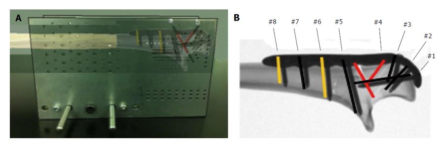
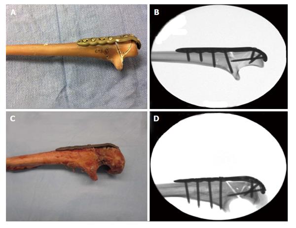
The ulna was positioned in a custom cutting jig and locked into position with K-wires, prior to osteotomy creation[11], of a simulated comminuted fracture. A pre-contoured olecranon plate (Stryker Trauma AG, Selzach, Switzerland) was fixed onto the posterior aspect of the ulna with five VariAx 3.5 mm locking and two VariAx 3.5 mm non-locking screws (Figures 1 and 2). In each ulna (composite and human), the screw application was kept consistent with respect to length and configuration (Figure 1 and Table 1) with one at the olecranon tip, two through the olecranon, and four distal to the fracture (Figure 2). Screw length variability was dictated by the anatomical variation of the human specimens. After fracture fixation with plate and screws, the K-wires were removed.
| Plate hole number | #1 | #2 | #3 | #4 | #5 | #6 | #7 | #8 |
| Screw types used in composite bones | ||||||||
| L/NL/- | L | L | L | - | L | NL | L | NL |
| Screw types used in cadaveric bones | ||||||||
| L/NL/- | L | L | L | - | L | NL | L | NL |
Tests were performed in a custom designed test apparatus (Figure 3) with the standardized osteotomy analogous to that described by Buijze et al[11] and Gordon et al[12] in their cadaveric models as well as others[13-15]. For cadaveric bones, the triceps tendons were fixed with a soft-tissue clamp, and the proximal ulna was also fixed with a bolt similar to the composite bones for more stability. For composite bones, the proximal ulna was fixed with an olecranon tip bolt only, simulating the attachment of the triceps tendon. Cantilever bending load was applied to the ulna with a lever-arm of 200 mm (measured from the elbow joint rotational axis to the point of load application). The starting position was 70° flexion[11], when the cantilever force was not in contact with the bone. A differential variable resistance transducer (DVRT; MicroStrain, United States), with a ± 1 μm accuracy, was attached across the bony fracture site (Figure 3). Fracture displacement, as determined by the DVRT, was recorded from the fracture site before, during, at, and after load to failure was achieved.
Force was applied at the rate of 1 mm/s (actuator speed) until each specimen failed catastrophically, determined both visually and via live data from the Instron graphic software. Failure load was recorded for each specimen and the mode of failure was noted. The load at ≥ 2 mm displacement was obtained from the data files.
Statistical review of the data was performed by a biomedical statistician prior to submission and peer review. The results were statistically analyzed with two-tailed Mann-Whitney-U test a nonparametric measure of statistical dependence between these 2 variables (10000 samples) with statistical significance ascribed a value of P < 0.05. In addition, power was calculated (2-tailed, alpha = 0.05) where 80% was considered as sufficient power (IBM SPSS Sample Power 3).
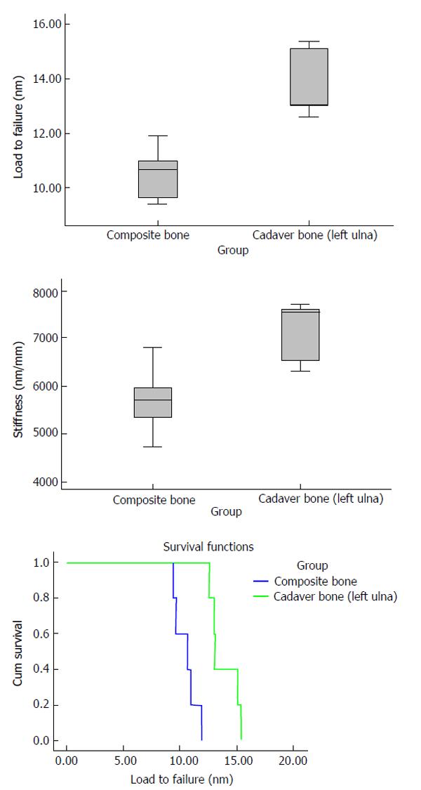
The Kaplan-Meier survival function was estimated for load to failure results. The result plot estimated the cumulative survival at a certain load value (estimated survival rate at certain load application). The correlation between stiffness or failure load and bone mineral density was analyzed with a two-tailed Spearman’s rank correlation coefficient.
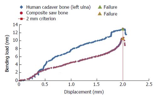
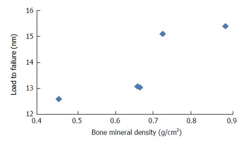
Failure was defined as either a 2 mm fracture gap or complete failure (Figure 4). The load to failure for the composite bones (n = 5) and human cadaver bones (n = 5) specimens were 10.67 nm (range 9.40-11.91 nm) and 13.05 nm (range 12.59-15.38 nm) respectively (Figure 5 and Table 1). This difference was statistically significant (P < 0.007, 97% power). Median stiffness for composite bones and human cadaver bones specimens were 5.69 nm/mm (range 4.69-6.80 nm/mm) and 7.55 nm/mm (range 6.31-7.72 nm/mm). There was a significant difference for stiffness (P < 0.033, 79% power) between composite bones and cadaveric bones (Figure 5). Displacements for these specimens were not compared, due to the 2 mm failure criterion. DEXA scans were used to assess cadaveric bone mineral density (Table 2 and Figure 6). Overall, the DEXA bone mineral density (BMD) for human cadaver specimens ranged from 0.456 to 0.883 g/cm2, with a mean of 0.677 g/cm2. BMD correlated with load to failure (P = 0.037), but did not correlate with stiffness (P = 0.188). Previous studies have shown that BMD significantly correlated with fracture loads in isolated human cadaveric pelvis[16] and femurs[17-19].
| Composite bones | Human cadaver bones | |||||||||
| 1 | 2 | 3 | 4 | 5 | 1 | 2 | 3 | 4 | 5 | |
| Load to failure criteria (nm) | 11.91 | 9.40 | 10.97 | 10.67 | 9.64 | 15.10 | 12.59 | 13.05 | 13.02 | 15.38 |
| Displacement (mm) | 1.76 | 2.00 | 1.85 | 2.00 | 1.70 | 2.00 | 2.00 | 1.71 | 2.00 | 2.00 |
| Stiffness (nm/mm) | 6.80 | 4.69 | 5.95 | 5.33 | 5.69 | 7.55 | 6.31 | 7.62 | 6.52 | 7.72 |
| Bone density (g/cm2) | - | - | - | - | - | 0.723 | 0.456 | 0.658 | 0.665 | 0.883 |
| Failure mode | Distal fracture | Fracture gap | Distal fracture | Fracture gap | Distal fracture | Fracture gap | Fracture gap | Triceps failure | Fracture gap | Fracture gap |
In the plated specimens, catastrophic failure occurred in 3/5 fourth generation composite bone specimens before the pre-defined 2 mm fracture gap was observed and in 2/5 cases when the fracture gap of 2 mm was observed. All complete failures (5/5) resulted from composite bone failure and not hardware failure. Failure occurred at the most distal screw.
There were no catastrophic fracture failures in the cadaveric specimens. Failure of 4/5 cadaveric specimens was defined when a fracture gap of 2 mm was observed, but 1/5 cadaveric specimens failed due to a failure of the triceps mechanism (the triceps tendon slipped in the soft-tissue clamp, and the augmentative trans-olecranon pin bent).
There were 2 primary objectives of this study. Firstly to ascertain whether there is acceptable fixation of a comminuted olecranon fracture with a pre-contoured locking plate and screw construct in an in-vitro model. Secondly to directly compare synthetic composite bones to human cadaveric bones under conditions of destructive loading to determine the construct strength, representing the first time this comparison has been performed. Options for fixation of olecranon fractures include casting, external fixation, and internal fixation with intramedullary nails or plates[20]. Plate-and-screw fixation has proved to be the most reliable and successful strategy and is widely used. Clinical series have demonstrated excellent union rates with few complications[8,21-25]. Successful plate fixation of these fractures allows for early return to function of the upper extremity.
Gordon et al[12] reported that load to failure and mean stiffness values of olecranon fracture fixation was significantly greater with a posterior plate with long intramedullary screw than with dual-plated. However, no statistically significant increase in load to failure in stiffness was demonstrated when compared to posterior plates alone. Buijze et al[11] compared the stiffness and strength of locking compression plate fixation to one-third tubular plate fixation in a cadaveric comminuted olecranon fracture model with a standardized osteotomy. Stiffness and load to failure values from those two studies were similar to those found in the current investigation (Table 3).
| Load to failure | Stiffness | |
| Gordon | 30-34.5 nm | 7-13 nm/mm |
| Buijze | 4.5-27 nm | 8.8-13.3 nm/mm |
| This study (cadaveric) | 12.59-15.38 nm | 7.55 nm/mm |
| This study (composite) | 9.40-11.91 nm | 5.69 nm/mm |
Our study demonstrates that the pre-contoured olecranon is adequate when controlling the fracture position against physiologically relevant forces. Bone mineral density affected load to failure in our model and should be considered when evaluating the results of cadaveric biomechanical studies. In addition, when comparing composite bones to human cadaveric bones, with load to failure, stiffness, and fracture gapping of 2 mm as objective criteria, composite bones were found to be inferior in their mechanical properties. This latter finding has implications when interpreting studies which utilize fourth generation composite bones.
Cantilever forces were 9.40-11.91 nm (for composite bones) and 12.59-15.38 nm (for human cadaver bones) for the fracture sites which would be considerably greater than those experienced in normal activities after fracture fixation[11]. We conclude the following: (1) The pre-contoured plate and screw construct investigated in this model appears to withstand a force greater than the expected physiologic load in the postoperative prior to fracture consolidation. It appears to be a viable option for in vivo treatment of comminuted olecranon fractures; (2) Fourth generation composite bones are not a suitable model for olecranon fracture and plate stiffness testing, in a comminution model, since the interface stresses, at the distal extent of the plate was the site of failure in 5/5 tests, prior to failure loads seen in the cadaveric specimens; (3) Pre-contoured plate and screw constructs are more than adequate to control fracture displacement, when tested in a small cadaveric cohort, with pre-defined failure (fracture gapping of > 2 mm). None of the cadaveric specimens underwent catastrophic failure; and (4) The current study is with a pre-contoured plate with locking and non-locking screws, to stabilize a comminuted olecranon fracture, whereas prior relevant literature studies utilize dual plates, one-third tubular plates, and plate/intramedullary screw constructs. Hence direct comparisons of these study results should be considered carefully.
Authors would like to thank Claudia Beimel from Stryker for her expertise in statistical analysis.
Olecranon fractures are common injuries. Simple fractures are amenable to fixation with a tension band device. Comminuted olecranon fractures pose a unique treatment challenge because tension band devices fail when the cortex opposite of the fixation construct is not intact. A pre-contoured locking plate is an option in the armamentarium for treating this challenging fracture pattern.
Previous biomechanical studies have evaluated various methods of fixation for comminuted olecranon fractures. However, the optimal plate and screw configuration is unclear.
To the authors’ knowledge, no biomechanical study investigating this pre-contoured locking plate has been pursued. Further, no study has investigated behavior of cadaveric vs composite bone substitute in a comminuted olecranon fracture model.
This study highlights the efficacy of a unique option for comminuted olecranon fracture fixation. It also gives context to biomechanical studies performed using composite bone substitute. Variation of results in cadaveric vs composite biomechanical may be applicable to broader applications other than a comminuted olecranon fracture model.
Sawbones-composite physiologic strength bone substitute.
The authors deal about an interesting topic, plating of comminuted olecranon fractures and fixation failure with cadaveric bones and composite bones. The article is well written.
P- Reviewer: Anazawa U, Carbone S, Garg B S- Editor: Ji FF L- Editor: A E- Editor: Li D
| 1. | Zuelzer WA. Fixation of small but important bone fragments with a hook plate. J Bone Joint Surg Am. 1951;33-A:430-436. [RCA] [PubMed] [DOI] [Full Text] [Cited by in Crossref: 1] [Cited by in RCA: 2] [Article Influence: 0.0] [Reference Citation Analysis (0)] |
| 2. | Hak DJ, Golladay GJ. Olecranon fractures: treatment options. J Am Acad Orthop Surg. 2000;8:266-275. [PubMed] |
| 3. | Fyfe IS, Mossad MM, Holdsworth BJ. Methods of fixation of olecranon fractures. An experimental mechanical study. J Bone Joint Surg Br. 1985;67:367-372. [PubMed] |
| 4. | Horner SR, Sadasivan KK, Lipka JM, Saha S. Analysis of mechanical factors affecting fixation of olecranon fractures. Orthopedics. 1989;12:1469-1472. [PubMed] |
| 5. | Hume MC, Wiss DA. Olecranon fractures. A clinical and radiographic comparison of tension band wiring and plate fixation. Clin Orthop Relat Res. 1992;229-235. [PubMed] |
| 6. | Baecher N, Edwards S. Olecranon fractures. J Hand Surg Am. 2013;38:593-604. [RCA] [PubMed] [DOI] [Full Text] [Cited by in Crossref: 62] [Cited by in RCA: 60] [Article Influence: 5.0] [Reference Citation Analysis (0)] |
| 7. | Anderson ML, Larson AN, Merten SM, Steinmann SP. Congruent elbow plate fixation of olecranon fractures. J Orthop Trauma. 2007;21:386-393. [RCA] [PubMed] [DOI] [Full Text] [Cited by in Crossref: 88] [Cited by in RCA: 72] [Article Influence: 4.0] [Reference Citation Analysis (0)] |
| 8. | Bailey CS, MacDermid J, Patterson SD, King GJ. Outcome of plate fixation of olecranon fractures. J Orthop Trauma. 2001;15:542-548. [PubMed] |
| 9. | Simpson NS, Goodman LA, Jupiter JB. Contoured LCDC plating of the proximal ulna. Injury. 1996;27:411-417. [PubMed] |
| 10. | Waddell G, Howat TW. A technique of plating severe olecranon fractures. Injury. 1973;5:135-140. [PubMed] |
| 11. | Buijze GA, Blankevoort L, Tuijthof GJ, Sierevelt IN, Kloen P. Biomechanical evaluation of fixation of comminuted olecranon fractures: one-third tubular versus locking compression plating. Arch Orthop Trauma Surg. 2010;130:459-464. [RCA] [PubMed] [DOI] [Full Text] [Full Text (PDF)] [Cited by in Crossref: 47] [Cited by in RCA: 48] [Article Influence: 3.2] [Reference Citation Analysis (0)] |
| 12. | Gordon MJ, Budoff JE, Yeh ML, Luo ZP, Noble PC. Comminuted olecranon fractures: a comparison of plating methods. J Shoulder Elbow Surg. 2006;15:94-99. [RCA] [PubMed] [DOI] [Full Text] [Cited by in Crossref: 62] [Cited by in RCA: 54] [Article Influence: 2.8] [Reference Citation Analysis (0)] |
| 13. | Dieterich J, Kummer FJ, Ceder L. The olecranon sled--a new device for fixation of fractures of the olecranon: a mechanical comparison of two fixation methods in cadaver elbows. Acta Orthop. 2006;77:440-444. [RCA] [PubMed] [DOI] [Full Text] [Cited by in Crossref: 12] [Cited by in RCA: 12] [Article Influence: 0.6] [Reference Citation Analysis (0)] |
| 14. | Hutchinson DT, Horwitz DS, Ha G, Thomas CW, Bachus KN. Cyclic loading of olecranon fracture fixation constructs. J Bone Joint Surg Am. 2003;85-A:831-837. [PubMed] |
| 15. | Nowak TE, Burkhart KJ, Mueller LP, Mattyasovszky SG, Andres T, Sternstein W, Rommens PM. New intramedullary locking nail for olecranon fracture fixation--an in vitro biomechanical comparison with tension band wiring. J Trauma. 2010;69:E56-E61. [RCA] [PubMed] [DOI] [Full Text] [Cited by in Crossref: 22] [Cited by in RCA: 28] [Article Influence: 2.0] [Reference Citation Analysis (0)] |
| 16. | Beason DP, Dakin GJ, Lopez RR, Alonso JE, Bandak FA, Eberhardt AW. Bone mineral density correlates with fracture load in experimental side impacts of the pelvis. J Biomech. 2003;36:219-227. [PubMed] |
| 17. | Courtney AC, Wachtel EF, Myers ER, Hayes WC. Effects of loading rate on strength of the proximal femur. Calcif Tissue Int. 1994;55:53-58. [PubMed] |
| 18. | Bouxsein ML, Courtney AC, Hayes WC. Ultrasound and densitometry of the calcaneus correlate with the failure loads of cadaveric femurs. Calcif Tissue Int. 1995;56:99-103. [PubMed] |
| 19. | Pinilla TP, Boardman KC, Bouxsein ML, Myers ER, Hayes WC. Impact direction from a fall influences the failure load of the proximal femur as much as age-related bone loss. Calcif Tissue Int. 1996;58:231-235. [PubMed] |
| 20. | Crow BD, Mundis G, Anglen JO. Clinical results of minimal screw plate fixation of forearm fractures. Am J Orthop (Belle Mead NJ). 2007;36:477-480. [PubMed] |
| 21. | Anderson LD, Sisk D, Tooms RE, Park WI. Compression-plate fixation in acute diaphyseal fractures of the radius and ulna. J Bone Joint Surg Am. 1975;57:287-297. [PubMed] |
| 22. | Chapman MW, Gordon JE, Zissimos AG. Compression-plate fixation of acute fractures of the diaphyses of the radius and ulna. J Bone Joint Surg Am. 1989;71:159-169. [PubMed] |
| 23. | Hertel R, Pisan M, Lambert S, Ballmer FT. Plate osteosynthesis of diaphyseal fractures of the radius and ulna. Injury. 1996;27:545-548. [PubMed] |
| 24. | Tejwani NC, Garnham IR, Wolinsky PR, Kummer FJ, Koval KJ. Posterior olecranon plating: biomechanical and clinical evaluation of a new operative technique. Bull Hosp Jt Dis. 2002;61:27-31. [PubMed] |
| 25. | Hewins EA, Gofton WT, Dubberly J, MacDermid JC, Faber KJ, King GJ. Plate fixation of olecranon osteotomies. J Orthop Trauma. 2007;21:58-62. [RCA] [PubMed] [DOI] [Full Text] [Cited by in Crossref: 52] [Cited by in RCA: 44] [Article Influence: 2.4] [Reference Citation Analysis (0)] |


