Copyright
©The Author(s) 2015.
World J Orthop. Jul 18, 2015; 6(6): 498-504
Published online Jul 18, 2015. doi: 10.5312/wjo.v6.i6.498
Published online Jul 18, 2015. doi: 10.5312/wjo.v6.i6.498
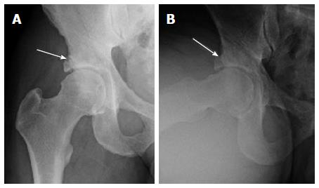
Figure 1 AP (A) and Dunn lateral (B) views of the right hip show right femoral head and neck bump and superolateral acetabular over coverage with suggestion of a rim fracture (arrows).
Notice mild subchondral acetabular sclerosis.
- Citation: Chhabra A, Nordeck S, Wadhwa V, Madhavapeddi S, Robertson WJ. Femoroacetabular impingement with chronic acetabular rim fracture - 3D computed tomography, 3D magnetic resonance imaging and arthroscopic correlation. World J Orthop 2015; 6(6): 498-504
- URL: https://www.wjgnet.com/2218-5836/full/v6/i6/498.htm
- DOI: https://dx.doi.org/10.5312/wjo.v6.i6.498


