Revised: October 29, 2013
Accepted: November 15, 2013
Published online: January 18, 2014
Processing time: 140 Days and 10 Hours
Here, we present the clinical and radiological results of three neglected volar metacarpophalangeal dislocations in 2 patients, which were treated with open reductions 10 and 24 mo after the dislocations. There was a mean of a 20° (range 10°-30°) limitation of extension and a 53.3° (range 30°-70°) limitation of flexion preoperatively. Postoperatively, there was no limitation of extension (at 8 and 12 mo) in any of the fingers. In terms of flexion, one finger had full function, one had a 10° and the last one had a 30° limitation of flexion. Two of the fingers presented anesthesia preoperatively, which improved to hypesthesia postoperatively. One finger had hypesthesia, which improved postoperatively. During surgery, a ruptured dorsal capsule was found to have interposed into the joint, making closed reduction impossible. Our experience with these two patients demonstrated that, even in neglected cases, open reduction using an isolated dorsal approach may result in satisfactory clinical and radiological outcomes.
Core tip: Irreducible complex metacarpophalangeal (MP) dislocations are rare. Volar MP dislocations are very rare. There are no data in the literature reporting volar MP dislocations. We also observed neglected volar MP dislocations in our study. A dorsal or volar approach can be applied toward the surgical treatment of MP joint dislocations. In this study, we observed that isolated dorsal open reduction and K wire fixation results in a successful functional outcome in neglected volar MP dislocations. Volar exposure was not necessary for reduction and stabilization.
- Citation: Başar H, İnanmaz ME, Köse K&, Tetik C. Isolated dorsal approach for the treatment of neglected volar metacarpophalangeal joint dislocations. World J Orthop 2014; 5(1): 62-66
- URL: https://www.wjgnet.com/2218-5836/full/v5/i1/62.htm
- DOI: https://dx.doi.org/10.5312/wjo.v5.i1.62
Irreducible complex metacarpophalangeal (MP) dislocations are rare. Among these, volar dislocations are even rarer[1,2]. The reduction in complex dislocations is usually achieved via open reductions[3]. To our knowledge, there are no data in the literature reporting the results of late open reductions of neglected volar MP dislocations. In this study, we aimed to report the results of the open reductions of 3 volar MP dislocations of 2 patients using an isolated dorsal approach.
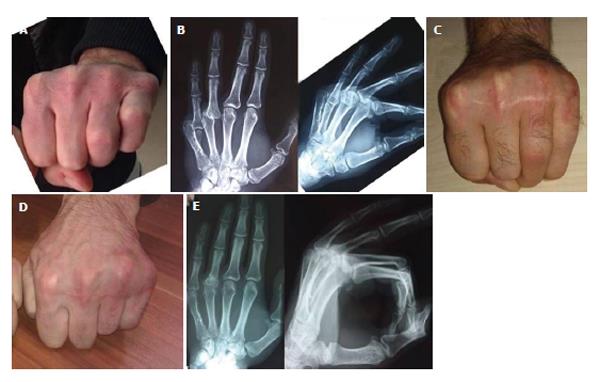
A 22-year-old male presented with complaints of pain, anesthesia of (and inability to move) the 4th and 5th fingers and deformity of the 4th and 5th MP joints of his left hand. He had suffered a dorsal laceration of his left hand during a traffic accident. The laceration had extended from the 2nd MP joint to the 5th. Six hours after the injury, a closed reduction was attempted at another center, and 24 h later, the 2nd, 3rd, 4th and 5th extensor tendons were repaired. He was followed with a below elbow cast for 2 wk. After 2 wk, the cast was removed, and passive and active exercises were started. Since removal of the cast, he noticed stiffness at the 4th and 5th MP joints and hypesthesia on the radial side of the 4th and 5th fingers. Despite these complaints, he did not seek further medical care. After 18 mo, he started a new job that required heavy manual labor, which increased the hypesthesia and eventually progressed to anesthesia of both the 4th and 5th fingers 23 mo after the injury. When he presented 24 mo after injury, he had anesthesia of the radial side of both fingers. The 4th MP joint had a 30° limitation of flexion and a 20° limitation of extension. The 5th MP joint had a 60° limitation of flexion and a 30° limitation of extension (Figure 1A). X-rays revealed a dislocated 5th MP joint and a bone fragment distal to the 5th metacarpal. The 4th MP joint was also dislocated together with a deformity of the dorsal joint surface of the proximal phalanx (Figure 1B).
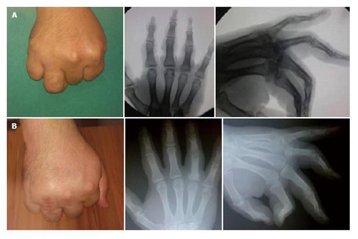
A 34-year-old male presented with complaints of pain, hypesthesia and an inability to move the 4th finger due to deformity of the 4th MP joint of his right hand. His complaints began after punching a wall 10 mo prior. He was given a below elbow splint for 10 d, followed by 4 wk of physical therapy, which improved his joint range of motion but worsened his hypesthesia. His pain and hypesthesia had increased over time because of his job (bus driver). When he presented 10 mo after the injury, the 4th MP joint had a 70° limitation of flexion and a 10° limitation of extension. The 4th finger was hypesthetic. His X-rays revealed a 4th MP joint dislocation with deformity dorsal to the proximal phalanx proximal joint surface (Figure 2A).
A dorsal approach was performed under regional anesthesia and tourniquet hemostasis. A longitudinal cut was made over the MP joint. The ulnar sagittal band of the extensor mechanism was cut longitudinally. When the capsule was exposed, it was found to have ruptured in all three fingers. Achieving exposure of the joint was difficult because of adhesions to the surrounding structures. The dorsal capsular adhesions were released to expose the joint (Figure 3). In the first patient, the radial digital arteries and nerves of the 4th and 5th fingers were compressed between the distal end of the metacarpal and the proximal end of the proximal phalanx. The bone fragment that had fused with the distal volar surface of the 5th metacarpal in the 1st patient was excised, and the MP joint was reduced. The reduction was maintained using a K wire (Figure 3). Because both patients presented a deformation at the dorsal end of both proximal phalanges, a shortening together with a volar closing wedge osteotomy was performed in each case to enhance the joint congruity (Figure 3). The reduction was maintained using a K wire. After reduction, the digital arteries and nerves were observed to have decompressed. The dorsal capsule and the ulnar sagittal band of the extensor mechanism were then repaired.
The 4th and 5th fingers were kept in a below-elbow splint for 4 wk. Subsequently, the K wires were removed. A removable split was applied, and passive and active assisted joint range of motion exercises were assigned for 2 wk. At the end of the 6th week, active exercises were assigned, and the removable splints were discontinued after 8 wk.
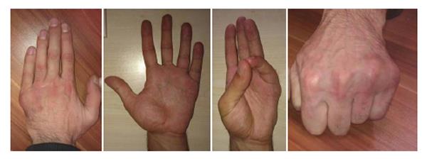
Improvements were observed in terms of pain, hypesthesia and function in all fingers in both patients (Figure 1C). At the 12th month control, the range of motion had improved with full flexion of the 4th MP joint and a 10° flexion limitation of the 5th MP joint (Figures 1D, 4). In both fingers, there was no extension limitation (Figure 4). During the early postoperative period (2 mo), the anesthesia of both fingers improved to hypesthesia, and this improvement persisted to the 12th month. There was no pain in the 3 affected fingers in either patient at the 12th month control visit. The 12th month X-rays revealed a maintenance of the reduction with a fully united osteotomy line (Figure 1E).
The 2nd patient achieved a 30° limitation of flexion at the 8th month control visit. There was no limitation of extension (Figure 2B). The hypesthesia of the 4th finger decreased. The 8-mo X-rays revealed a maintenance of the reduction with a fully united osteotomy line (Figure 2B).
Both patients were able to return to their former jobs after surgery with an improved functional status.
Complex volar MP dislocation is a frequent consequence of a proximal translational force acting on the proximal phalanx while the MP joint is in hyperflexion[2,4]. This phenomenon was the mechanism of injury in our patients. In addition, the extensor mechanisms of the 4th and 5th fingers were lacerated.
When the MP joint is dislocated during extension, the volar plate can avulse and interpose into the joint, thereby precluding a closed reduction[5].
A dorsal or volar approach can be applied during the surgical treatment of MP joint dislocations. The dorsal approach enables an evaluation of the dorsal capsule, the osteochondral injury on the metacarpal head and even the volar plate. The risk of neurovascular injury is extremely low with this approach. If a volar plate rupture is detected, it can be repaired through a separate volar incision. The dorsal capsule cannot be repaired using a volar approach, and the risk of neurovascular injury is higher[6,7]. In our patients, we used an isolated dorsal approach, dissected the avulsed dorsal capsule from the surrounding structures, reduced the joint and repaired the capsule. Typically, the volar plate is not ruptured during volar MP dislocations, and no further approach seems to be necessary.
Complex dorsal MP joint dislocations are frequently observed in the 2nd and 5th fingers because of the lack of a deep transverse metacarpal ligament, which is a natural stabilizer[8]. In our patients, 2 injuries were observed on the 4th finger and 1 was observed on the 5th finger.
There are no data in the literature regarding neglected volar MP dislocations. In this study, we have demonstrated that isolated dorsal open reduction and K wire fixation result in a successful functional outcome in neglected volar MP dislocations.
Main clinical findings of neglected volar metacarpophalangeal (MP) joint dislocations were pain, stiffness of MP joint, anesthesia or hypesthesia of the finger.
Neglected volar MP joint dislocations must be differentiated from flexor tenosynovitis, metacarpal and phalangeal fractures.
Radiographies reveal neglected volar MP joint dislocation with disruption of joint sequence and deformity dorsal to the proximal phalanx proximal joint surface.
Dorsal surgical approach was choiced to reduce the MP joint dislocation and provide MP joint congruency.
The authors gave informations about related reports in discussion but in literature there are a few study over MP joint dislocations.
The ulnar sagittal bands arise from the palmar plate of the finger MP joint and the adjacent intermetacarpal ligaments, which attach to the meta-carpal neck. They pass around the sides of the meta-carpophalangeal joints and attach to zone 5 of the extensor tendon. It is through the sagittal bands that the finger extensor tendons extend the MP joints and also stabilize the extensor tendons on the dorsum of the MP joints and prevent them from subluxing ulnarly, into the intermetacarpal spaces.
The authors experience with these two patients demonstrated that, even in neglected cases, open reduction using an isolated dorsal approach may result in satisfactory clinical and radiological outcomes.
The authors present the clinical and radiological results of open reduction using an isolated dorsal approach in only three neglected volar MP dislocations. If the study consisted more cases and compared two different surgical approach, power of the study have increased.
P- Reviewers: Grote S, Kumar P, Kutscha-Lissberg F S- Editor: Ma YJ L- Editor: A E- Editor: Liu SQ
| 1. | Green DP, Terry GC. Complex dislocation of the metacarpophalangeal joint. Correlative pathological anatomy. J Bone Joint Surg Am. 1973;55:1480-1486. [PubMed] |
| 2. | Wood MB, Dobyns JH. Chronic, complex volar dislocation of the metacarpophalangeal joint. J Hand Surg Am. 1981;6:73-76. [RCA] [PubMed] [DOI] [Full Text] [Cited by in Crossref: 26] [Cited by in RCA: 21] [Article Influence: 0.5] [Reference Citation Analysis (0)] |
| 3. | Bousselmame N, Galuia F, Jaafar A, Lazrak K, Taobane H, Moulay I. [Metacarpophalangeal dislocation of the index finger. Apropos of 7 operated cases]. Chir Main. 1998;17:338-347. [PubMed] |
| 4. | Shah SS, Techy F, Mejia A, Gonzalez MH. Ligamentous and capsular injuries to the metacarpophalangeal joints of the hand. J Surg Orthop Adv. 2012;21:141-146. [PubMed] |
| 5. | Betz RR, Browne EZ, Perry GB, Resnick EJ. The complex volar metacarpophalangeal-joint dislocation. A case report and review of the literature. J Bone Joint Surg Am. 1982;64:1374-1375. [RCA] [PubMed] [DOI] [Full Text] [Cited by in Crossref: 2] [Cited by in RCA: 2] [Article Influence: 0.0] [Reference Citation Analysis (1)] |
| 6. | Andersen JA, Gjerløff CC. Complex dislocation of the metacarpophalangeal joint of the little finger. J Hand Surg Br. 1987;12:264-266. [RCA] [PubMed] [DOI] [Full Text] [Cited by in Crossref: 8] [Cited by in RCA: 9] [Article Influence: 0.2] [Reference Citation Analysis (0)] |
| 7. | Barry K, McGee H, Curtin J. Complex dislocation of the metacarpo-phalangeal joint of the index finger: a comparison of the surgical approaches. J Hand Surg Br. 1988;13:466-468. [RCA] [PubMed] [DOI] [Full Text] [Cited by in Crossref: 19] [Cited by in RCA: 21] [Article Influence: 0.6] [Reference Citation Analysis (0)] |
| 8. | Viegas SF, Heare TC, Calhoun JH. Complex fracture-dislocation of a fifth metacarpophalangeal joint: case report and literature review. J Trauma. 1989;29:521-524. [RCA] [PubMed] [DOI] [Full Text] [Cited by in Crossref: 5] [Cited by in RCA: 6] [Article Influence: 0.2] [Reference Citation Analysis (0)] |


