Copyright
©2011 Baishideng Publishing Group Co.
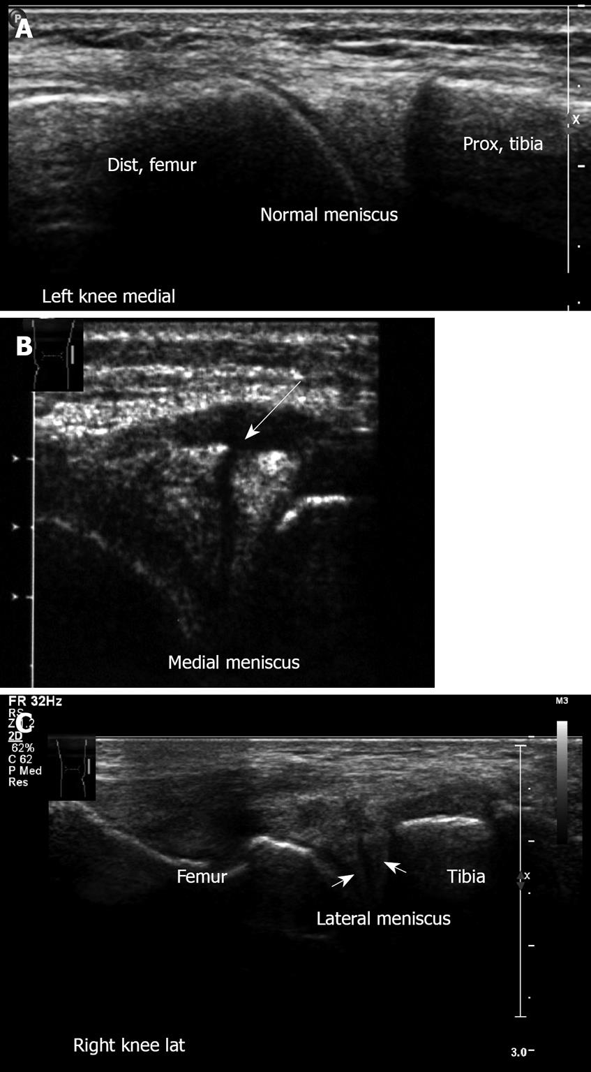
Figure 18 Meniscal lesions.
A: Left knee medial aspect, longitudinal sonogram. Medial meniscus anterior horn. Note the triangle-shaped hyperechoic structure of the normal medial meniscus; B: Medial meniscus lesion. Note the cleft and irregularity of the torn meniscus. This young football player suffered an acute twisting injury. Clinically he had pain with mild swelling on the medial aspect, there was a joint effusion with medial line tenderness. Mac-Murray and Apley tests were positive over the medial meniscus; Right knee lateral meniscus lesion. Note the irregularity of the torn lateral meniscus.
- Citation: Blankstein A. Ultrasound in the diagnosis of clinical orthopedics: The orthopedic stethoscope. World J Orthop 2011; 2(2): 13-24
- URL: https://www.wjgnet.com/2218-5836/full/v2/i2/13.htm
- DOI: https://dx.doi.org/10.5312/wjo.v2.i2.13


