Chen QQ, Liu Y, Yang JH, Yang B. Postural correction training improves chronic pain, nerve function, and inflammation in knee osteoarthritis: A retrospective cohort study. World J Orthop 2025; 16(8): 110332 [DOI: 10.5312/wjo.v16.i8.110332]
Corresponding Author of This Article
Bo Yang, Associate Chief Physician, Dean, Department of Orthopedics, Haian Hospital of Traditional Chinese Medicine Affiliated to Nanjing University of Chinese Medicine, No. 55 Ninghai Middle Road, Nantong 226600, Jiangsu Province, China. yangbo_12138@163.com
Research Domain of This Article
Orthopedics
Article-Type of This Article
Retrospective Cohort Study
Open-Access Policy of This Article
This article is an open-access article which was selected by an in-house editor and fully peer-reviewed by external reviewers. It is distributed in accordance with the Creative Commons Attribution Non Commercial (CC BY-NC 4.0) license, which permits others to distribute, remix, adapt, build upon this work non-commercially, and license their derivative works on different terms, provided the original work is properly cited and the use is non-commercial. See: http://creativecommons.org/licenses/by-nc/4.0/
Qing-Qing Chen, Bo Yang, Department of Orthopedics, Haian Hospital of Traditional Chinese Medicine Affiliated to Nanjing University of Chinese Medicine, Nantong 226600, Jiangsu Province, China
Yang Liu, Department of Clinical Nutrition, Haian Hospital of Traditional Chinese Medicine Affiliated to Nanjing University of Chinese Medicine, Nantong 226600, Jiangsu Province, China
Ju-Hui Yang, Department of Emergency, Haian Hospital of Traditional Chinese Medicine Affiliated to Nanjing University of Chinese Medicine, Nantong 226600, Jiangsu Province, China
Author contributions: Chen QQ, Liu Y and Yang B conceived and designed the study; Chen QQ performed the literature search; Yang JH and Chen QQ acquired data and drafted the manuscript; Liu Y assisted in revising the manuscript. Yang B and Yang JH wrote the original draft; Chen QQ wrote, reviewed and edited the manuscript; Yang B and Chen QQ ensured the authenticity of all the raw data. All authors have read and approved the final manuscript. Chen QQ and Liu Y contributed equally to this work as co-first authors.
Institutional review board statement: The study was approved by the Medical Ethics Committee of Haian Hospital of Traditional Chinese Medicine Affiliated to Nanjing University of Chinese Medicine (HZYLL2022061).
Informed consent statement: Informed consent was obtained from patients or their guardians.
Conflict-of-interest statement: The authors declared that they have no conflicts of interest regarding this work.
STROBE statement: The authors have read the STROBE Statement-checklist of items, and the manuscript was prepared and revised according to the STROBE Statement-checklist of items.
Data sharing statement: Data are provided within the manuscript or supplementary information files.
Open Access: This article is an open-access article that was selected by an in-house editor and fully peer-reviewed by external reviewers. It is distributed in accordance with the Creative Commons Attribution NonCommercial (CC BY-NC 4.0) license, which permits others to distribute, remix, adapt, build upon this work non-commercially, and license their derivative works on different terms, provided the original work is properly cited and the use is non-commercial. See: https://creativecommons.org/Licenses/by-nc/4.0/
Corresponding author: Bo Yang, Associate Chief Physician, Dean, Department of Orthopedics, Haian Hospital of Traditional Chinese Medicine Affiliated to Nanjing University of Chinese Medicine, No. 55 Ninghai Middle Road, Nantong 226600, Jiangsu Province, China. yangbo_12138@163.com
Received: June 6, 2025 Revised: June 21, 2025 Accepted: July 15, 2025 Published online: August 18, 2025 Processing time: 65 Days and 7.2 Hours
Abstract
BACKGROUND
Knee osteoarthritis (KOA) is a prevalent degenerative joint disorder characterized by complex neuroinflammatory mechanisms involving peripheral-central nervous system crosstalk. Current research gaps exist regarding the modulatory effects of biomechanical interventions such as postural correction training (PCT) on these pathways, particularly its impact on neurogenic inflammation and associated nerve dysfunction.
AIM
To examine the effect of PCT on chronic pain related to KOA, nerve function, and inflammatory factors and further assess the influencing factors.
METHODS
This study included 100 patients with chronic pain related to KOA admitted to our hospital from March 2022 to March 2024 who were selected as research subjects, and divided into a control group (conventional treatment, n = 50) and observation group (combined treatment with PCT, n = 50). Efficacy, pain [visual analog scale (VAS)], nerve function [the National Institute of Health Stroke Scale (NIHSS)] and inflammatory factors [interleukin (IL)-1β, IL-6, tumor necrosis factor-alpha (TNF-α), C-reactive protein (CRP)] were assessed and compared. Moreover, the factors influencing efficacy were assessed according to clinical efficacy.
RESULTS
The clinical effectiveness rate of 90.00% in the observation group was higher than that of 72.00% in the control group (P < 0.05). VAS scores at 14 and 30 days of the intervention were lower than those before the intervention (P < 0.05). Moreover, VAS scores in the observation group at 14 and 30 days after the intervention were lower than those in the control group (P < 0.05). The NIHSS scores were lower after the intervention than those before the intervention for both groups (P < 0.05). The improvement in NIHSS score in the observation group was higher than that in the control group (P < 0.05). Inflammatory factors such as IL-1β, IL-6, TNF-α, and CRP in both groups among patients with osteoarthritis-related chronic pain were lower after the intervention than before the intervention (P < 0.05). After the intervention, all inflammatory factors in the observation group were lower than those in the control group (P < 0.05). The proportion of ineffective treatment combined with joint effusion, Kellgren-Lawrence (K-L) staging grade III-IV, fixed flexion contracture with varus and valgus deformity > 5°, was higher in the control group than in the observation group (P < 0.05), while the joint compartment involvement in the observation group was higher than that in the control group (P < 0.05). The logistic regression results demonstrated that relevant joint effusion, K-L staging grade III-IV, fixed flexion contracture with varus and valgus deformity > 5°, and intervention mode of PCT were higher in the control group than in the observation group (P < 0.05) and were influencing factors on clinically ineffective treatment (P < 0.05).
CONCLUSION
PCT can improve the treatment effect on chronic pain related to KOA, nerve function and inflammatory response. Joint effusion, joint stiffness, and KOA are factors for y ineffective treatment. Joint effusion, higher K-L stage, and larger flexion contracture were risk factors, while PCT was a protective factor for ineffective treatment.
Core Tip: This retrospective analysis demonstrated that postural correction training (PCT) combined with conventional treatment significantly improves outcomes in knee osteoarthritis (KOA) beyond pain relief. This intervention uniquely reduced neurological impairment (measured by the National Institute of Health Stroke Scale) and key inflammatory biomarkers (interleukin-1β, interleukin-6, tumor necrosis factor-α, C-reactive protein) compared to conventional treatment alone. Crucially, joint effusion, advanced Kellgren-Lawrence stage (III-IV), and > 5° flexion contracture were identified as independent risk factors for treatment failure. Conversely, PCT served as a protective factor against inefficacy. These findings position mechanical alignment correction as a disease-modifying strategy addressing neuro-inflammatory mechanisms in KOA.
Citation: Chen QQ, Liu Y, Yang JH, Yang B. Postural correction training improves chronic pain, nerve function, and inflammation in knee osteoarthritis: A retrospective cohort study. World J Orthop 2025; 16(8): 110332
Knee osteoarthritis (KOA) is the most common clinical joint disorder[1], and is characterized by articular cartilage degeneration and osteophyte formation, with patients typically experiencing chronic pain, stiffness, and functional impairment. Its prevalence escalates with age[2], affecting 10%-30% of older adults[3], while chronic pain-initially exercise-induced but progressing to rest pain-significantly compromises quality of life[4]. Current management includes physical therapy, exercise, weight control, and pharmacotherapy[5], although emerging biomechanical interventions show promise: Gait retraining reduces medial compartment loading by 18% (P < 0.01) through toe-out angle modification[6]; proprioceptive training improves postural stability (Berg Balance Scale +32%, P = 0.003)[7]; and neuromuscular exercises lower serum interleukin (IL)-6 Levels by 27% (P = 0.02) via optimized joint kinematics[8]. Despite these advances, critical gaps persist regarding postural correction training's (PCT) simultaneous modulation of neuro-immune pathways and compartment-specific biomechanical factors. Therefore, this study introduces three pioneering innovations: Firstly, it is the inaugural investigation quantitatively evaluating PCT's concurrent effects on neurological function National Institute of Health Stroke Scale (NIHSS), systemic inflammation [IL-1β/IL-6/tumor necrosis factor-alpha (TNF-α)/C-reactive protein (CRP)], and compartmental load redistribution. Secondly, it establishes PCT's disease-modifying mechanism through neuro-immune axis regulation. Thirdly, it identifies Kellgren-Lawrence (K-L) grade > II and varus/valgus deformity > 5° as key predictors of PCT responsiveness-addressing unmet needs in precision rehabilitation beyond conventional symptom management[9]. Our findings provide mechanistic insights into PCT's therapeutic efficacy for KOA-related chronic pain.
MATERIALS AND METHODS
Patients
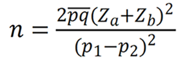
Patients with chronic pain associated with KOA admitted to our hospital from March 2022 to March 2024 were selected as study participants. Inclusion criteria were as follows: (1) Patients who met the diagnostic criteria of KOA[10], and with knee pain symptoms existing within 1 month of selection; (2) Males and females aged > 40 years; (3) Those who were able to walk independently and completed the training maneuvers; (4) Those with normal cognitive function, and no communication barriers; and (5) Those who did not receive other treatments within 1 month of selection. Exclusion criteria were: (1) Patients with a combination of knee tumors and tuberculosis; (2) Those with a history of previous KOA surgery or received intra-articular pulse treatment; (3) pregnant or breastfeeding women; (4) Those with a combination of central and peripheral neuropathy; (5) Those with a combination of acute/chronic infection; and (6) Those with foot and ankle deformity and other joint trauma affecting the function of the lower limbs. The sample size was calculated using the following formula:
Based on visual analog scale (VAS) score differences (α = 0.05, β = 0.2, σ = 1.2, δ = 1.0) from prior gait intervention trials[11]. The final sample size was determined to be 100 cases. The patients were divided into the conventional treatment (control group) and PCT treatment groups (observation group) with 50 patients in each group. Outcome assessors were masked to group assignment. The control group consisted of 39 men and 11 women, 23 cases were aged < 70 years, and 27 cases were aged ≥ 70 years. Forty-one cases were K-L grade I-II and 9 cases were grade III-IV. The observation group consisted of 30 men and 10 women, 30 patients were aged < 70 years, and 20 were aged ≥ 70 years. Forty cases were K-L grade I-II and 10 cases were grade III-IV. Moreover, the differences between the two groups in terms of sex, K-L staging, and other general information were comparable and not statistically significant (P > 0.05). This study strictly adhered to the guidelines of the Declaration of Helsinki and was approved by Medical Ethics Committee of Haian Hospital of Traditional Chinese Medicine Affiliated to Nanjing University of Chinese Medicine (HZYLL2022061). Informed consent was obtained from patients or their guardians.
Methods
All patients received oral celecoxib capsules (200 mg/day for 30 days) alongside identical routine rehabilitation: Days 1-7 included ankle pump exercises (15 min/session, 3 times/day) and power cycling (20 min/day); from Day 7, multi-angle straight leg raises (10 repetitions/set, 3 sets/day) were added. The observation group underwent additional PCT (Figure 1). Pre-intervention static posture assessment (Global Positioning System-based evaluation of shoulders/hips/knees/ankles/spine alignment, stress load, plantar pressure, and center of gravity) guided individualized sitting/standing/walking correction protocols. Orthotic selection criteria: (1) Insoles were customized for plantar pressure redistribution (e.g., medial/Lateral imbalance > 15%); and (2) Braces were prescribed for joint malalignment correction (e.g., varus/valgus > 5°). The position, stress load, plantar pressure, and distribution of the center of gravity of the patient’s shoulders, hips, knees, ankles, spine, and other parts were assessed by a professional physician. The evaluation was conducted according to the results of targeted corrective training for sitting, standing, and walking postures, combined with orthopedic insoles, supports, and other auxiliary corrections. This was combined with core/weak muscle training (30 minutes/session, 2 times/day, 5 days/week for 4 weeks). Methodological safeguards: Randomization used sealed opaque envelopes and outcome assessors were blinded. For inflammatory factors (IL-1β, IL-6, TNF-α, CRP), venous blood (5 mL) was drawn from fasting patients pre-/post-intervention (Day 30), centrifuged (3000 rpm, 10 minutes), and the serum analyzed via enzyme-linked immunosorbent assay (ELISA) by a single technician during unified assay sessions.
Figure 1 Schematic diagram of postural correction training.
A: Supine position with knee joint suspension; B: Lateral decubitus position with knee joint suspension; C: Prone position with knee joint suspension; D: Supine position with ankle joint suspension; E: Lateral decubitus position with ankle joint suspension; F: Prone position with ankle joint suspension.
Observation indicators
Cure was defined as no knee joint activity limitation and no pain; improvement was defined as no limitation of knee joint activity but slight pain during activity; effective was defined as some limitation of knee joint activity and slight pain during activity; and ineffective was defined as knee joint activity limitation and obvious pain during activity. Cured, improved, and effective were recorded as clinically effective. Pain. A VAS was used to assess the degree of pain in the two groups of patients before the intervention, 14 days after the intervention, and 30 days after the intervention, The VAS score ranged from 0 to 10 points, with a high score indicating that the degree of pain was more critical. Neurological function. Neurological function before and after the intervention (30 days of intervention) was assessed using the neurological deficit degree (the NIHSS)[12]. Inflammatory factors. ELISA was used to detect IL-1β and IL-6 (kits purchased from Wuhan Fion Biotechnology Co., Ltd.), TNF-α (kit purchased from Shenzhen Xinbosheng Biotechnology Co., Ltd.), CRP (kit purchased from Hangzhou Link Biotechnology Co., Ltd.), and other inflammatory factors in patients of both groups before and after the intervention. Influencing factors on efficacy. The patient data were collected and influencing factors on efficacy were analyzed according to the clinical efficacy as the basis of grouping. Involved knee compartments. The number of affected compartments (medial tibiofemoral, lateral tibiofemoral, patellofemoral) was quantified using weight-bearing radiographs and magnetic resonance imaging according to standardized compartmental assessment protocols[13-15]. Involvement was defined as the presence of osteophytes and/or ≥ 50% joint space narrowing in any compartment.
Statistical analysis
Statistical analysis was conducted using SPSS 20.0 software. The count data are expressed as a rate (%) and non-ranked count data are expressed using the χ² test. The rank sum test was used to determine the ranked data. The count data that conformed to normality are expressed as mean ± SD. The overall comparisons were made by repeated measures analysis of variance, while the t-sample test was used for post-hoc multiple comparisons and comparisons between the two groups. The influencing factors were analyzed using logistic regression analysis. Statistical significance for all differences was set at P < 0.05.
RESULTS
Efficacy
The clinical effective rate in the observation group was 90.00%, which was higher than that of 72.00% in the control group (P < 0.05) (Table 1).
Table 1 Comparison of therapeutic efficacy between the two groups, n (%).
Groups
Curable
Improved
Validity
Null
Clinical efficiency
Observation group (n = 50)
18 (36.00)
19 (38.00)
8 (16.00)
5 (10.00)
90.00 (45/50)
Control group (n = 50)
8 (16.00)
17 (34.00)
11 (22.00)
14 (28.00)
72.00 (36/50)
χ2 value
-
-
-
-
5.263
P value
-
-
-
-
0.022
Pain
The VAS scores of the two groups were assessed using Mauchly’s W. P = 0.001 < 0.05. The VAS score differences in the two groups were statistically significant (P group < 0.05), and the trend differed with the change in time (P time point < 0.05). Moreover, an interaction between time and group existed (P interaction < 0.05). Following post-hoc multiple comparisons, the VAS scores in the two groups were lower than those in the control group at 14 days and 30 days of the intervention; these were lower than those at pre-intervention (P < 0.05). The VAS scores in the observation group were lower than those in the control group at 14 days and 30 days of the intervention (P < 0.05). These results are shown in Table 2 and Figure 2.
Figure 2 Graph of trend in changes of visual analog scale scores in the two groups.aP < 0.05, within-group vs pre-intervention; bP < 0.05, within-group vs 14 days of intervention; cP < 0.05, between-group comparison at the same time point. VAS: Visual analog scale.
Table 2 Comparison of visual analog scale scores between the two groups, mean ± SD.
The patients in both groups had lower NIHSS scores than those before the intervention (P < 0.05), while the improvement effect in NIHSS scores was higher in the observation group than in the control group (P < 0.05) (Table 3).
Table 3 Comparison of the National Institute of Health Stroke Scale scores between the two groups (mean ± SD, points).
Groups
Pre-intervention
Post-intervention
Difference (the result of subtraction)
Observation group (n = 50)
18.20 ± 2.52
5.56 ± 1.20
11.98 ± 3.15
Control group (n = 50)
18.72 ± 2.95
9.90 ± 1.43
8.82 ± 3.26
t value
0.947
16.436
4.931
P value
0.346
0.000
0.000
Inflammatory factors
In patients with osteoarthritis-associated chronic pain after the intervention in both groups, the inflammatory factors IL-1β, IL-6, TNF-α, and CRP were lower than those before the intervention (P < 0.05); however, no difference was observed when the inflammatory factors between the two groups were compared before the intervention (P > 0.05). Moreover, all the inflammatory factors in the observation group were lower than those in the control group after the intervention (P < 0.05) (Table 4).
Table 4 Comparison of inflammatory factors between the two groups (mean ± SD).
The results of univariate analysis of patient data in the clinically effective and ineffective subgroups demonstrated that the percentages of patients in the ineffective group with joint effusion, K-L staging grade III-IV, varus and valgus deformities with flexion contracture > 5°, and the conventional intervention mode were 31.58%, 42.11%, 36.84%, and 73.68%, respectively, which were higher than those in the effective group, which were 7.41%, 13.58%, 11.11%, and 44.44% (P < 0.05). In addition, joint compartment involvement in the ineffective group was higher than that in the control group (P < 0.05). These details are presented in Table 5.
Table 5 Comparison of treatment effectiveness, n (%)/mean ± SD.
Characteristics
Ineffective (n = 19)
Effective (n = 81)
Statistical value
P value
Gender
0.102
0.750
Male
14 (73.68)
65 (80.25)
Female
5 (26.32)
16 (19.75)
Age
0.226
0.635
< 70 years
11 (57.89)
42 (51.85)
≥ 70 years
8 (42.11)
39 (48.15)
Body mass index (kg/m2)
23.68 ± 0.94
23.47 ± 1.06
0.807
0.421
Duration of disease (months)
33.27 ± 5.18
34.05 ± 5.17
0.588
0.558
Diabetes
0.000
1.000
Present
3 (15.79)
13 (16.05)
Absent
16 (84.21)
68 (83.95)
Hypertension
0.000
1.000
Present
3 (15.79)
12 (14.81)
Absent
16 (84.21)
69 (85.19)
Osteoporosis
0.033
0.856
Present
5 (26.32)
23 (28.40)
Absent
14 (73.68)
58 (71.60)
Smoking
0.014
0.906
Present
2 (10.53)
12 (14.81)
Absent
17 (89.47)
69 (85.19)
Alcohol abuse
0.022
0.883
Present
4 (21.05)
21 (25.93)
Absent
15 (78.95)
60 (74.07)
Joints involved
0.000
1.000
Single knee
4 (21.05)
16 (19.75)
Both knees
15 (78.95)
65 (80.25)
Joint effusion
6.380
0.012
Present
6 (31.58)
6 (7.41)
Absent
13 (68.42)
75 (92.56)
K-L staging
6.389
0.011
I-II
11 (57.89)
70 (86.42)
III-IV
8 (42.11)
11 (13.58)
Flexion contracture of varus deformity
5.788
0.016
≤ 5°
12 (63.16)
72 (88.89)
> 5°
7 (36.84)
9 (11.11)
Intervention mode
5.263
0.022
Routine intervention
14 (73.68)
36 (44.44)
Postural correction training
5 (26.32)
45 (55.56)
Number of lesions (n)
2.89 ± 0.57
2.65 ± 0.50
1.827
0.071
Number of joint spaces (n)
2.58 ± 0.51
1.57 ± 0.50
7.931
0.000
Baseline VAS score (points)
6.37 ± 0.90
6.38 ± 1.01
0.053
0.958
Baseline IL-1β (ng/L)
18.95 ± 2.99
18.35 ± 2.69
0.859
0.392
Baseline IL-6 (ng/L)
26.60 ± 4.36
26.01 ± 3.19
0.670
0.505
Baseline TNF-α (ng/L)
35.23 ± 5.23
35.98 ± 5.40
0.505
0.615
Baseline CRP (mg/L)
12.92 ± 2.54
12.81 ± 2.44
0.181
0.857
The dependent variable for the study was whether the treatment was effective or not (ineffective = 1, effective = 0). Moreover, the independent variables were established based on the statistically significant differences in the aforementioned table of joint effusion (yes = 1, no = 0); K-L staging (grade III-IV = 1, I-II = 0); varus and valgus deformity flexion contracture ( > 5° = 1, ≤ 5° = 0); the mode of intervention (postural corrective training = 1, routine intervention = 0); and the high number of interstitial compartments. In addition, a logistic regression analysis model was established. Logistic regression results demonstrated that joint effusion (OR = 5.769, P = 0.007); K-L staging of grade III-IV (OR = 4.628, P = 0.007); varus and valgus deformity flexion contracture > 5° (OR = 4.667, P = 0.009); and the intervention mode of PCT (OR = 0.286, P = 0.027) were all influential factors in the ineffectiveness of clinical treatment. Moreover, the number of intercompartments was not an influential factor in clinical treatment ineffectiveness (P > 0.05). These results are shown in Table 6.
Table 6 Logistic regression analysis of clinical treatment ineffectiveness.
Variant
β
SE
Wald χ2
P value
OR (95%CI)
Joint effusion (with reference to yes)
1.753
0.651
7.251
0.007
5.769 (1.611-20.659)
K-L staging (based on I-II reference)
1.532
0.567
7.311
0.007
4.628 (1.524-14.052)
Flexion contracture of varus deformity (≤ 5° as reference)
1.54
0.593
6.757
0.009
4.667 (1.461-14.909)
Intervention mode (referenced to conventional intervention)
-1.253
0.567
4.883
0.027
0.286 (0.094-0.868)
Compartments
21.357
5213.902
0.000
0.997
-
DISCUSSION
KOA is a joint disease commonly observed in the elderly, which is characterized by clinical manifestations such as pain, stiffness, and dysfunction. The prevalence of the disease can be up to 80% in individuals aged over 70 years[16]. Data demonstrate[17] that the global prevalence of KOA increased by approximately 10% between 1999 and 2010. The prevalence of KOA is also increasing in China due to accelerated aging and lifestyle changes. It is anticipated that the number of patients with KOA will reach approximately 50 million by 2030[18]. Thus, in this context, clinical attention needs to be paid to the treatment of KOA. Chronic pain associated with KOA is generally centered on the medial, lateral, or posterior aspect of the knee, presenting as a dull, aching pain, with a stinging, burning sensation in some patients[19]. The pain is sufficiently severe to reduce the ability of the patient to perform daily activities and to have poor quality of life. Currently, the treatment of chronic pain associated with KOA can be categorized into pharmacologic and non-pharmacologic modalities[20,21], with pharmacologic treatment including nonsteroidal anti-inflammatory drugs, acetaminophen, and steroid injections, while non-pharmacologic treatment includes physical therapy, weight loss, and training. Chen et al[22] demonstrated that plyometric training improved functional knee activities and alleviated pain symptoms in patients with KOA, while the study by Simic et al[23] showed that an exercise intervention program helped to alleviate pain and improve biomechanical parameters of the lower extremity. Our study results demonstrated that the VAS scores of patients in both groups were lower at 14 and 30 days of intervention than those pre-intervention (P < 0.05), which was similar to previous studies.
The results of this study revealed that post-intervention neurological function NIHSS scores and inflammatory factors such as IL-1β, IL-6, TNF-α, and CRP were lower than those pre-intervention in both groups (P < 0.05). The source of pain in patients with KOA is primarily twofold[4,24]. In contrast, abnormally elevated levels of inflammatory cytokines (TNF-α and IL-6) will promote apoptosis and matrix degradation of chondrocytes and aggravate further damage to articular cartilage. Moreover, cartilage abrasion and joint inflammation will cause pain by releasing pain mediators; in contrast, chronic pain can lead to neurological changes and increased pain sensitivity, making the pain sensation more pronounced. The study results showed that the NIHSS score and inflammatory factors in the observation group were lower than those in the control group after the intervention, while the clinical effectiveness rate of 90.00% in the observation group was higher than that of the control group, which was 72.00% (P < 0.05), indicating that PCT can enhance the therapeutic effect in chronic pain associated with KOA. PCT is a systematic assessment of the incorrect posture of patients by clinicians and the development of personalized interventions, combined with orthopedic braces, to achieve restoration of the normal biomechanical axis of the human body and rehabilitation. PCT in patients with chronic pain associated with KOA can strengthen the muscles that support the knee joint such as the quadriceps and biceps femoris, thus reducing the burden on the joint and decelerating wear and tear of the cartilage. Simultaneously, PCT can improve joint flexibility and stability, which can help reduce cartilage damage due to the abnormal movement patterns caused by joint instability. Additionally, PCT can promote the production of synovial fluid to improve joint lubrication, which in turn assists in cartilage protection. PCT not only reduces cartilage wear and the inflammatory response and promotes adaptive changes in the nervous system but also improves joint function and reduces pain[25,26]. Moreover, the observation group had better neurological function and inflammatory response than the control group after the intervention in this study, thus confirming these findings. Some patients also had clinical symptoms that did not improve significantly post-intervention. These patients must be analyzed to develop a scientific and personalized intervention program. In this study, the presence of joint effusion was 5.769 times more common than the absence of joint effusion, K-L grade III-IV was 4.628 times more common than grade I-II, and the varus and valgus deformity of flexion contracture > 5° was 4.667 times more common than that of ≤ 5°, while the likelihood of ineffective treatment after PCT was reduced to 28.6% of that in the conventional intervention through the initial screening of differences using univariate and logistic regression analyses. Therefore, the therapeutic efficacy of patients with joint effusion, K-L grade III-IV, and varus and valgus deformity with flexion contracture > 5° should be monitored, and PCT should be adopted as much as possible. Although this study demonstrated significant reductions in systemic inflammatory biomarkers (IL-1β, IL-6, TNF-α, CRP) following PCT, we acknowledge potential confounders including the concurrent use of celecoxib-a COX-2 inhibitor with established systemic anti-inflammatory effects that may have contributed to biomarker reductions in both groups[27]. Furthermore, the clinical utility of systemic biomarkers in KOA remains controversial due to their poor correlation with localized joint pathology and symptom severity[28], highlighting the need for future studies to incorporate synovial fluid analysis which provides more direct evidence of joint-specific inflammatory changes[29]; nevertheless, the significantly greater reduction in these biomarkers in the PCT group vs controls (Table 4) suggests a potential synergistic effect between mechanical correction and pharmacotherapy, possibly mediated through reduced mechanical stress on inflamed joints. While systemic inflammation reductions aligned with neuro-immune pathway modulation, we acknowledge celecoxib's potential confounding anti-inflammatory effects and biomarker limitations in KOA. Future studies should incorporate synovial fluid analysis to validate joint-specific effects, although PCT's synergistic benefits with pharmacotherapy highlight its value as a disease-modifying strategy for advanced KOA where biomechanical correction is critical per Richards et al's framework[30].
However, there are some limitations in this study. We were unable to assess the role and location of brain function in patients receiving PCT using functional magnetic resonance imaging, which might supplement our understanding of chronic pain in KOA. Additionally, the study included a small sample size. These issues should be investigated in future research.
CONCLUSION
PCT can augment the treatment effect in chronic pain associated with KOA and improve nerve function and inflammatory response. Moreover, the risk factors for ineffective treatment include the combination of joint effusion, higher K-L staging, and larger fixed flexion contracture with varus and valgus deformity, whereas PCT is a protective factor against ineffective treatment.
Footnotes
Provenance and peer review: Unsolicited article; Externally peer reviewed.
Peer-review model: Single blind
Specialty type: Orthopedics
Country of origin: China
Peer-review report’s classification
Scientific Quality: Grade A, Grade A, Grade B, Grade B, Grade B, Grade B
Novelty: Grade A, Grade A, Grade B, Grade B, Grade B, Grade B
Creativity or Innovation: Grade A, Grade A, Grade B, Grade B, Grade B, Grade B
Scientific Significance: Grade A, Grade A, Grade A, Grade A, Grade A, Grade A
P-Reviewer: Jeong T; Wang Y; Zhou YQ S-Editor: Qu XL L-Editor: A P-Editor: Yu HG
Zhou P, Chen F, Dai ZY. [Progress in magnetic resonance imaging of brain structure and resting-state function in patients with knee osteoarthritis pain].Cigongzhen Chengxiang. 2024;15:140-145.
[PubMed] [DOI] [Full Text]
Merritt A, Roemer F, Heiss R, Jarraya M, Guermazi D, Engebretsen L, Crema M, Guermazi A. Frequency of osteoarthritis in athletes participating in summer olympics in Rio 2016.Osteoarthritis Cartilage. 2020;28:S297-298.
[PubMed] [DOI] [Full Text]
Cross M, Smith E, Hoy D, Nolte S, Ackerman I, Fransen M, Bridgett L, Williams S, Guillemin F, Hill CL, Laslett LL, Jones G, Cicuttini F, Osborne R, Vos T, Buchbinder R, Woolf A, March L. The global burden of hip and knee osteoarthritis: estimates from the global burden of disease 2010 study.Ann Rheum Dis. 2014;73:1323-1330.
[RCA] [PubMed] [DOI] [Full Text][Cited by in Crossref: 2496][Cited by in RCA: 2306][Article Influence: 209.6][Reference Citation Analysis (0)]
Li YT, Niu XM, Hong KD, Chen J, Qiu ZW, Jin LL, Zhong WH. [Study on the brain mechanism of Yi Jin Jing regulating emotions to relieve chronic pain in knee osteoarthritis].Zhongguo Kangfu Yixue Zazhi. 2022;37:1478-1484.
[PubMed] [DOI] [Full Text]
Chen LL, Kang H, Cai J, Wu GX. [Clinical efficacy observation of manipulation combined with muscle training in the treatment of knee osteoarthritis].Hubei Zhongyiyao Daxue Xuebao. 2023;25:75-78.
[PubMed] [DOI] [Full Text]
Liu LY, Sun ML. [Meta-analysis of whole-body vibration training to improve pain and joint function in patients with knee osteoarthritis].Zhongguo Kangfu Yixue Zazhi. 2023;38:1133-1137.
[PubMed] [DOI] [Full Text]
Messier SP, Mihalko SL, Beavers DP, Nicklas BJ, DeVita P, Carr JJ, Hunter DJ, Lyles M, Guermazi A, Bennell KL, Loeser RF. Effect of High-Intensity Strength Training on Knee Pain and Knee Joint Compressive Forces Among Adults With Knee Osteoarthritis: The START Randomized Clinical Trial.JAMA. 2021;325:646-657.
[RCA] [PubMed] [DOI] [Full Text][Cited by in Crossref: 37][Cited by in RCA: 110][Article Influence: 27.5][Reference Citation Analysis (0)]