Published online Aug 18, 2024. doi: 10.5312/wjo.v15.i8.754
Revised: June 5, 2024
Accepted: June 24, 2024
Published online: August 18, 2024
Processing time: 168 Days and 20.3 Hours
The sphericity of the femoral head is a metric used to evaluate hip pathologies and is associated with the development of osteoarthritis and femoral-acetabular impingement.
To analyze the three-dimensional asphericity of the femoral head of asymptomatic pediatric hips. We hypothesized that femoral head asphericity will vary significantly between male and female pediatric hips and increase with age in both sexes.
Computed tomography scans were obtained on 158 children and adolescents from a single institution in the United States (8-18 years; 50% male) without hip pain. Proximal femoral measurements including the femoral head diameter, femoral head volume, residual volume, asphericity index, and local diameter difference were used to evaluate femoral head sphericity.
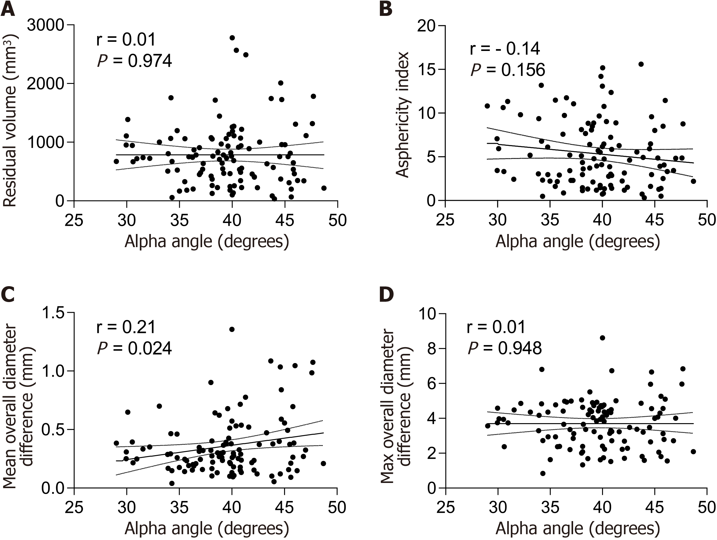
In both sexes, the residual volume increased by age (P < 0.05). Despite significantly smaller femoral head size in older ages (> 13 years) in females, there were no sex-differences in residual volume and aspherity index. There were no age-related changes in mean diameter difference in both sexes (P = 0.07) with no significant sex-differences across different age groups (P = 0.06). In contrast, there were significant increases in local aspherity (maximum diameter difference) across whole surface of the femoral head and all quadrants except the inferior regions in males (P = 0.03). There were no sex-differences in maximum diameter difference at any regions and age group (P > 0.05). Increased alpha angle was only correlated to increased mean diameter difference across overall surface of the femoral head (P = 0.024).
There is a substantial localized asphericity in asymptomatic hips which increases with age in. While 2D measured alpha angle can capture overall asphericity of the femoral head, it may not be sensitive enough to represent regional asphericity patterns.
Core Tip: Femoral head asphericity changes with age demonstrated by significant femoral head asphericity in asymptomatic hips via increased residual volume with age for both sexes. However, no significant overall asphericity differences existed between males and females. While the overall femoral head asphericity was small, there were substantial local divergencies from perfect sphericity in both males and females, with maximum diameter difference correlating with age overall and across all quadrants except for inferior quadrants in males.
- Citation: Hassan MM, Feroe AG, Douglass BW, Jimenez AE, Kuhns B, Mitchell CF, Parisien RL, Maranho DA, Novais EN, Kim YJ, Kiapour AM. Three-dimensional analysis of age and sex differences in femoral head asphericity in asymptomatic hips in the United States. World J Orthop 2024; 15(8): 754-763
- URL: https://www.wjgnet.com/2218-5836/full/v15/i8/754.htm
- DOI: https://dx.doi.org/10.5312/wjo.v15.i8.754
Femoral head sphericity is a common metric used to evaluate the severity of various hip pathologies, including symptomatic femoroacetabular impingement (FAI) and Legg-Calve-Perthes disease[1,2]. Asphericity of the femoral head has been associated with the development of early osteoarthritis (OA) and cam-type FAI, particularly when the deformity involves the frontal plane[1]. A spherical femoral head articulates smoothly within the spherical acetabulum. In contrast, an aspherical contour of the femoral head-neck junction leads to decreased head-neck offset and the risk of contacting the acetabular rim during terminal range of motion[1-3]. When observed radiographically, this morphologic feature has been termed a “pistol grip” deformity[4]. While its exact etiology has yet to be elucidated, means of assessing FAI still heavily rely on examining the sphericity of the femoral head.
The alpha angle of the femoral head, which acts as a proxy for sphericity[5-8], has similarly been associated with the development of OA, especially in cases of cam-type FAI[9-11]. The alpha angle’s utility and consistency, however, is influenced by radiographic view and femoral rotation at time of image acquisition[7,12-15], Furthermore, the repeatability between viewers remains controversial[16-19], and solely radiographic imaging limits the ability to consider local differences in three dimensional (3D) femoral head morphology[5,8,13,20-22]. To address these limitations, many other studies have attempted to create a new means of evaluating femoral head sphericity using both two and three dimensional imaging techniques[6,21-24]. Despite these limitations, the alpha angle remains as the dominant proxy for evaluating femoral head asphericity.
The three-dimensional variability of femoral head sphericity, particularly as it relates to patient age and sex, has not been formally evaluated in an asymptomatic population. A recent study using computed tomography (CT) scans on asymptomatic adolescent patients found alpha angles to be higher in asymptomatic adolescent males, particularly in the anterior, anterosuperior, and superior planes, and overall increased with age[25]. Given the relevance of abnormal femoral head morphology to clinical hip pathology, a method to comprehensively evaluate femoral head sphericity, in addition to alpha angles in a healthy population, may provide insight into normal vs abnormal hip morphology. The purpose of this study was to investigate the asphericity of the femoral head during skeletal growth and maturation in children and adolescents with asymptotic hips using 3D image analysis and to propose a new 3D metric for asphericity analysis termed the asphericity index. We hypothesized that femoral head asphericity will change with age with different trends in males compared to age matched females. We also hypothesized that alpha angle correlates with quantified measures of femoral head asphericity with higher alpha angles correlating to increased 3D asphericity.
Following our institutional review board’s approval, we searched our institutional database for patients from a single institution in the United States who underwent pelvic CT scans for evaluation of suspected appendicitis from 2008 to 2010 who were 8 to 18 years old. Patients were scanned from the third lumbar to the level of the proximal femur at 120 kilovolts (peak) and 50 to 180 mA based on weight and girth. Patients were positioned supine with the pelvis in a neutral position with their hips extended. The CT was acquired helically with a collimation of 0.625 mm or 1.25 mm. Exclusion criteria included patients with a known history of hip pain, injury or disease; the presence of a neuromuscular, developmental, or genetic condition; and low-quality images which would obstruct segmentation.
Using a previously reported patient cohort[3,25] that was extracted from our institution’s database, a total of 176 patients were included for initial review. Seventeen patients were excluded, one for a history of hip pain, eight for unsuitable CT images and another eight were excluded due to associated comorbidities (six with genetic/neurologic diseases, one with osteochondromatosis, and one with craniosynostosis). The remaining 158 patients were randomly selected to have five males and five females in each age from 8 to 18 years. Each patient’s left or right hip was selected using a random number generator. Some patients included in this study were subjects of previous studies evaluating the development of capital femoral epiphysis morphology and femoral head-neck offset in children and adolescents[3,25].
Hip morphology was assessed via measuring the acetabular index angle in the coronal plane, alpha angle[12], and epiphyseal tilt angle[26] from the oblique axial plane. These values were compared to normal references. All assessments were performed by an orthopedic surgeon (DAM) using Osirix Viewer (v8.5, Pixmeo SARL, Bernex, Switzerland).
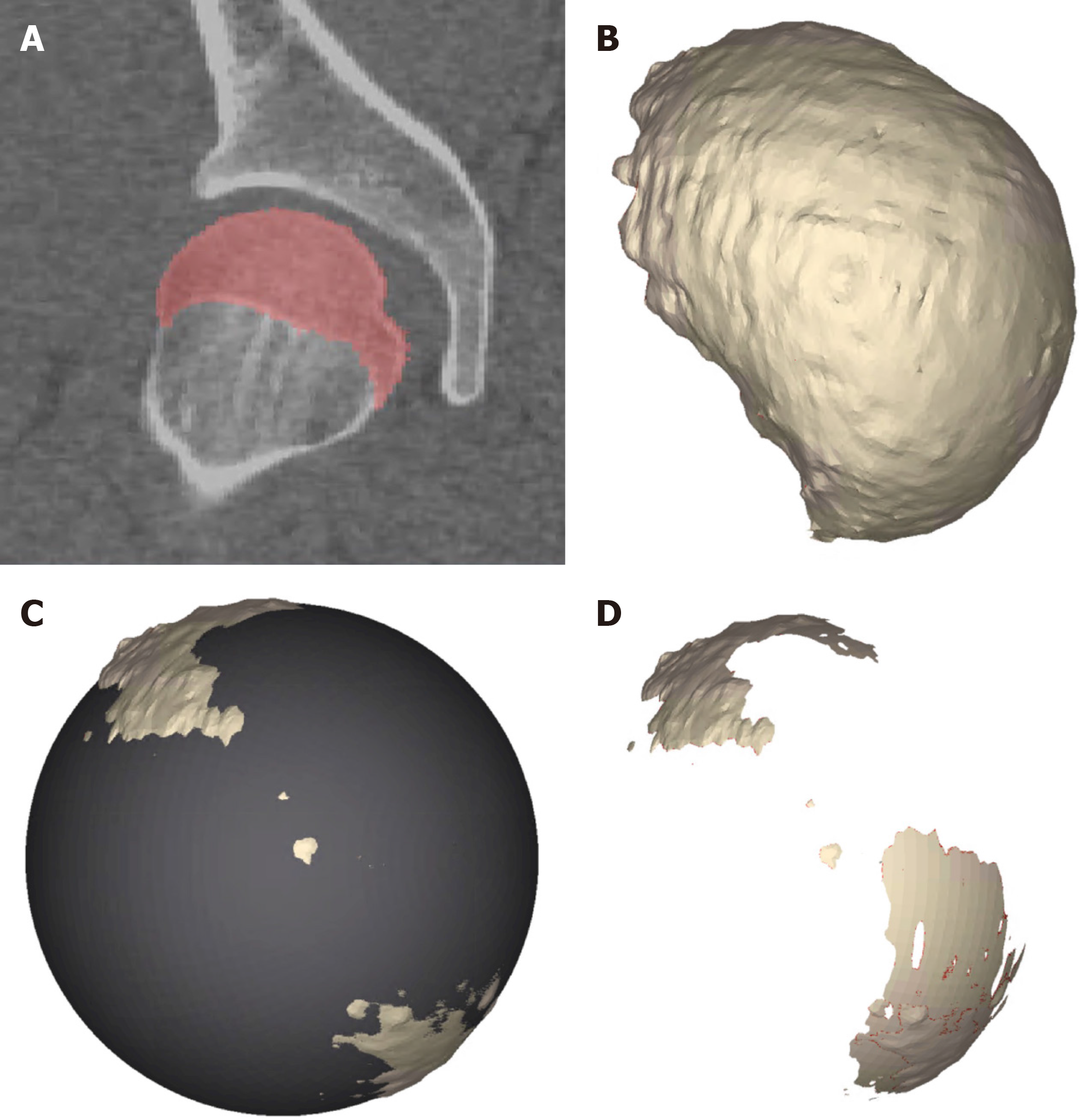
3D models of the femoral head were manually segmented from CT scans by an orthopedic surgeon (DAM) using a commercial image processing software (Mimics v19.0; Materialise, Belgium). The epiphysis was segmented as an independent body from the rest of the proximal femur, with the sclerotic line of the physeal scar being included in the epiphysis for patients with closed growth plates (Figure 1A). 3D models were constructed from each segmented mask then transferred to 3-matics software (v9.0; Materialise, Belgium) to align the 3D models to an anatomic coordinate system and to define the four anatomical quadrants on the femoral head, as described before (Figure 1B)[3]. The aligned 3D models were then exported as point cloud. A custom code (Matlab, MathWorks, Natick, MA, United States) was used to find the center and radius of the best-fit sphere to the femoral head point cloud (Figure 1C). The Boolean subtraction between the femoral head and the best-fit sphere was used to identify regions of the femoral head which were outside the best-fit sphere (Figure 1D).
The following metrics were calculated to evaluate the extent of overall and local femoral head asphericity: (1) Residual volume (mm3): The volume of the leftover regions from the Boolean subtraction between the femoral head and best-fit sphere. The measurements were calculated across the whole femoral head and across each individual quadrant; (2) Asphericity index (ratio): Defined as the ratio between the residual volume and femoral head volume across the whole femoral head and across each quadrant. Higher ratios indicate more asphericity; and (3) Local diameter difference (mm): The local radius, calculated as the distance between each residual point on the surface of the femoral head and the center of the best-fit sphere, was then used to find the local differences in femoral head diameter compared to the best-fit sphere diameter. Higher differences in diameter (local femoral head radius × 2 - best-fit sphere diameter) indicated a higher degree of asphericity. All the segmentations, 3D modeling, and measurements were done blinded to age and sex on deidentified images.
The measures of femoral head size (volume and diameter), asphericity (residual volume, asphericity index and diameter difference) and alpha angle were defined as continuous dependent variables. The diameter difference was reported as maximum (largest difference in diameter between the edge of the perfect sphere and the largest protrusion of the femoral head away from the sphere’s edge) and mean (the average of the differences in diameter between the edges of the perfect sphere and the protrusions of the femoral head away from the sphere’s edge). Higher differences in diameter (local femoral head radius × 2 - best-fit sphere diameter) indicated a higher degree of asphericity. Pearson correlation was used to investigate the effect changes in the dependent variable by age (continuous independent variable) for each sex. To further investigate the sex differences, the dependent variables were divided into 4 age groups for each sex (8-10, 11-13, 14-16, and 17-18). Two-way analysis of variance (ANOVA) with age group and sex as independent variables were used to compare the femoral head size and asphericity between males and females at each age group (four pairwise comparisons). Holm-Sidak post hoc correction was used to adjust the P values to account for the potential increases in type 1 errors due to multiple comparisons. Pearson correlation was also used to investigate the correlations between alpha angle and measures of overall femoral head asphericity (i.e., residual volume, asphericity index, mean overall diameter difference and maximum overall diameter difference). All analyses were completed using Prism (v9.0; GraphPad Software Inc).
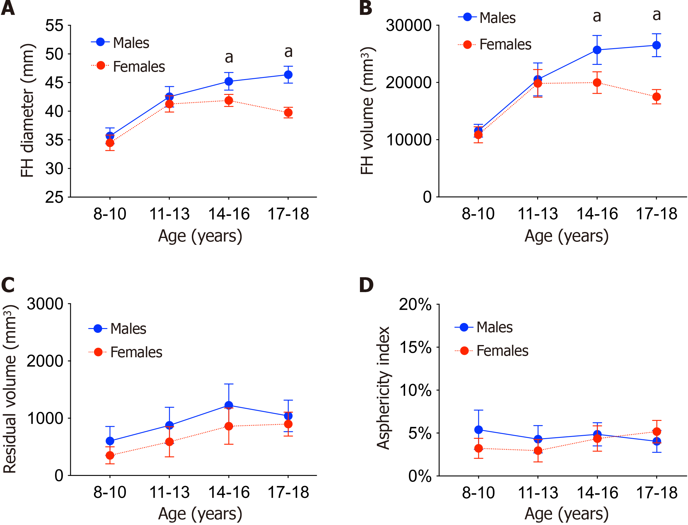
The age-related changes in quantified anatomical features are presented in Table 1. Femoral head diameter and volume correlated with increasing age for both males and females (r > 0.2, P < 0.05; Table 1). Compared to females, males had significantly larger femoral head diameter at 14-16 years (45.2 ± 2.8 mm vs 41.9 ± 1.9 mm, P = 0.001) and at 17-18 years (46.4 ± 2.1 mm vs 39.7 ± 1.3 mm, P < 0.001) age groups (Figure 2A). Similarly, males had significantly larger femoral head volume at 14-16 year (25690 ± 4569 mm3vs 19979 ± 3396 mm3, P < 0.001) and at 17-18 year (26503 ± 2794 mm3vs 17521 ± 1752 mm3, P < 0.001) age groups (Figure 2B). There were positive correlations between residual volume and age for both sexes (r > 0.3, P < 0.02; Table 1), with no significant sex differences in any age group (P > 0.1; Figure 2C). There were no significant correlations between asphericity index and age for either sex (r < 0.3, P > 0.05; Table 1). There were no sex differences in asphericity index at any age group (P > 0.1, Figure 2D).
| Males | Females | |||||
| r | 95%CI | P value | r | 95%CI | P value | |
| FH diameter | 0.79 | 0.67 to 0.88 | < 0.001a | -0.27 | -0.5 to 0 | 0.047a |
| FH volume | 0.80 | 0.68 to 0.88 | < 0.001a | 0.55 | 0.34 to 0.71 | < 0.001a |
| Residual volume | 0.32 | 0.06 to 0.54 | 0.016a | 0.42 | 0.17 to 0.61 | 0.002a |
| Asphericity index | 0.20 | -0.01 to 0.49 | 0.16 | 0.24 | 0.4 to 0.75 | 0.058 |
| Mean overall diameter difference | -0.25 | -0.48 to 0.02 | 0.067 | 0.15 | -0.12 to 0.4 | 0.275 |
| Mean anterosuperior diameter difference | -0.14 | -0.35 to -0.08 | 0.412 | 0.17 | -0.1 to 0.42 | 0.211 |
| Mean posterosuperior diameter difference | 0.14 | -0.13 to 0.39 | 0.294 | 0.25 | -0.02 to 0.48 | 0.072 |
| Mean posteroinferior diameter difference | -0.10 | -0.36 to 0.17 | 0.450 | 0.12 | -0.15 to 0.37 | 0.378 |
| Mean anteroinferior diameter difference | -0.19 | -0.42 to -0.03 | 0.231 | 0.03 | -0.24 to 0.29 | 0.843 |
| Max overall diameter difference | 0.30 | 0.04 to 0.52 | 0.026a | 0.59 | 0.38 to 0.74 | < 0.001a |
| Max anterosuperior diameter difference | 0.34 | 0.09 to 0.56 | 0.01a | 0.58 | 0.37 to 0.74 | < 0.001a |
| Max posterosuperior diameter difference | 0.52 | 0.29 to 0.69 | < 0.001a | 0.64 | 0.45 to 0.77 | < 0.001a |
| Max posteroinferior diameter difference | 0.22 | -0.05 to 0.45 | 0.116 | 0.55 | 0.33 to 0.71 | < 0.001a |
| Max anteroinferior diameter difference | -0.15 | -0.4 to 0.13 | 0.292 | 0.32 | 0.06 to 0.54 | 0.016a |
| Alpha angle | -0.48 | -0.66 to -0.24 | < 0.001a | -0.43 | -0.62 to -0.19 | 0.001a |
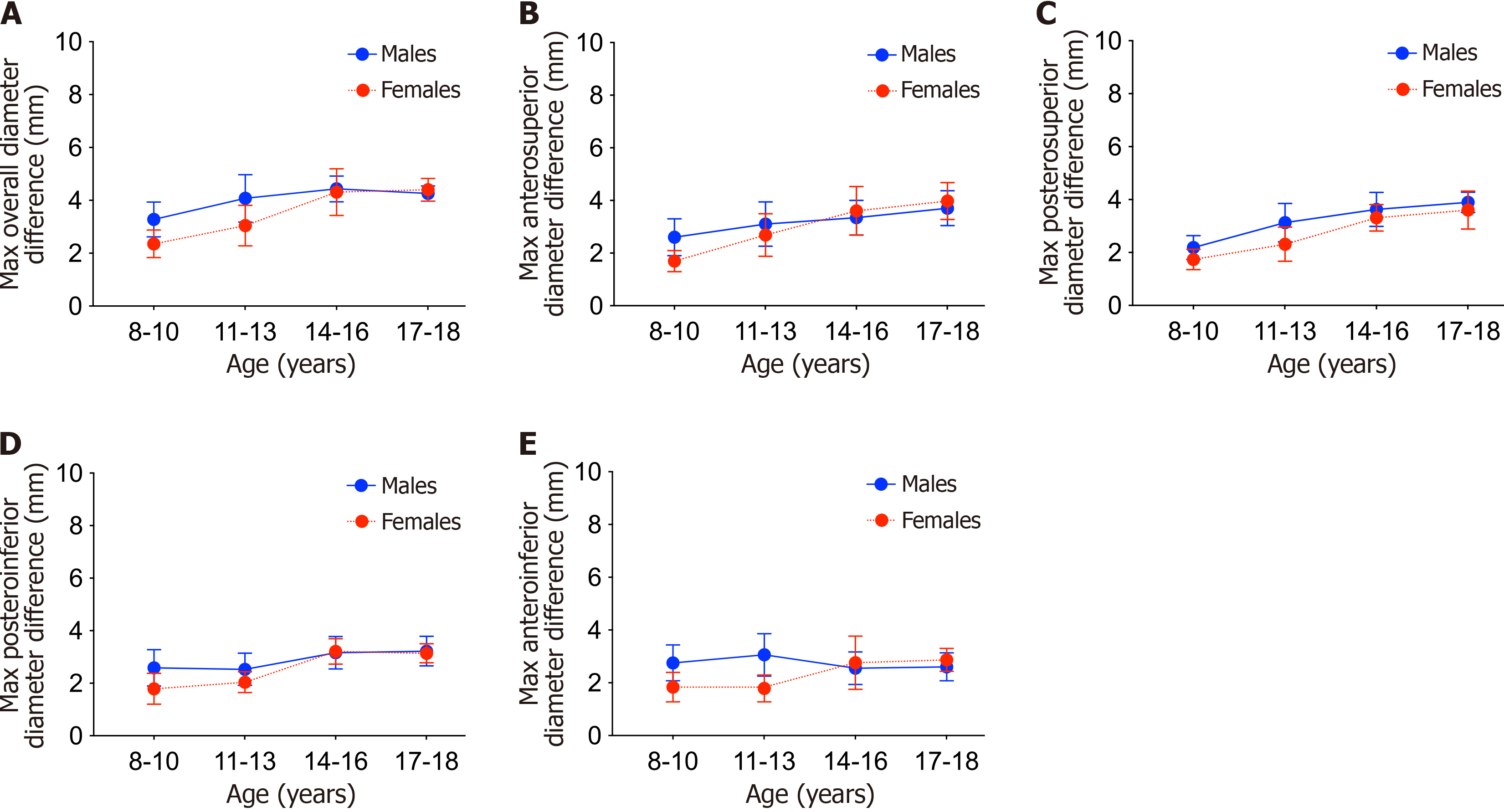
With regards to maximum diameter difference, there were significant positive correlations with age across overall femoral head as well as anterosuperior and the posterosuperior quadrants in both sexes (r > 0.3, P < 0.03; Table 1). There were significant positive correlations between age and the maximum diameter difference across posteroinferior and anteroinferior quadrants (r > 0.5; P < 0.001; Table 1). There were no significant differences in maximum diameter difference between males and females across any regions at any age group (P > 0.05; Figure 3).
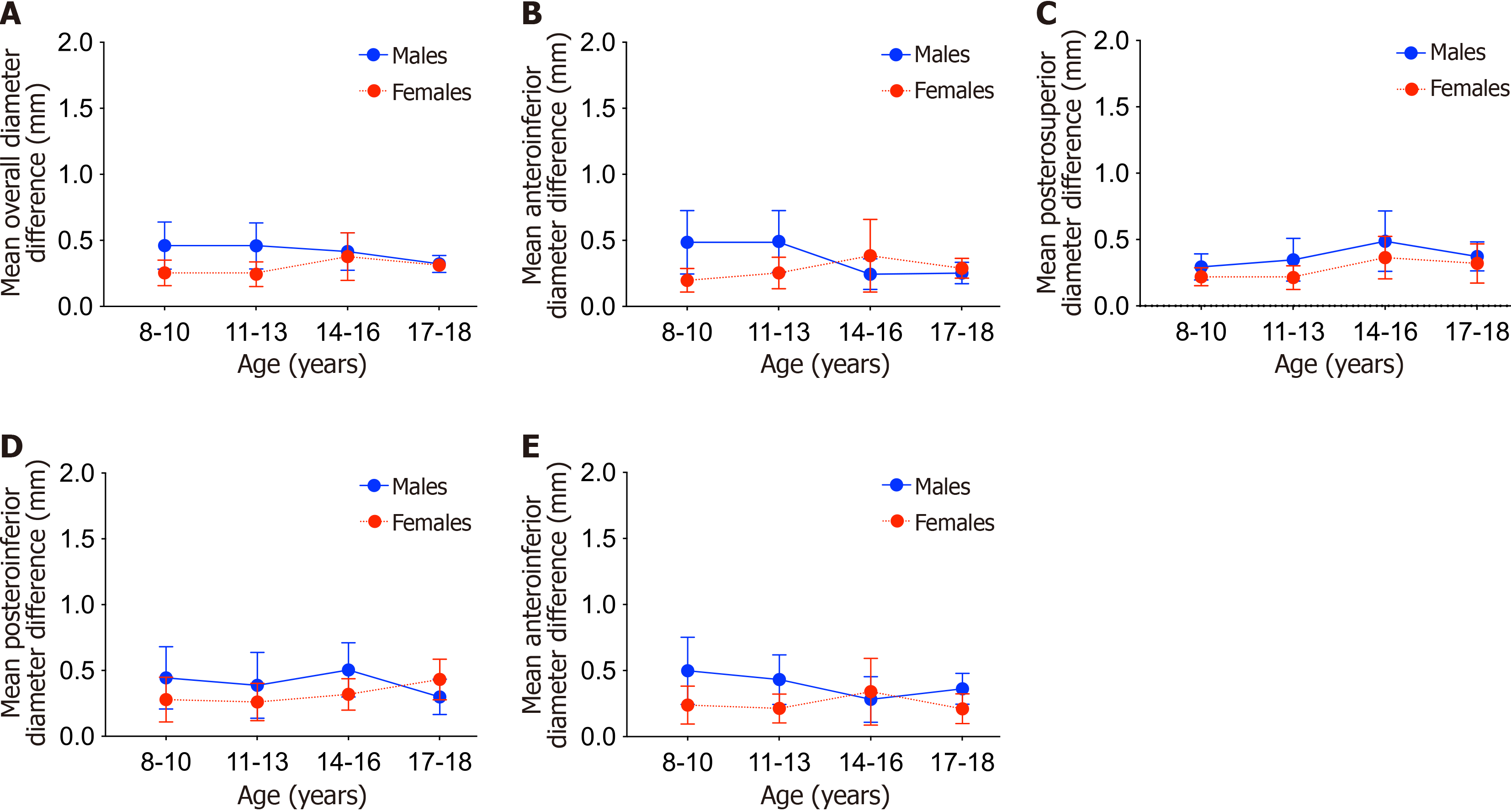
With regards to mean diameter difference, there were no significant correlations to age across any regions in males and females (P > 0.07; Table 1). There were no significant differences in mean diameter difference between males and females across any regions at any age group (P > 0.06; Figure 4). There were significant decreases in alpha angle by age in both sexes (r < -0.4; P < 0.01; Table 1). There were no significant correlations between alpha angle and overall measures of femoral head asphericity except for mean diameter difference (r = 0.21, P = 0.024; Figure 5).
The most important finding of this study was that femoral head asphericity changes with age demonstrated by significant femoral head asphericity in asymptomatic hips via increased residual volume with age for both sexes. However, no significant overall asphericity differences existed between males and females, and thus, the primary hypothesis of this study is partially supported. While the overall femoral head asphericity was small (e.g., average asphericity index of 4.3%), there were substantial local divergencies from perfect sphericity in both males and females (up to a 9 mm difference in diameter), with maximum diameter difference correlating with age overall and across all quadrants except for inferior quadrants in males. The second hypothesis is partially supported by significant relationships between average overall measures of femoral head asphericity (maximum diameter difference) and alpha angle.
Previous studies have attempted to improve upon asphericity evaluation techniques in various hip pathologies. Okano et al[21] developed a roundness index to evaluate the sphericity of femoral heads in patients with developmental hip dysplasia (DDH). Steppacher et al[22] measured femoral head sphericity using multiple radial views obtained from magnetic resonance imaging arthrography in DDH patients. Sankar et al[24] compared differences in measured radii from elliptical and perfect spheres using anteroposterior and lateral radiographs in DDH patients. Audenaert et al[5] sought to improve alpha angle’s utility in determining sphericity in FAI patients utilizing CTs of randomized dry cadavers and found that AAs on 3D radial images and axial oblique radiographs were equivocal. While all these new methods of assessing sphericity showed utility within their specific study and disease state, alpha angle measured on Dunn view remains the predominant method of evaluating asphericity.
Only one other study to date has examined femoral head asphericity regionally in three-dimensions for asymptomatic hips. Harris et al[23] compared CT constructed 3D models of a cam-type FAI hip cohort to normal controls. Similar to our study, Harris’s models created a best fit sphere around the femoral head and then calculated the maximum deviations from the actual CT generated 3D model. Their results found the largest deviations in sphericity in the antero-lateral region of the femoral head for both cohorts, with significantly increased maximum deviations for the FAI hips in the antero-lateral, antero-medial, and postero-lateral regions, and increased mean deviations in the postero-lateral region. Their study, however, did not stratify by age or sex.
The present study represents the first to examine the asphericity of native hips with respect to age and sex. Femoral head diameter, volume, and residual volume overall increased with age with practically no differences between sexes. This would imply that asphericity increases similarly with age for both sexes. Asphericity index, however, did not change with age in males or females and without any sex differences at any age group. This would imply that pediatric male and female femoral heads grow similarly from an overall geometric perspective.
While there were no significant differences observed in overall asphericity of the femoral head between adolescent males and females, local morphological differences over time differed by sex. These morphological differences can have a huge impact on hip biomechanics. In female adolescent hips, local asphericity (maximum diameter difference) increased with age overall and across all quadrants. However, in males the changes were prominently in the superior aspect of the femoral head. The demonstrated age- and sex-based differences in femoral head asphericity may provide insight into epidemiological trends of various hip pathologies. Subsequent studies are necessary to determine the relationship between femoral head sphericity, femoral head-neck morphology, and the development and progression of hip pathologies.
Alpha angle has been widely used to study the role of abnormalities of femoral head-neck morphology in the development of hip pathologies[2,3,27]. Such morphologies, including epiphyseal cupping and extension, are thought to be either pathologic or adaptive responses to increased mechanical stress at the femoral head-neck junction[2,3,28]. This clinical utility, however, does not necessary indicate that alpha angle is a valid measurement of asphericity. Our observed correlations between alpha angle and average asphericity (mean diameter difference) support the alpha angle as a global measure of femoral head asphericity. However, the lack of correlations between alpha angle and local measures of apshericity (maximum diameter difference) suggest limited ability of alpha angle to depict the local femoral head asphericity. The analysis of asphericity by Harris et al[13] found that alpha angles from eight of nine common 2D radiographic views strongly correlated with their 3D measurements of asphericity. Their alpha angle measurements did not, however, correlate with maximum deviations in any region, which they agreed meant that alpha angle may not be capable of determining the magnitude of aspherical deformities. Additionally, the present study found that alpha angle had an inverse relationship with age, which is contrary to previous literature findings suggesting that alpha angle has a positive relationship to age[29]. Given this discrepancy and the trend in the literature to revalidate or replace alpha angle, more sensitive methods of quantifying asphericity in the 2D plane are needed.
Our intention with developing the asphericity index was to create a clinically adoptable 3D evaluation of asphericity which, in conjunction with regional data, can determine specific regional asphericity magnitudes that can help guide surgical management. Our results, however, found that asphericity index does not increase with age despite the fact that diameter differences do. This discrepancy could be due to biomechanical and growth factors that lead to local asphericity in specific quadrants of the femoral head which may have counter active effects in a different region of the femoral head, thus keeping the asphericity index statistically the same over time. Overall asphericity may not change with age even though local quadrants have increases in deviation. In other words, these local deviations may not be significantly large enough to affect the overall asphericity of the femoral head. Regardless, asphericity index will need to be reevaluated and tested in future asphericity studies.
Findings must be considered in the setting of limitations to the present study. First, our analysis of age was based on chronological age, which may not correspond to skeletal maturity. However, chronological age provided a more generalizable measure than other methods commonly used to assess bone maturity. Second, we did not examine the relationship between femoral head sphericity and physical activity, despite the known relationship between athlete status and femoral head-neck morphology. We felt that this was beyond the scope of the present study, but it is an important question to guide subsequent work. Third, this is CT study and does not take into account the contour of the articular cartilage which may have resulted in different asphericity patterns. Another limitation of this study is the sample size of patients that was randomly selected with age range of 8-18 that was not controlled for even distribution of ages included. Additionally, specific movements and activities were not controlled for between patients. Finally, this is not a longitudinal study, and patients were not followed over time. Thus, future symptom presentation could not be determined. Overall, we believe this study contributes valuable quantitative data characterizing femoral head morphology in the skeletally maturing hip to inform further investigation of the role of femoral head asphericity in the natural history of hip pathologies.
Though often modelled as a perfect sphere, normal asymptotic hips contain some amounts of asphericity with significant age-related increased in regional asphericity in both males and females from the United States throughout different stages of skeletal maturity. These regional age-related changes could have significant influence on hip biomechanics and disease pathology. Despite the ability of alpha angle in capturing overall asphericity of the femoral head, it could not capture the regional deviations from perfect sphere. Subsequent longitudinal studies are necessary to determine the relationship between femoral head sphericity and the development and progression of hip disease.
| 1. | Ganz R, Parvizi J, Beck M, Leunig M, Nötzli H, Siebenrock KA. Femoroacetabular impingement: a cause for osteoarthritis of the hip. Clin Orthop Relat Res. 2003;112-120. [RCA] [PubMed] [DOI] [Full Text] [Cited by in Crossref: 724] [Cited by in RCA: 1182] [Article Influence: 53.7] [Reference Citation Analysis (0)] |
| 2. | Siebenrock KA, Wahab KH, Werlen S, Kalhor M, Leunig M, Ganz R. Abnormal extension of the femoral head epiphysis as a cause of cam impingement. Clin Orthop Relat Res. 2004;54-60. [RCA] [PubMed] [DOI] [Full Text] [Cited by in Crossref: 311] [Cited by in RCA: 273] [Article Influence: 13.0] [Reference Citation Analysis (0)] |
| 3. | Novais EN, Maranho DA, Kim YJ, Kiapour A. Age- and Sex-Specific Morphologic Variations of Capital Femoral Epiphysis Growth in Children and Adolescents Without Hip Disorders. Orthop J Sports Med. 2018;6:2325967118781579. [RCA] [PubMed] [DOI] [Full Text] [Full Text (PDF)] [Cited by in Crossref: 12] [Cited by in RCA: 17] [Article Influence: 2.4] [Reference Citation Analysis (0)] |
| 4. | Stulberg S. Unrecognized childhood hip disease: a major cause of idiopathic osteoarthritis of the hip. In: Cordell LD, Harris WH, Ramsey PL, MacEwen GD. The Hip: Proceedings of the Third Open Scientific Meeting of the Hip Society. City of Saint Louis: Mosby, 1975: 212-228. |
| 5. | Audenaert EA, Baelde N, Huysse W, Vigneron L, Pattyn C. Development of a three-dimensional detection method of cam deformities in femoroacetabular impingement. Skeletal Radiol. 2011;40:921-927. [RCA] [PubMed] [DOI] [Full Text] [Cited by in Crossref: 41] [Cited by in RCA: 39] [Article Influence: 2.8] [Reference Citation Analysis (0)] |
| 6. | Gosvig KK, Jacobsen S, Palm H, Sonne-Holm S, Magnusson E. A new radiological index for assessing asphericity of the femoral head in cam impingement. J Bone Joint Surg Br. 2007;89:1309-1316. [RCA] [PubMed] [DOI] [Full Text] [Cited by in Crossref: 145] [Cited by in RCA: 144] [Article Influence: 8.5] [Reference Citation Analysis (0)] |
| 7. | Meyer DC, Beck M, Ellis T, Ganz R, Leunig M. Comparison of six radiographic projections to assess femoral head/neck asphericity. Clin Orthop Relat Res. 2006;445:181-185. [RCA] [PubMed] [DOI] [Full Text] [Cited by in Crossref: 396] [Cited by in RCA: 345] [Article Influence: 18.2] [Reference Citation Analysis (0)] |
| 8. | Tannast M, Kubiak-Langer M, Langlotz F, Puls M, Murphy SB, Siebenrock KA. Noninvasive three-dimensional assessment of femoroacetabular impingement. J Orthop Res. 2007;25:122-131. [RCA] [PubMed] [DOI] [Full Text] [Cited by in Crossref: 185] [Cited by in RCA: 193] [Article Influence: 10.7] [Reference Citation Analysis (0)] |
| 9. | Agricola R, Waarsing JH, Thomas GE, Carr AJ, Reijman M, Bierma-Zeinstra SM, Glyn-Jones S, Weinans H, Arden NK. Cam impingement: defining the presence of a cam deformity by the alpha angle: data from the CHECK cohort and Chingford cohort. Osteoarthritis Cartilage. 2014;22:218-225. [RCA] [PubMed] [DOI] [Full Text] [Cited by in Crossref: 112] [Cited by in RCA: 133] [Article Influence: 12.1] [Reference Citation Analysis (0)] |
| 10. | Thomas GE, Palmer AJ, Batra RN, Kiran A, Hart D, Spector T, Javaid MK, Judge A, Murray DW, Carr AJ, Arden NK, Glyn-Jones S. Subclinical deformities of the hip are significant predictors of radiographic osteoarthritis and joint replacement in women. A 20 year longitudinal cohort study. Osteoarthritis Cartilage. 2014;22:1504-1510. [RCA] [PubMed] [DOI] [Full Text] [Cited by in Crossref: 129] [Cited by in RCA: 139] [Article Influence: 12.6] [Reference Citation Analysis (0)] |
| 11. | Wylie JD, Kim YJ. The Natural History of Femoroacetabular Impingement. J Pediatr Orthop. 2019;39:S28-S32. [RCA] [PubMed] [DOI] [Full Text] [Cited by in Crossref: 32] [Cited by in RCA: 34] [Article Influence: 5.7] [Reference Citation Analysis (0)] |
| 12. | Domayer SE, Ziebarth K, Chan J, Bixby S, Mamisch TC, Kim YJ. Femoroacetabular cam-type impingement: diagnostic sensitivity and specificity of radiographic views compared to radial MRI. Eur J Radiol. 2011;80:805-810. [RCA] [PubMed] [DOI] [Full Text] [Cited by in Crossref: 94] [Cited by in RCA: 99] [Article Influence: 6.6] [Reference Citation Analysis (0)] |
| 13. | Harris MD, Kapron AL, Peters CL, Anderson AE. Correlations between the alpha angle and femoral head asphericity: Implications and recommendations for the diagnosis of cam femoroacetabular impingement. Eur J Radiol. 2014;83:788-796. [RCA] [PubMed] [DOI] [Full Text] [Cited by in Crossref: 69] [Cited by in RCA: 78] [Article Influence: 7.1] [Reference Citation Analysis (0)] |
| 14. | Monazzam S, Bomar JD, Agashe M, Hosalkar HS. Does femoral rotation influence anteroposterior alpha angle, lateral center-edge angle, and medial proximal femoral angle? A pilot study. Clin Orthop Relat Res. 2013;471:1639-1645. [RCA] [PubMed] [DOI] [Full Text] [Cited by in Crossref: 12] [Cited by in RCA: 9] [Article Influence: 0.8] [Reference Citation Analysis (0)] |
| 15. | Pfirrmann CW, Mengiardi B, Dora C, Kalberer F, Zanetti M, Hodler J. Cam and pincer femoroacetabular impingement: characteristic MR arthrographic findings in 50 patients. Radiology. 2006;240:778-785. [RCA] [PubMed] [DOI] [Full Text] [Cited by in Crossref: 387] [Cited by in RCA: 360] [Article Influence: 18.9] [Reference Citation Analysis (0)] |
| 16. | Barton C, Salineros MJ, Rakhra KS, Beaulé PE. Validity of the alpha angle measurement on plain radiographs in the evaluation of cam-type femoroacetabular impingement. Clin Orthop Relat Res. 2011;469:464-469. [RCA] [PubMed] [DOI] [Full Text] [Cited by in Crossref: 238] [Cited by in RCA: 269] [Article Influence: 19.2] [Reference Citation Analysis (0)] |
| 17. | Carlisle JC, Zebala LP, Shia DS, Hunt D, Morgan PM, Prather H, Wright RW, Steger-May K, Clohisy JC. Reliability of various observers in determining common radiographic parameters of adult hip structural anatomy. Iowa Orthop J. 2011;31:52-58. [PubMed] |
| 18. | Clohisy JC, Carlisle JC, Beaulé PE, Kim YJ, Trousdale RT, Sierra RJ, Leunig M, Schoenecker PL, Millis MB. A systematic approach to the plain radiographic evaluation of the young adult hip. J Bone Joint Surg Am. 2008;90 Suppl 4:47-66. [RCA] [PubMed] [DOI] [Full Text] [Cited by in Crossref: 783] [Cited by in RCA: 882] [Article Influence: 51.9] [Reference Citation Analysis (0)] |
| 19. | Konan S, Rayan F, Haddad FS. Is the frog lateral plain radiograph a reliable predictor of the alpha angle in femoroacetabular impingement? J Bone Joint Surg Br. 2010;92:47-50. [RCA] [PubMed] [DOI] [Full Text] [Cited by in Crossref: 47] [Cited by in RCA: 47] [Article Influence: 3.1] [Reference Citation Analysis (0)] |
| 20. | Almoussa S, Barton C, Speirs AD, Gofton W, Beaulé PE. Computer-assisted correction of cam-type femoroacetabular impingement: a Sawbones study. J Bone Joint Surg Am. 2011;93 Suppl 2:70-75. [RCA] [PubMed] [DOI] [Full Text] [Cited by in Crossref: 17] [Cited by in RCA: 17] [Article Influence: 1.2] [Reference Citation Analysis (0)] |
| 21. | Okano K, Enomoto H, Osaki M, Takahashi K, Shindo H. Femoral head deformity after open reduction by Ludloff's medial approach. Clin Orthop Relat Res. 2008;466:2507-2512. [RCA] [PubMed] [DOI] [Full Text] [Cited by in Crossref: 17] [Cited by in RCA: 15] [Article Influence: 0.9] [Reference Citation Analysis (0)] |
| 22. | Steppacher SD, Tannast M, Werlen S, Siebenrock KA. Femoral morphology differs between deficient and excessive acetabular coverage. Clin Orthop Relat Res. 2008;466:782-790. [RCA] [PubMed] [DOI] [Full Text] [Cited by in Crossref: 112] [Cited by in RCA: 132] [Article Influence: 7.8] [Reference Citation Analysis (0)] |
| 23. | Harris MD, Reese SP, Peters CL, Weiss JA, Anderson AE. Three-dimensional quantification of femoral head shape in controls and patients with cam-type femoroacetabular impingement. Ann Biomed Eng. 2013;41:1162-1171. [RCA] [PubMed] [DOI] [Full Text] [Cited by in Crossref: 35] [Cited by in RCA: 38] [Article Influence: 3.2] [Reference Citation Analysis (0)] |
| 24. | Sankar WN, Neubuerger CO, Moseley CF. Femoral head sphericity in untreated developmental dislocation of the hip. J Pediatr Orthop. 2010;30:558-561. [RCA] [PubMed] [DOI] [Full Text] [Cited by in Crossref: 20] [Cited by in RCA: 24] [Article Influence: 1.6] [Reference Citation Analysis (0)] |
| 25. | Bixby SD, Kienle KP, Nasreddine A, Zurakowski D, Kim YJ, Yen YM. Reference values for proximal femoral anatomy in adolescents based on sex, physis, and imaging plane. Am J Sports Med. 2013;41:2074-2082. [RCA] [PubMed] [DOI] [Full Text] [Cited by in Crossref: 26] [Cited by in RCA: 29] [Article Influence: 2.4] [Reference Citation Analysis (0)] |
| 26. | Albers CE, Steppacher SD, Haefeli PC, Werlen S, Hanke MS, Siebenrock KA, Tannast M. Twelve percent of hips with a primary cam deformity exhibit a slip-like morphology resembling sequelae of slipped capital femoral epiphysis. Clin Orthop Relat Res. 2015;473:1212-1223. [RCA] [PubMed] [DOI] [Full Text] [Cited by in Crossref: 42] [Cited by in RCA: 42] [Article Influence: 4.2] [Reference Citation Analysis (0)] |
| 27. | Horenstein RE, Meslier Q, Spada JA, Halverstadt A, Lewis CL, Gimpel M, Birchall R, Wedatilake T, Fernquest S, Palmer A, Glyn-Jones S, Shefelbine SJ. Measuring 3D growth plate shape: Methodology and application to cam morphology. J Orthop Res. 2021;39:2398-2408. [RCA] [PubMed] [DOI] [Full Text] [Reference Citation Analysis (0)] |
| 28. | Miyasaka D, Sakai Y, Ibuchi S, Suzuki H, Imai N, Endo N. Sex- and age-specific differences in femoral head coverage and acetabular morphology among healthy subjects-derivation of normal ranges and thresholds for abnormality. Skeletal Radiol. 2017;46:523-531. [RCA] [PubMed] [DOI] [Full Text] [Cited by in Crossref: 15] [Cited by in RCA: 19] [Article Influence: 2.4] [Reference Citation Analysis (0)] |
| 29. | Rhyu KH, Chun YS, Jung GY, Cho YJ. Age and sex-related distribution of alpha angles and the prevalence of the cam morphology of the hip in Asians do not differ from those of other ethnicities. Knee Surg Sports Traumatol Arthrosc. 2019;27:3125-3132. [RCA] [PubMed] [DOI] [Full Text] [Cited by in Crossref: 1] [Cited by in RCA: 1] [Article Influence: 0.2] [Reference Citation Analysis (0)] |


