Published online Jul 18, 2024. doi: 10.5312/wjo.v15.i7.618
Revised: April 26, 2024
Accepted: June 5, 2024
Published online: July 18, 2024
Processing time: 152 Days and 5.1 Hours
Pes cavovarus has an estimated incidence of 8%-17% in patients with spina bifida (SB). The majority of the current literature on surgical treatment of cavovarus feet in children and adolescents includes a variety of diagnoses. There are currently no case series describing a treatment algorithm for deformity correction in this specific patient population. The authors of this study present the results of a retrospective case series performed to assess the radiographic outcomes of two-stage corrective surgery in patients with SB.
To assess the radiographic outcomes of a staged operation consisting of radical plantar release followed by osteotomy for pes cavovarus in patients with SB.
Retrospective chart review was performed on patients with SB with a diagnosis of pes cavovarus at a freestanding children’s hospital who underwent surgical correction of the deformity. Patients were excluded for lack of two-stage corrective operation, nonambulatory status, lack of at least six months follow-up, and age > 18 years at the time of surgery. This resulted in a cohort of 19 patients. Radiographic analysis was performed on 11 feet that had a complete series of preoperative and postoperative weightbearing X-rays. Preoperative and postoperative radiographic outcome measurements were compared using a two-sample t-test.
Significant changes between the preoperative and postoperative measurements were seen in Meary’s angle, the anteroposterior talo-first metatarsal (AP TMT1) angle, and the talonavicular coverage. Mean values of Meary’s angle were 17.9 ± 13.1 preoperatively and 4.7 ± 10.3 postoperatively (P = 0.016). Mean AP TMT1 angle was 20.6 ± 15.1 preoperatively and 9.3 ± 5.5 postoperatively (P = 0.011). Mean talonavicular coverage values were -10.3 ± 9.6 preoperatively and -3.8 ± 10.1 postoperatively (P = 0.025).
The two-stage corrective procedure demonstrated efficacy in correcting cavovarus deformity in patients with SB. Providers should strongly consider employing the staged surgical algorithm presented in this manuscript for management of these patients.
Core Tip: Currently, there is a paucity of literature on the treatment of pes cavovarus in patients with spina bifida. Cavovarus and cavus are common foot deformities in this patient population. In this article, the authors demonstrate that a two-stage operation consisting of plantar release with subsequent osteotomy demonstrates good correction of the deformity. Outcomes were evaluated by preoperative and postoperative values of six common radiographic measurements.
- Citation: Padgett AM, Kothari E, Conklin MJ. Two-stage corrective operation for the treatment of pes cavovarus in patients with spina bifida. World J Orthop 2024; 15(7): 618-626
- URL: https://www.wjgnet.com/2218-5836/full/v15/i7/618.htm
- DOI: https://dx.doi.org/10.5312/wjo.v15.i7.618
Pes cavovarus is a foot deformity defined by a varus hindfoot and a cavus midfoot. The primary deformity results from an elevated longitudinal arch of the foot, also known as cavus. The varus deformity subsequently develops either due to muscle imbalance between the tibialis posterior and the peroneal muscles[1] or due to the tripod effect as the plantar flexed first metatarsal drives the heel into varus[2]. In the pediatric population, cavovarus most commonly occurs secondary to neuropathic disorders[3-5].
Mosca previously elucidated general principles for treatment of the cavus foot[6]. Central to these principles is the need to correct the deformity and balance muscular forces[6,7]. Selected treatment of pes cavovarus largely depends on whether the hindfoot varus deformity is flexible or rigid. Ankle foot orthosis or radical plantar release can be used in the management of flexible cavus deformities, depending on severity[1,8]. If the hindfoot varus is rigid, then the hindfoot will need to be specifically addressed. Muscle forces are balanced with tendon transfers. Tendon transfers should not be performed until the deformity is corrected, so that they are under appropriate tension. The first stage of the two-stage correction, as elucidated by Mosca, involves plantar or plantar-medial release followed by postoperative casting to achieve correction. Casting is performed to achieve adequate soft tissue relaxation so that at the second stage, residual bony deformity is corrected with a plantar opening wedge osteotomy of the medial cuneiform (+/- intermediate cuneiform) and residual hindfoot varus is corrected with calcaneal osteotomy. Tendon transfers are performed at the second stage once the corrected position of the foot is attained[6].
Multiple studies have demonstrated the utility of osteotomies in the management of pes cavovarus[9-11]. It is important to note that fusions should be avoided in patients with neuropathic disorders, except as a salvage procedure in severe cases, due to the high incidence of future skin breakdown[1,8,12,13].
Cavovarus has an estimated incidence of 8%-17% in patients with spina bifida (SB)[1,14,15]. Even though this is a common foot deformity in SB, published studies on surgical management of cavovarus feet in pediatric patients includes variable underlying pathologies[4,5,16-20]. We found no case series specifically addressing the treatment of cavovarus in SB. The authors of this study present the results of a retrospective single-institutional case series designed to evaluate the radiographic outcomes of a two-stage operation consisting of radical plantar release and osteotomy in the treatment of cavovarus in patients with SB.
Retrospective chart review was performed on SB patients managed at a freestanding children’s hospital who underwent surgical intervention performed by a single fellowship-trained pediatric orthopaedic surgeon for a primary diagnosis of cavovarus foot deformity between 2012 and 2021. Patients who underwent two-stage correction for their foot deformity were identified via current procedural terminology codes. Patients were excluded for lack of two-stage corrective operation with osteotomy, lack of at least six months follow-up, and age > 18 years at the time of first surgery. This resulted in a cohort of 19 patients. Typical indications for surgery were history of pressure sores or impending pressure sores (hyperkeratotic skin) in the typical location for a cavovarus foot such as the base of the 5th metatarsal, the head of the 5th metatarsal, or the head of the 1st metatarsal. Brace intolerance or gait instability due to the varus hindfoot were additional indications. Functional motor levels of the patients are shown in Table 1.
| Functional motor level | Number of patients |
| Mid-lumbar | 9 |
| Low-lumbar | 2 |
| Sacral | 8 |
Radiographic analysis was performed on 11 of 19 (57.9%) patients that had a complete series of preoperative and postoperative weightbearing X-rays. 8 of 19 (42.1%) patients were unable to be included in radiographic analysis due to lack of acquisition of follow-up weightbearing radiographs. These patients had their last radiographs performed non-weightbearing but continue to be followed in the multidisciplinary SB clinic.
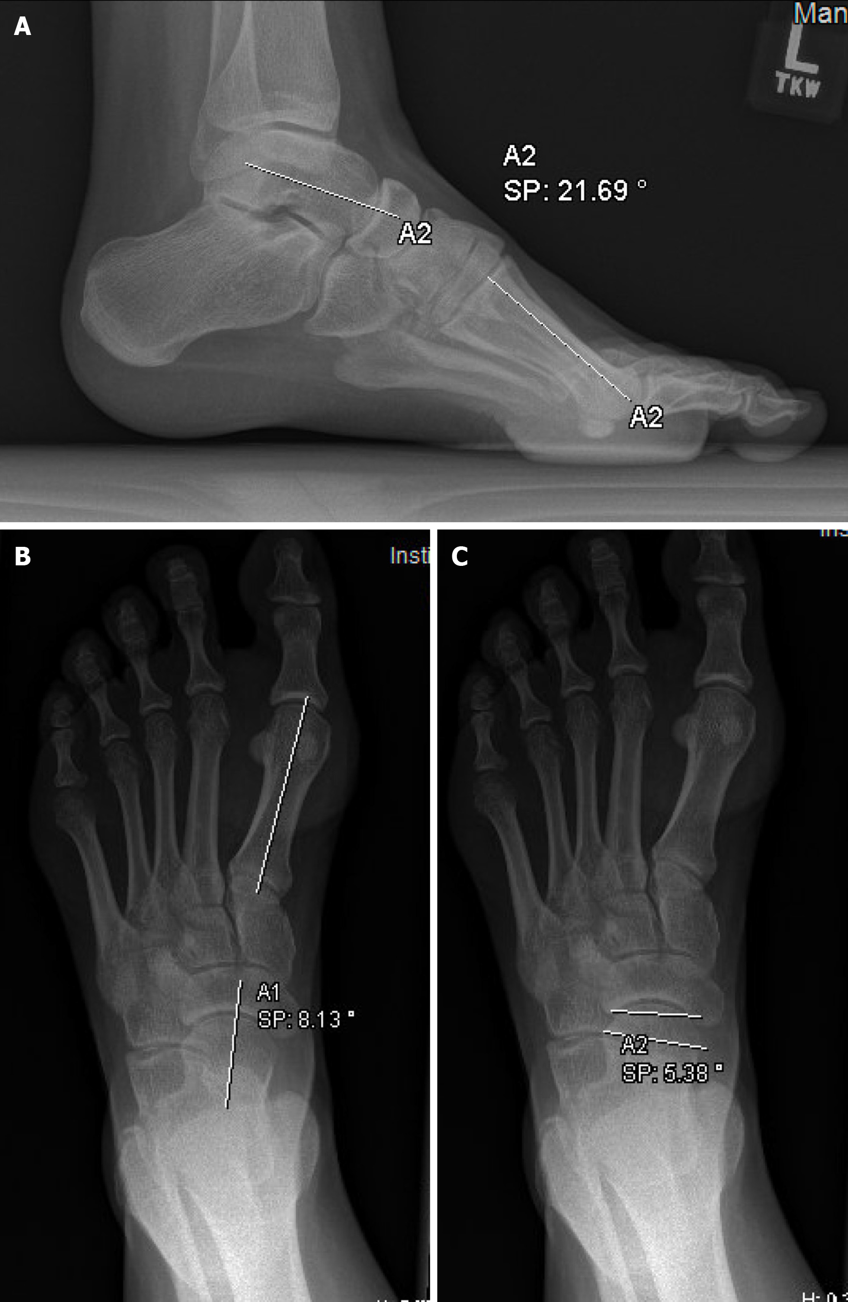
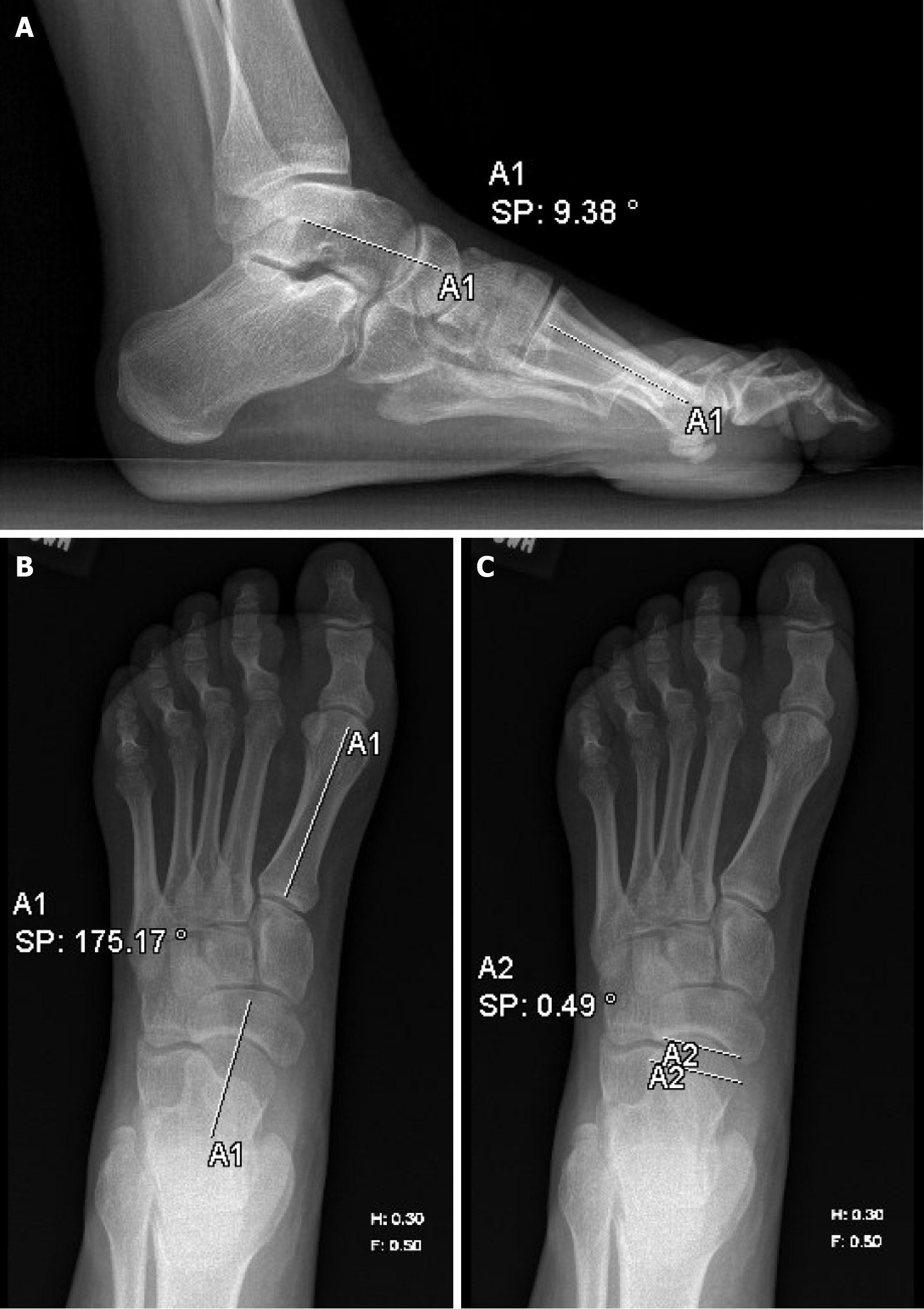
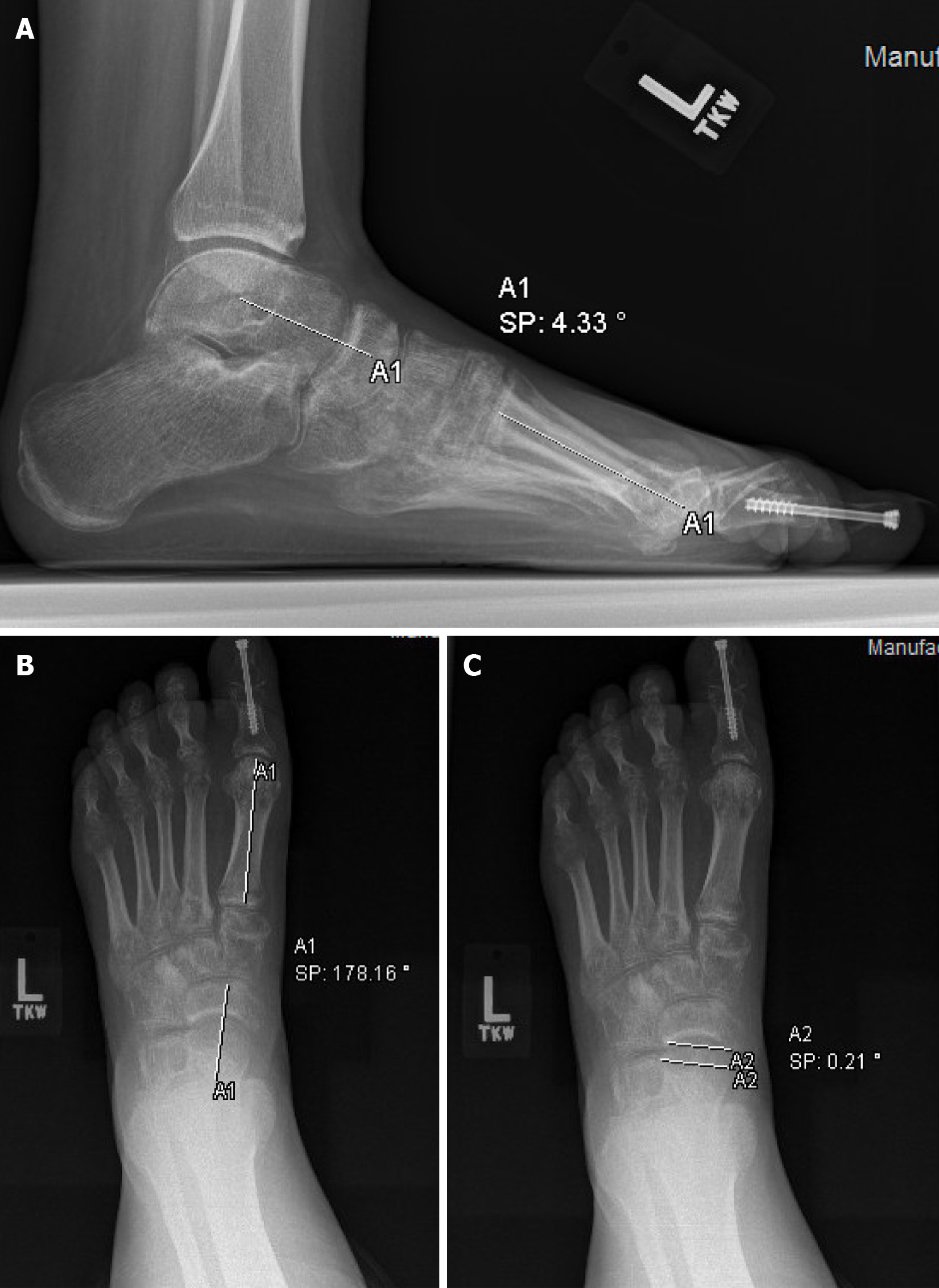
Preoperative measurements were based on radiographs obtained prior to the first stage of the operation, while postoperative measurements were based on radiographs obtained after both the first and second stages of the operation. The postoperative radiographic measurements were based on radiographs taken at least 6 months after the second stage of the surgery to ensure that the reported results reflect a lasting correction. 7 of the 11 patients included in the radiographic analysis had X-rays taken between the first and second stage of the operation, while 4 did not. Six radiographic angles were measured on both the preoperative and postoperative weightbearing radiographs. Meary’s angle was defined as positive in the case of midfoot cavus and negative in the case of midfoot planus. The anteroposterior talo-first metatarsal (AP TMT1) angle was defined as positive in the case of forefoot adduction and negative in the case of forefoot abduction. The talonavicular coverage angle was defined as positive in the case of midfoot abduction and negative in the case of midfoot adduction. All X-ray imaging analyses were verified by a fellowship-trained pediatric orthopaedic surgeon. PACS software was utilized to analyze all radiographs.
Charts were also reviewed for patient age at the time of surgery, gender, race, and postoperative complications. Preoperative and postoperative radiographic outcome measurements were compared using a two-sample t-test. A P value of 0.05 was considered significant for all analyses. All statistical analysis was performed in R-4.1.2.
A total of 19 patients were included in the analysis. 7 (36.8%) patients were male, and 12 (63.2%) were female. 13 (68.4%) patients were Caucasian, 3 (15.8%) were African American, and 3 (15.8%) were Asian. The average age at the time of surgery was 9.9 years (SD = 3.3; range 4 to 17).
All patients in the study population underwent two-stage correction of the cavovarus deformity with radical plantar fascia release and osteotomy. Superficial and deep plantar medial release as described by Mosca were performed at the first stage[6]. This involved release of the three origins of the abductor hallucis, the plantar fascia, the plantar and medial talonavicular capsule and release or lengthening (if innervated) of the posterior tibial tendon. This was followed by one month of weekly serial casting. A plantar opening wedge (with allograft) osteotomy of the medial cuneiform bone and/or a calcaneal slide osteotomy was then performed to correct residual cavus or varus, respectively. Tendon transfers were performed at the second stage and were dependent on functional motor level and the presence or absence of an exaggerated windlass mechanism. Additional soft tissue and osseous procedures are summarized in Table 2. Of the 19 feet analyzed, 13 (68.4%) cases underwent cuneiform osteotomy, and 6 (31.6%) cases underwent calcaneal osteotomy. The cumulative average number of cast changes throughout the postoperative course of the two-stage operation was 4.0 ± 1.2 casts. Average follow-up time was 43.8 ± 33.8 months (range 9-118 months).
| Soft tissue and osseous procedures | Number of patients undergoing procedure |
| Talonavicular joint capsule release | 19 |
| Abductor hallucis release | 19 |
| Tibialis posterior lengthening/release | 18 |
| Peroneus longus to peroneus brevis transfer | 13 |
| Jones transfer (extensor hallucis longus transfer to first metatarsal neck) | 10 |
| Flexor hallucis longus lengthening/release | 9 |
| Flexor digitorum longus lengthening/release | 9 |
| Great toe interphalangeal joint fusion | 7 |
| Achilles tendon lengthening | 6 |
| Calcaneocuboid joint capsule release | 4 |
| Extensor hallucis longus to extensor hallucis brevis transfer | 2 |
| Tibialis anterior lengthening | 1 |
| Split posterior tibial tendon transfer to peroneus brevis | 1 |
| Anterior tibial tendon transfer to lateral cuneiform | 1 |
Significant changes between preoperative and postoperative radiographic angle measurements were seen in Meary’s angle, the AP first tarsometatarsal angle, and the talonavicular coverage angle. The average Meary’s angle changed from 17.9 ± 13.1 preoperatively to 4.7 ± 10.3 following the second stage of the operation (P = 0.016). The mean AP TMT1 angle changed from 20.6 ± 15.1 preoperatively and 9.3 ± 5.5 following the second stage of the operation (P = 0.011). The mean talonavicular coverage changed from -10.3 ± 9.6 preoperatively to -3.8 ± 10.1 following the second stage of the operation
| Angle measurement | Average preoperative value (range) | Average value after stage 11 (range) | Average value after stage 2 (range) | P value preoperative to post-stage 12 | P value post-stage 1 to post-stage 22 | P value preoperative to post-stage 2 |
| Meary’s | 17.9 ± 13.1 (3.0-50.1) | 13.8 ± 7.1 (4.4-25.3) | 4.7 ± 10.3 (-8.0 to 21.) | 0.47 | 0.056 | 0.016a |
| Calcaneal pitch | 22.4 ± 4.0 (16.4-29.6) | 25.0 ± 6.3 (15.8-32.7) | 21.3 ± 7.6 (12.3-32.4) | 0.30 | 0.30 | 0.53 |
| AP TMT1 | 20.6 ± 15.1 (6.2-40.8) | 8.4 ± 7.4 (1.4-21.5) | 9.3 ± 5.5 (1.5-22.9) | 0.066 | 0.78 | 0.011a |
| Lateral talocalcaneal | 43.6 ± 9.8 (22.9-57.9) | 42.6 ± 10.7 (26.2-53.0) | 41.7 ± 9.2 (27.5-51.1) | 0.85 | 0.86 | 0.46 |
| AP talocalcaneal | 15.8 ± 6.7 (6.3-25.0) | 16.7 ± 9.8 (3.5-29.7) | 18.9 ± 8.6 (8.7-33.9) | 0.81 | 0.62 | 0.32 |
| Talonavicular coverage | -10.3 ± 9.6 (-28.3 to 1.2) | -4.0 ± 7.2 (-11.0 to 9.3) | -3.8 ± 10.1 (-22.1 to 16) | 0.16 | 0.97 | 0.025a |
Within the first nine months of follow-up from the second stage of the operation, only 1 (5.3%) patient in our cohort developed a post-operative complication. The patient required reoperation with Z-lengthening of the Achilles tendon due to an equinus contracture. There were no cases of postoperative infections, neuropathic ulcers, nerve injury, or non-union.
Pes cavus or cavovarus is a common foot deformity in children and adults with various neurologic disorders[3-5]. The orthopaedic literature discussing the treatment of cavus or cavovarus feet has typically grouped individuals with various diagnoses[4,5,16-20]. However, there is a paucity of literature addressing the correction of cavovarus specifically in children with SB. The two-stage procedure for pes cavovarus correction in pediatric patients was refined by Mosca[6]. Based on the results of the current study, the authors advocate for the use of a two-stage, rather than single-stage, operation for the correction of these deformities in patients with SB. While muscle imbalance leading to soft tissue contracture may be the primary factor precipitating the deformity, bony deformity develops over time[1]. The results of this study demonstrate the importance of addressing both the soft tissue and osseous elements of the deformity. Radical plantar release, the primary component of the first stage of our treatment algorithm, has been previously validated as an effective procedure for cavus correction[8]. Plantar opening wedge osteotomy of the medial cuneiform may not be feasible at the time of the first stage as relaxation of the soft tissues with serial casting needs to occur to gain length[6]. The second stage addresses residual bony deformity that does not resolve with serial casting after the first stage. This approach substantiates previous literature that has shown positive outcomes when utilizing a combination of soft tissue release and osteotomy[11].
Weightbearing radiographic analysis is the primary determinant of clinical management in pediatric foot deformities[21,22]. In addition to helping guide clinical decision making, analysis of these angles can be used to assess outcomes following corrective procedures for malalignment, including pes cavovarus. Davids et al[23] described weight bearing radiographic measurements that assess segmental alignment of the foot. Both intraobserver and interobserver reliability of these radiographic measurements is excellent. Utilizing the Wicart grading system for the assessment of therapeutic efficacy of cavovarus foot correction[2] (Table 4), our results demonstrate good correction of the deformity, with the average Meary’s angle decreasing from 17.9 ± 13.1 degrees preoperatively to 4.7 ± 10.3 degrees postoperatively (P = 0.016). The results also demonstrate significant improvement of both the AP TMT1 angle (P = 0.011) and talonavicular coverage (P = 0.025), respectively. Furthermore, in 7 of the 11 feet included for radiographic follow-up that had radiographs obtained between the first and second stages, correction of Meary’s, AP TMT1 and talonavicular coverage angles were not significant after the first stage but reached statistical significance after the second stage.
| Correction score | M |
| Very good | 0 ≤ M < 5 |
| Good | 5 ≤ M < 15 |
| Fair | 15 ≤ M < 20 |
| Poor | M ≥ 20 or need for revision surgery |
The radiological outcomes of this study compare favorably to previous literature assessing these parameters following corrective surgery for pes cavovarus[11,19,20]. In a case series of 24 cavovarus feet undergoing correction via plantar fascia release and first metatarsal dorsal hemiepiphysiodesis, Sanpera et al[20] report a mean difference of -6.2 and -8.2 degrees in Meary’s angle and the AP TMT1 angle, respectively. Our data shows mean differences of -13.2 degrees in Meary’s angle and -11.3 degrees in the AP TMT1 angle. Additionally, Chen et al[11] report an average postoperative Meary’s angle of 6.36 ± 1.810 following a single-stage operation consisting of soft tissue release and osteotomy. The results of our study show an average postoperative Meary’s angle of 4.7 ± 10.3 after the two-stage technique described here.
There was a low incidence of post-operative complications among the patients in our study. Pressure sore development in patients with SB has a reported incidence up to 60%, and the feet are a commonly affected anatomic location[24,25]. Within the first nine months of follow-up after the second stage of the operation, no patients in our cohort developed neuropathic ulceration. There was one patient that required re-operation for correction of an equinus contracture.
To the authors’ knowledge, this is the first paper to specifically describe a treatment algorithm for cavus and cavovarus in patients with SB. Limitations of the study include its retrospective nature and relatively small sample size. Also, due to the treatment algorithm, it is impossible to blind stage of treatment in the radiographic analyses. As exemplified in Figure 3, osseous procedures such as the corrective osteotomy and hallux interphalangeal joint arthrodesis are easily identifiable. Therefore, the senior author recognizes that such images must have been obtained after the second stage of the operation. Additionally, the study does not specifically evaluate clinical outcome measures such as the American Orthopedic Foot and Ankle Society score or pedobarographic data. However, efficacy of the operation was evaluated through radiographic parameters which have been shown to correlate with correction[2]. Lastly, a portion of our patients did not have final weightbearing films and thus were not included in the radiographic analysis. Future studies could include a prospective design that would allow for functional scoring both pre- and postoperatively with a larger sample size to better characterize the clinical outcomes of cavovarus correction.
Two-stage operation consisting of plantar fascia release followed by osteotomy has been previously described as an effective treatment option for the surgical correction of pediatric pes cavovarus. No study exists, to date, describing the treatment for children and adolescents with SB. The results of this study demonstrate that a two-stage treatment algorithm consisting of plantar fascia release and osteotomy produced good correction of the cavovarus deformity in these patients. The two-stage procedure promoted the sequential correction of residual bony or soft tissue contributions to the deformity that may not be adequately corrected by a single-stage operation. When surgical intervention becomes necessary, the authors advocate for consideration of the two-stage operation presented here.
| 1. | Swaroop VT, Dias L. Orthopaedic management of spina bifida-part II: foot and ankle deformities. J Child Orthop. 2011;5:403-414. [RCA] [PubMed] [DOI] [Full Text] [Cited by in Crossref: 50] [Cited by in RCA: 52] [Article Influence: 3.7] [Reference Citation Analysis (1)] |
| 2. | Wicart P. Cavus foot, from neonates to adolescents. Orthop Traumatol Surg Res. 2012;98:813-828. [RCA] [PubMed] [DOI] [Full Text] [Cited by in Crossref: 45] [Cited by in RCA: 38] [Article Influence: 2.9] [Reference Citation Analysis (0)] |
| 3. | Laurá M, Singh D, Ramdharry G, Morrow J, Skorupinska M, Pareyson D, Burns J, Lewis RA, Scherer SS, Herrmann DN, Cullen N, Bradish C, Gaiani L, Martinelli N, Gibbons P, Pfeffer G, Phisitkul P, Wapner K, Sanders J, Flemister S, Shy ME, Reilly MM; Inherited Neuropathies Consortium. Prevalence and orthopedic management of foot and ankle deformities in Charcot-Marie-Tooth disease. Muscle Nerve. 2018;57:255-259. [RCA] [PubMed] [DOI] [Full Text] [Full Text (PDF)] [Cited by in Crossref: 24] [Cited by in RCA: 51] [Article Influence: 6.4] [Reference Citation Analysis (0)] |
| 4. | Olney B. Treatment of the cavus foot. Deformity in the pediatric patient with Charcot-Marie-Tooth. Foot Ankle Clin. 2000;5:305-315. [PubMed] [DOI] [Full Text] |
| 5. | Ziebarth K, Krause F. Updates in Pediatric Cavovarus Deformity. Foot Ankle Clin. 2019;24:205-217. [RCA] [PubMed] [DOI] [Full Text] [Cited by in Crossref: 5] [Cited by in RCA: 5] [Article Influence: 0.8] [Reference Citation Analysis (0)] |
| 6. | Mosca VS. The cavus foot. J Pediatr Orthop. 2001;21:423-424. [PubMed] [DOI] [Full Text] |
| 7. | Sanpera I, Villafranca-Solano S, Muñoz-Lopez C, Sanpera-Iglesias J. How to manage pes cavus in children and adolescents? EFORT Open Rev. 2021;6:510-517. [RCA] [PubMed] [DOI] [Full Text] [Full Text (PDF)] [Reference Citation Analysis (0)] |
| 8. | Schwend RM, Drennan JC. Cavus foot deformity in children. J Am Acad Orthop Surg. 2003;11:201-211. [RCA] [PubMed] [DOI] [Full Text] [Cited by in Crossref: 89] [Cited by in RCA: 67] [Article Influence: 3.0] [Reference Citation Analysis (0)] |
| 9. | Wicart P, Seringe R. Plantar opening-wedge osteotomy of cuneiform bones combined with selective plantar release and dwyer osteotomy for pes cavovarus in children. J Pediatr Orthop. 2006;26:100-108. [RCA] [PubMed] [DOI] [Full Text] [Cited by in Crossref: 47] [Cited by in RCA: 38] [Article Influence: 2.0] [Reference Citation Analysis (0)] |
| 10. | Mubarak SJ, Van Valin SE. Osteotomies of the foot for cavus deformities in children. J Pediatr Orthop. 2009;29:294-299. [RCA] [PubMed] [DOI] [Full Text] [Cited by in Crossref: 41] [Cited by in RCA: 32] [Article Influence: 2.0] [Reference Citation Analysis (0)] |
| 11. | Chen ZY, Wu ZY, An YH, Dong LF, He J, Chen R. Soft tissue release combined with joint-sparing osteotomy for treatment of cavovarus foot deformity in older children: Analysis of 21 cases. World J Clin Cases. 2019;7:3208-3216. [RCA] [PubMed] [DOI] [Full Text] [Full Text (PDF)] [Cited by in CrossRef: 4] [Cited by in RCA: 7] [Article Influence: 1.2] [Reference Citation Analysis (0)] |
| 12. | Wukich DK, Bowen JR. A long-term study of triple arthrodesis for correction of pes cavovarus in Charcot-Marie-Tooth disease. J Pediatr Orthop. 1989;9:433-437. [PubMed] [DOI] [Full Text] |
| 13. | Maynard MJ, Weiner LS, Burke SW. Neuropathic foot ulceration in patients with myelodysplasia. J Pediatr Orthop. 1992;12:786-788. [RCA] [PubMed] [DOI] [Full Text] [Cited by in Crossref: 29] [Cited by in RCA: 27] [Article Influence: 0.8] [Reference Citation Analysis (0)] |
| 14. | Frawley PA, Broughton NS, Menelaus MB. Incidence and type of hindfoot deformities in patients with low-level spina bifida. J Pediatr Orthop. 1998;18:312-313. [PubMed] [DOI] [Full Text] |
| 15. | Tubbs RS, Winters RG, Naftel RP, Acharya VK, Conklin M, Shoja MM, Loukas M, Oakes WJ. Predicting orthopedic involvement in patients with lipomyelomeningoceles. Childs Nerv Syst. 2007;23:835-838. [RCA] [PubMed] [DOI] [Full Text] [Cited by in Crossref: 2] [Cited by in RCA: 2] [Article Influence: 0.1] [Reference Citation Analysis (0)] |
| 16. | Lee MC, Sucato DJ. Pediatric issues with cavovarus foot deformities. Foot Ankle Clin. 2008;13:199-219, v. [RCA] [PubMed] [DOI] [Full Text] [Cited by in Crossref: 17] [Cited by in RCA: 13] [Article Influence: 0.8] [Reference Citation Analysis (0)] |
| 17. | Dreher T, Beckmann NA, Wenz W. Surgical Treatment of Severe Cavovarus Foot Deformity in Charcot-Marie-Tooth Disease. JBJS Essent Surg Tech. 2015;5:e11. [RCA] [PubMed] [DOI] [Full Text] [Full Text (PDF)] [Cited by in Crossref: 10] [Cited by in RCA: 10] [Article Influence: 1.0] [Reference Citation Analysis (0)] |
| 18. | Lin T, Gibbons P, Mudge AJ, Cornett KMD, Menezes MP, Burns J. Surgical outcomes of cavovarus foot deformity in children with Charcot-Marie-Tooth disease. Neuromuscul Disord. 2019;29:427-436. [RCA] [PubMed] [DOI] [Full Text] [Cited by in Crossref: 12] [Cited by in RCA: 17] [Article Influence: 2.8] [Reference Citation Analysis (0)] |
| 19. | Simon AL, Seringe R, Badina A, Khouri N, Glorion C, Wicart P. Long term results of the revisited Meary closing wedge tarsectomy for the treatment of the fixed cavo-varus foot in adolescent with Charcot-Marie-Tooth disease. Foot Ankle Surg. 2019;25:834-841. [RCA] [PubMed] [DOI] [Full Text] [Cited by in Crossref: 8] [Cited by in RCA: 13] [Article Influence: 2.2] [Reference Citation Analysis (0)] |
| 20. | Sanpera I Jr, Frontera-Juan G, Sanpera-Iglesias J, Corominas-Frances L. Innovative treatment for pes cavovarus: a pilot study of 13 children. Acta Orthop. 2018;89:668-673. [RCA] [PubMed] [DOI] [Full Text] [Full Text (PDF)] [Cited by in Crossref: 4] [Cited by in RCA: 4] [Article Influence: 0.6] [Reference Citation Analysis (0)] |
| 21. | Mosca VS. Calcaneal lengthening for valgus deformity of the hindfoot. Results in children who had severe, symptomatic flatfoot and skewfoot. J Bone Joint Surg Am. 1995;77:500-512. [RCA] [PubMed] [DOI] [Full Text] [Cited by in Crossref: 362] [Cited by in RCA: 278] [Article Influence: 9.3] [Reference Citation Analysis (0)] |
| 22. | Sutherland DH. Varus foot in cerebral palsy: an overview. Instr Course Lect. 1993;42:539-543. [PubMed] |
| 23. | Davids JR, Gibson TW, Pugh LI. Quantitative segmental analysis of weight-bearing radiographs of the foot and ankle for children: normal alignment. J Pediatr Orthop. 2005;25:769-776. [RCA] [PubMed] [DOI] [Full Text] [Cited by in Crossref: 161] [Cited by in RCA: 161] [Article Influence: 8.5] [Reference Citation Analysis (0)] |
| 24. | Akbar M, Bresch B, Seyler TM, Wenz W, Bruckner T, Abel R, Carstens C. Management of orthopaedic sequelae of congenital spinal disorders. J Bone Joint Surg Am. 2009;91 Suppl 6:87-100. [RCA] [PubMed] [DOI] [Full Text] [Cited by in Crossref: 21] [Cited by in RCA: 21] [Article Influence: 1.3] [Reference Citation Analysis (0)] |
| 25. | Conklin MJ, Hopson B, Arynchyna A, Atchley T, Trapp C, Rocque BG. Skin breakdown of the feet in patients with spina bifida: Analysis of risk factors. J Pediatr Rehabil Med. 2018;11:237-241. [RCA] [PubMed] [DOI] [Full Text] [Cited by in Crossref: 1] [Cited by in RCA: 2] [Article Influence: 0.3] [Reference Citation Analysis (0)] |


