Published online May 18, 2024. doi: 10.5312/wjo.v15.i5.477
Revised: January 24, 2024
Accepted: April 7, 2024
Published online: May 18, 2024
Processing time: 132 Days and 21.9 Hours
Congenital absence of the menisci is a rare anatomical variation characterized by the absence or underdevelopment of one or both menisci in the knee joint. The menisci are crucial in load distribution, joint stability, and shock absorption. Understanding the clinical presentation, diagnosis, and management of this condition is important for optimal patient care.
A 27-year-old male with a long-standing history of knee pain underwent diagn
This case of congenital absence of the menisci highlights the diagnostic challenges posed by rare anomalies. The diagnostic arthroscopy played a crucial role in iden
Core Tip: Congenital absence of the menisci is a rare anatomical variation characterized by the absence or underdevelopment of one or both menisci in the knee joint. We present a young male patient with a long-standing history of knee pain who underwent diagnostic arthroscopy, revealing the congenital absence of the lateral meniscus. The patient's clinical findings, imaging results, and surgical procedure are presented, along with relevant images. This case is notable for the congenital absence of the meniscus, a finding that contributes to the literature on such rare anomalies.
- Citation: Alkhunayfir HA, AlQahtani AA, Korkoman AJ. Congenital absence of the lateral meniscus: A case report. World J Orthop 2024; 15(5): 477-482
- URL: https://www.wjgnet.com/2218-5836/full/v15/i5/477.htm
- DOI: https://dx.doi.org/10.5312/wjo.v15.i5.477
Congenital absence of the menisci represents a rare anatomical variation where one or both menisci are absent or und
Clinically, patients diagnosed with congenital absence of the menisci often present with knee pain, instability, and limited range of motion. The absence of these significant structures compromises the load-bearing capacity of the knee joint, leading to altered biomechanics and increased stress on the articular surfaces[3]. In some cases, patients may experience mechanical symptoms such as catching, clicking, or locking of the knee.
Diagnosing congenital absence of the menisci can be challenging, as the condition may remain unnoticed until symp
Management strategies for congenital absence of the menisci depend on the severity of symptoms and associated findings. Non-operative approaches, including physical therapy, analgesic medications, and activity modification, may initially alleviate pain and improve knee function[4]. However, in cases where symptoms persist or significant structural abnormalities are present, surgical intervention may be necessary.
Surgical options range from meniscal repair or transplantation to partial or total meniscectomy. The choice of proce
A 27-year-old male with no significant medical history presented with a complaint of left knee pain that started 15 years ago.
The patient first experienced pain in his left knee during childhood, and it has been intermittent ever since, localized to his left knee without involvement of any other joints. The pain progressively aggravated with activity and improved with rest. The patient reported no recent history of trauma to the knee and denied any catching or giving way sensation. However, he did mention experiencing clicking sounds during knee movements. Furthermore, no accompanying symptoms such as fever, night sweats, weight loss, or loss of appetite were observed. Over the past year, the pain has become constant, even without any recent traumatic event.
The patient recalled experiencing similar symptoms when he was 12 years old, including catching and clicking sensations in the knee and an inability to fully flex it. At that time, the symptoms improved with time and physical therapy. The patient had no known allergies and was not ingesting any analgesic medications. He had undergone physical therapy on multiple occasions, which had provided some improvement in his symptoms.
The patient had no significant past medical, surgical, or family history.
Upon examination, no visible swelling, erythema, or signs of infection were observed around the knee. The contour of the knee appeared normal, with no tenderness along the joint line. Palpation of the knee elicited neither pain nor discomfort. Range of motion was not limited, with extension measuring -5° up to 130° flexion. Tests performed, including the Lachmann test, anterior drawer test, and posterior drawer test, yielded negative results. No sag sign was observed, and the valgus and varus tests demonstrated stability. The dial test and grinding test were also negative. Furthermore, X-ray imaging displayed no signs of arthritis or widening of the joint line.
No abnormalities were revealed in the laboratory examinations.
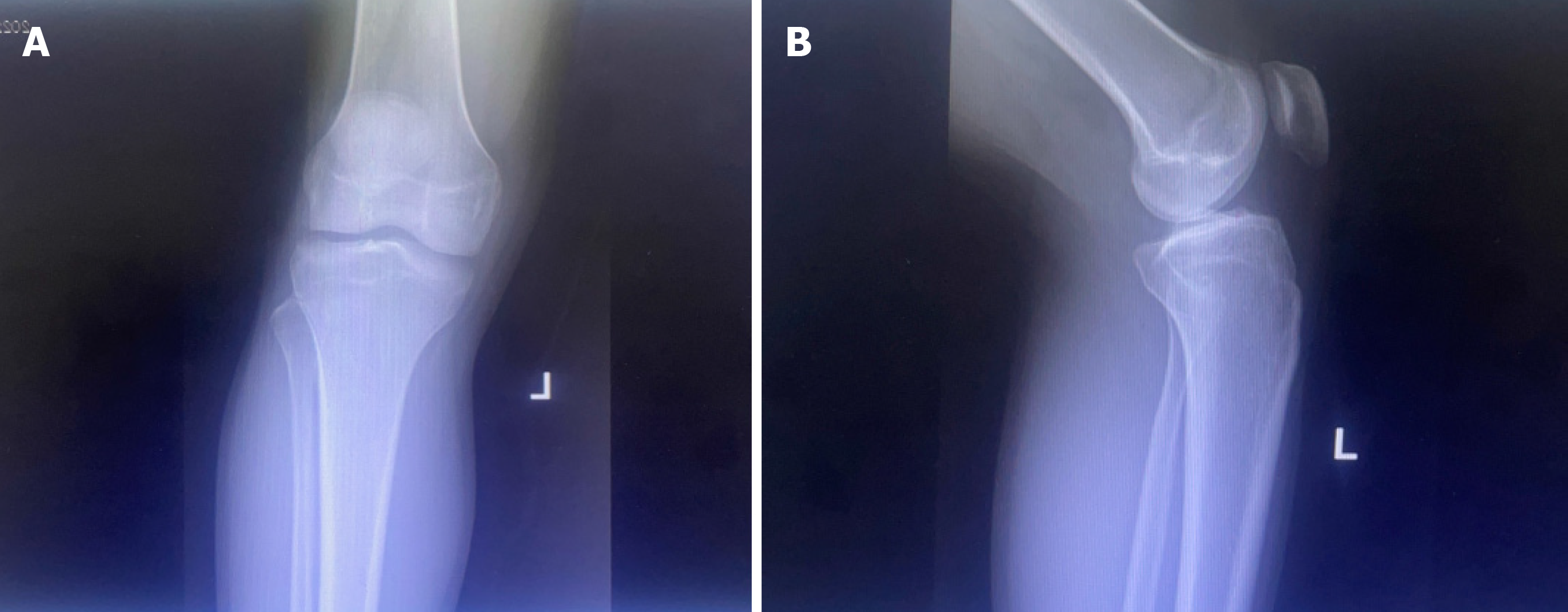
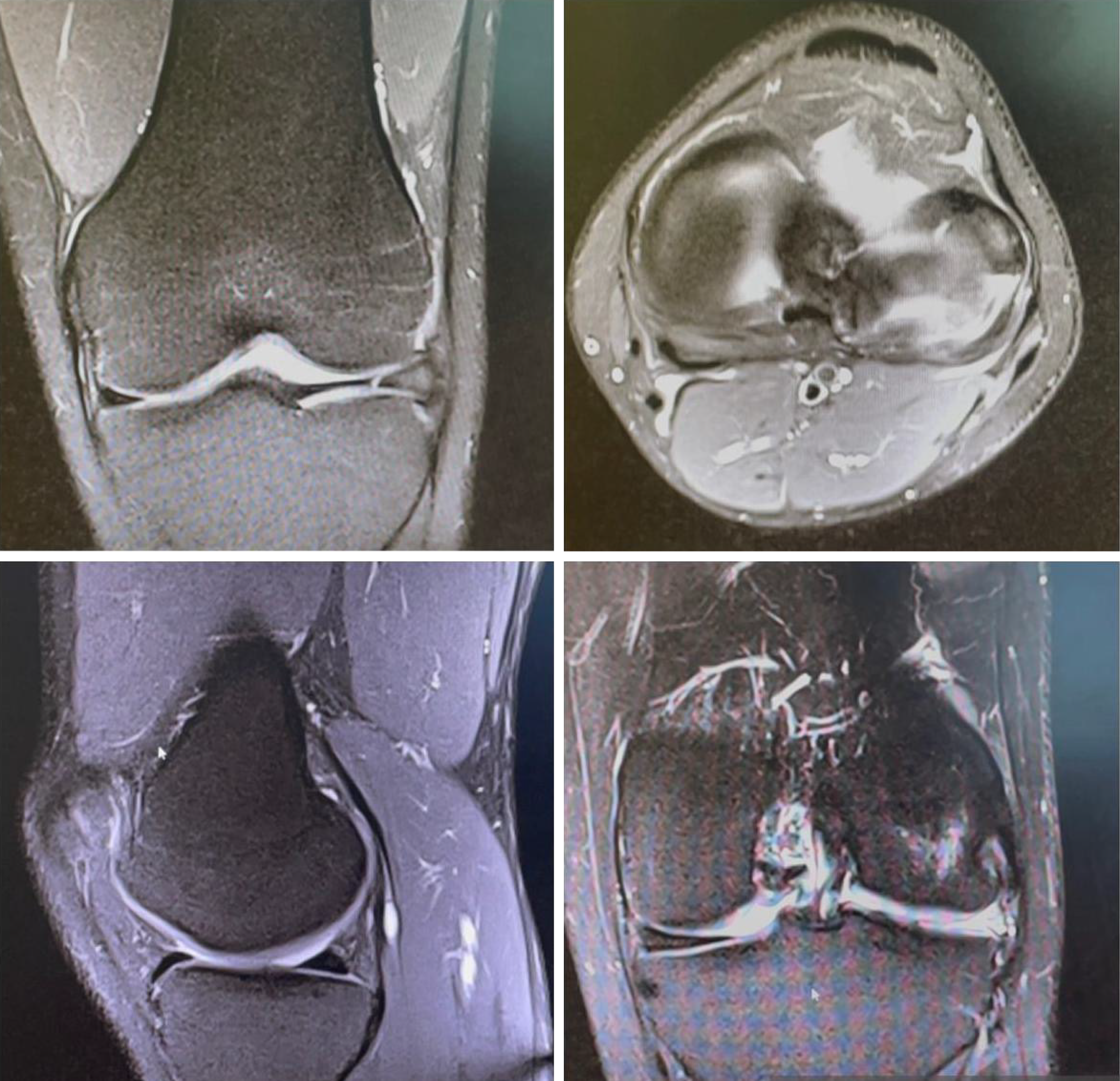
Plain X-ray imaging of the left knee joint demonstrated normal visualized bone densities with no evidence of fracture or dislocation (Figure 1A). The joint spaces were preserved, and the soft tissues appeared unremarkable, as displayed in Figure 1B. MRI of the patient confirmed that the anterior and posterior cruciate ligaments were intact. Significant attenuation and abnormal morphology of the posterior aspect of the body and posterior horn of the lateral meniscus were observed, along with thickening of the anterior meniscal tissue. The integrity of the medial meniscus was preserved. The collateral ligaments and extensor mechanisms were intact, with no evidence of joint effusion. A small popliteal cyst was observed. Furthermore, focal deep cartilage fissuring was noted at the posterior aspect of the lateral femoral condyle (Figure 2).
Based on the history, physical examination, imaging, and diagnostic arthroscopy, the patient was diagnosed with cong
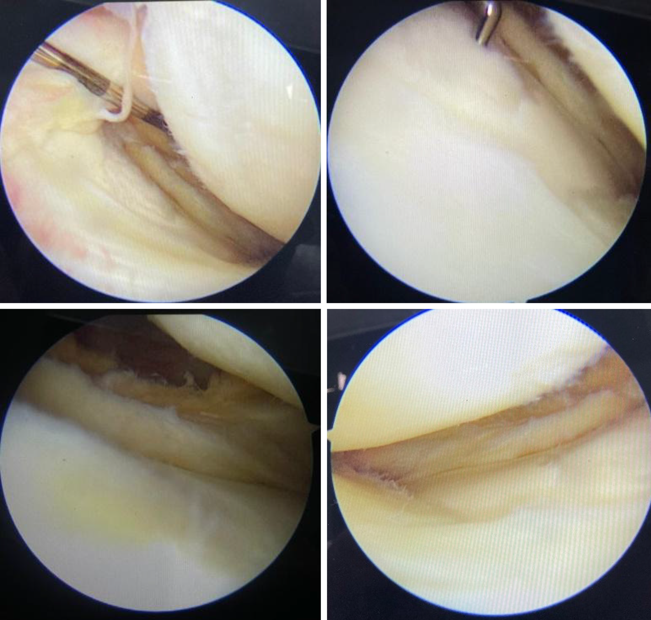
During the diagnostic arthroscopy, the absence of the lateral meniscus was discovered in the patient (Figure 3). No other surgical intervention was required. The patient continued with a range of motion and strengthening physical therapy for six months.
Clinically, the patient demonstrated improvement on a one-year follow-up with a pain score of zero and an Oxford knee score of 48.
The meniscus, one of the contributors to knee stability, is composed of fibrous cartilage[5]. The characteristic meniscus shape is formed by the eighth week of gestation when the knee is fully formed. The lateral femoral condyle is located at a high position with significant lateral long-axis deviation. The lateral tibial plateau is more circular than the medial tibial plateau. This normal anatomical variation causes asymmetric knee movement. The lateral meniscus shape, which is more widely variable than the medial meniscus, seems to adapt to the asymmetric knee motion. Such morphological variation could be involved in lateral meniscus anomalies[6].
The presented case is of a 27-year-old male with a long history of knee pain, tracing back to childhood, without any significant traumatic events. One striking finding is the congenital absence of the meniscus, discovered during diagnostic arthroscopy. This finding aligns with the rare occurrence of congenital meniscal anomalies as discussed in the initial literature.
The anomalies typically affect the lateral side of the knee, as supported by the significant attenuation of the posterior aspect of the body and posterior horn of the lateral meniscus in this patient. Contrary to Sachleben et al's[7] findings, no notable abnormalities were identified in the patient's articular surfaces or cruciate ligaments.
In the discussed literature, it is mentioned that the absence of the meniscus should lead to some degree of wear on the articular surfaces. This aligns with the finding of focal deep cartilage fissuring at the posterior aspect of the lateral femoral condyle in our patient, suggesting a certain level of cartilage damage despite the absence of typical signs of arthritis on the X-ray[7].
A unique aspect of this case is the congenital absence of the meniscus, which adds valuable insights to the literature on this uncommon anomaly. Although meniscal hypoplasia has previously been reported, isolated bilateral hypoplasia of the medial meniscus remains a first, as per available knowledge[8]. The case also emphasizes the importance of exploring the cause of knee pain via diagnostic arthroscopy, especially when symptoms persist despite conservative treatments like physical therapy.
Generally, lateral knee anomalies commonly involve a discoid meniscus, with only a few described cases of bilateral lateral meniscus hypoplasia and partial deficiency[9].
Finally, the natural history of these anomalies and their effect on normal knee function remains unclear. The patient's significant improvement post-arthroscopy and physical therapy is encouraging. The improvement suggests that even with rare meniscal anomalies, adequate diagnosis and management can lead to favorable clinical outcomes, including the reduction of pain and improvement of function.
In summary, this case presentation contributes to our understanding of congenital meniscal anomalies, particularly the rare occurrence of the absence of the lateral meniscus. Diagnostic arthroscopy played a crucial role in uncovering the congenital absence of the meniscus and identifying associated structural changes. The successful management of the patient's symptoms through physical therapy underscores the significance of individualized treatment approaches. In the future, delving deep into the management strategies for such rare anomalies. This will help improve patient outcomes and optimize the care of individuals with congenital meniscal anomalies.
Provenance and peer review: Unsolicited article; Externally peer-reviewed.
Peer-review model: Single-blind
Specialty type: Orthopedics
Country/Territory of origin: Saudi Arabia
Peer-review report’s classification
Scientific Quality: Grade B
Novelty: Grade B
Creativity or Innovation: Grade B
Scientific Significance: Grade B
P-Reviewer: Al-Mnayyis A, Jordan S-Editor: Lin C L-Editor: A P-Editor: Zhao YQ
| 1. | Barrett GR, Tomasin JD. Bilateral congenital absence of the anterior cruciate ligament. Orthopedics. 1988;11:431-434. [RCA] [PubMed] [DOI] [Full Text] [Cited by in Crossref: 18] [Cited by in RCA: 14] [Article Influence: 0.4] [Reference Citation Analysis (0)] |
| 2. | Fithian DC, Kelly MA, Mow VC. Material properties and structure-function relationships in the menisci. Clin Orthop Relat Res. 1990;19-31. [PubMed] |
| 3. | Maheshwer B, Williams BT, Polce EM, LaPrade RF, Chahla J. Role of the Meniscus in Cartilage Injury: Basic Science. In: Cartilage Injury of the Knee. Cham: Springer International Publishing, 2021: 131-142. |
| 4. | Zhang Y, Chen X, Tong Y, Luo J, Bi Q. Development and Prospect of Intra-Articular Injection in the Treatment of Osteoarthritis: A Review. J Pain Res. 2020;13:1941-1955. [RCA] [PubMed] [DOI] [Full Text] [Full Text (PDF)] [Cited by in Crossref: 12] [Cited by in RCA: 37] [Article Influence: 7.4] [Reference Citation Analysis (0)] |
| 5. | Fukazawa I, Hatta T, Uchio Y, Otani H. Development of the meniscus of the knee joint in human fetuses. Congenit Anom (Kyoto). 2009;49:27-32. [RCA] [PubMed] [DOI] [Full Text] [Cited by in Crossref: 25] [Cited by in RCA: 27] [Article Influence: 1.7] [Reference Citation Analysis (0)] |
| 6. | Finnilä MAJ, Das Gupta S, Turunen MJ, Hellberg I, Turkiewicz A, Lutz-Bueno V, Jonsson E, Holler M, Ali N, Hughes V, Isaksson H, Tjörnstrand J, Önnerfjord P, Guizar-Sicairos M, Saarakkala S, Englund M. Mineral Crystal Thickness in Calcified Cartilage and Subchondral Bone in Healthy and Osteoarthritic Human Knees. J Bone Miner Res. 2022;37:1700-1710. [RCA] [PubMed] [DOI] [Full Text] [Full Text (PDF)] [Cited by in Crossref: 1] [Cited by in RCA: 13] [Article Influence: 4.3] [Reference Citation Analysis (0)] |
| 7. | Sachleben BC, Nasreddine AY, Nepple JJ, Tepolt FA, Kasser JR, Kocher MS. Reconstruction of Symptomatic Congenital Anterior Cruciate Ligament Insufficiency. J Pediatr Orthop. 2019;39:59-64. [RCA] [PubMed] [DOI] [Full Text] [Cited by in Crossref: 7] [Cited by in RCA: 7] [Article Influence: 1.2] [Reference Citation Analysis (0)] |
| 8. | Yang S, Zhang S, Li R, Yang C, Zheng J, Wang C, Lu J, Zhang Z, Shang X, Zhang H, Wang W, Li W, Huang J, Zhang Y, Wang J, Wang Y, Zheng X, Chen G, Hua Y, Chen S, Li J. Chinese Experts Consensus and Practice Guideline on Discoid Lateral Meniscus. Orthop Surg. 2023;15:915-929. [RCA] [PubMed] [DOI] [Full Text] [Cited by in RCA: 11] [Reference Citation Analysis (0)] |
| 9. | Tetik O, Doral MN, Atay OA, Leblebicioğlu G, Türker S. Partial deficiency of the lateral meniscus. Arthroscopy. 2003;19:E42. [RCA] [PubMed] [DOI] [Full Text] [Cited by in Crossref: 21] [Cited by in RCA: 21] [Article Influence: 1.0] [Reference Citation Analysis (0)] |


