Editorial Open Access
Copyright ©The Author(s) 2024. Published by Baishideng Publishing Group Inc. All rights reserved.
World J Orthop. Dec 18, 2024; 15(12): 1118-1123
Published online Dec 18, 2024. doi: 10.5312/wjo.v15.i12.1118
Specific approach to total hip arthroplasty in patients with childhood hip disorders sequelae
Katarina Barbaric Starcevic, Department of Orthopedic Surgery, Special Hospital for Orthopedics and Traumatology “Akromion”, Zagreb 10020, Grad, Croatia
Goran Bicanic, Department for Orthopedic Surgery, Emirates Hospital Jumeirah Dubai, Dubai 00000, Dubayy, United Arab Emirates
Luka Bicanic, Department for Science, Dubai College, Dubai 00000, Dubayy, United Arab Emirates
ORCID number: Katarina Barbaric Starcevic (0000-0002-2814-4044); Goran Bicanic (0000-0003-2884-7775); Luka Bicanic (0009-0003-8061-8272).
Author contributions: Barbaric Starcevic K outlined the manuscript; Bicanic G designed the concept of the study; Bicanic L contributed to writing and editing the manuscript; Barbaric Starcevic K, Bicanic G, and Bicanic L contributed to this paper, reviewed the literature and prepared the final manuscript submission; all of the authors read and approved the final version of the manuscript to be published.
Conflict-of-interest statement: All authors declare no conflict of interest in publishing the manuscript.
Open-Access: This article is an open-access article that was selected by an in-house editor and fully peer-reviewed by external reviewers. It is distributed in accordance with the Creative Commons Attribution NonCommercial (CC BY-NC 4.0) license, which permits others to distribute, remix, adapt, build upon this work non-commercially, and license their derivative works on different terms, provided the original work is properly cited and the use is non-commercial. See: https://creativecommons.org/Licenses/by-nc/4.0/
Corresponding author: Goran Bicanic, PhD, Consultant Physician-Scientist, Doctor, Surgeon, Department for Orthopedic Surgery, Emirates Hospital Jumeirah Dubai, Jumeirah Beach Road Near Dubai Canal Jumeirah 2, Dubai 00000, Dubayy, United Arab Emirates. goran@bicanic.eu
Received: August 8, 2024
Revised: October 19, 2024
Accepted: November 14, 2024
Published online: December 18, 2024
Processing time: 130 Days and 21.6 Hours

Abstract

Hip arthroplasty in patients with a history of paediatric hip disorders presents a significant challenge for orthopaedic surgeons. These patients are typically younger and have greater functional demands. Therefore, achieving optimal biomechanical conditions is crucial, involving placement of the acetabulum at the ideal centre of rotation and securing a stable femoral component with good offset to preserve abductor muscle function and restore leg length. The altered anatomy in these cases makes total hip arthroplasty more complex, necessitating thorough preoperative imaging and an individualised surgical approach. Various techniques may be employed to optimise biomechanical outcomes. We propose a modified lateral hip approach, offering exceptional visualisation of the acetabulum and femur while preserving the continuity of the abductor muscles without requiring trochanteric osteotomy. To achieve the most biomechanically advantageous acetabular position, cotyloplasty is our preferred method.

Key Words: Total hip arthroplasty; Pediatric hip sequelae; Center of rotation; Cotyloplasty; Hip abductor muscles

Core Tip: Total hip arthroplasty (THA) in patients with sequelae from childhood hip diseases is often more complex than standard THA. Various techniques are required to restore optimal biomechanical conditions, including restoring the centre of rotation, leg length, and abductor muscle length. Common methods include femoral shortening and medialisation of the acetabular cup. Our preferred techniques are proximal femoral shortening and cotyloplasty.


INTRODUCTION

In patients with paediatric hip disorders, such as hip dysplasia, septic arthritis, Legg-Calvé-Perthes, slipped capital femoral epiphysis, or post-traumatic conditions, total hip arthroplasty (THA) presents a significant challenge for orthopaedic surgeons[1]. These patients are generally younger than the typical population undergoing THA and have greater functional demands. Therefore, achieving optimal biomechanical conditions is of paramount importance. This includes positioning the acetabulum at the ideal centre of rotation and ensuring a stable femoral component with sufficient offset to preserve abductor muscle function and restore leg length because life expectancy and the need for longevity of THA is greater[2-4]. The altered anatomy resulting from these disorders, coupled with previous reconstructive procedures performed during childhood, complicates the THA procedure. As a result, thorough preoperative imaging and planning, alongside an individualised surgical approach, are essential to success in these cases[5,6]. In this paper, we present possible treatment options and outline our preferred techniques for THA in young patients with sequelae from paediatric hip diseases.

SPECIFIC OPERATIVE TECHNIQUES IN PATIENTS WITH PEDIATRIC HIP SEQUELAE

The primary consideration when performing THA in young patients with paediatric hip sequelae is to ensure best possible long-term outcome of the prosthesis, which is accomplished by restoring proper anatomical and biomechanical alignment. This can be challenging because of insufficient acetabular bone thickness, irregular femoral anatomy, altered femoral head shape, changes in femoral neck length, variations in neck-shaft angle and issues such as increased or decreased anteversion, muscle contracture, and leg-length discrepancy[7,8]. Additionally, leg-length discrepancy is a critical issue that must be addressed, and maintaining good soft tissue balance while retaining the continuity of the abductor muscles is essential for a favourable postoperative functional outcome[9,10].

Detailed preoperative imaging is crucial for effective surgical planning. Standard radiographs are often insufficient to gather all the necessary information. Computed tomography should be the gold standard in diagnostics, as it provides more comprehensive data on acetabular bone stock, orientation of the acetabulum, femoral head and neck alignment, and the shape of the femoral canal. Preoperative planning software offers precise information regarding the size and orientation of the acetabular and femoral components and, most importantly, helps determine the ideal centre of rotation[11].

Positioning the acetabular component can be challenging due to the loss of anatomical landmarks. Achieving the correct balance of cup size, inclination, anteversion, and coverage is essential for stability. But for longevity, most important is to place the acetabulum at the ideal centre of rotation. This is because for every 1 mm of lateral placement of the cup, hip load increases by 0.7%, while every millimetre of proximalization raises the load by 0.1%[12]. Often, in these patients, bone stock at the position of the true centre of rotation is insufficient and acetabular component must be additionally augmented superolateral with the bone graft or metal augments. Un-coverage of cementless acetabular cups for more than 30% must be covered[13,14]. In this cases grafts can be used (structural allografts, free or vascularized autografts)[15-19]. Insufficient acetabular bone stock can also be improved with trabecular metal augments or custom shaped acetabular components[20,21]. There are potential problems related to bone grafts like delayed healing or non-healing. The main issue with metal augments is deficit of bone stock for future revision or difficulties in removal of the component in case of infection[21].

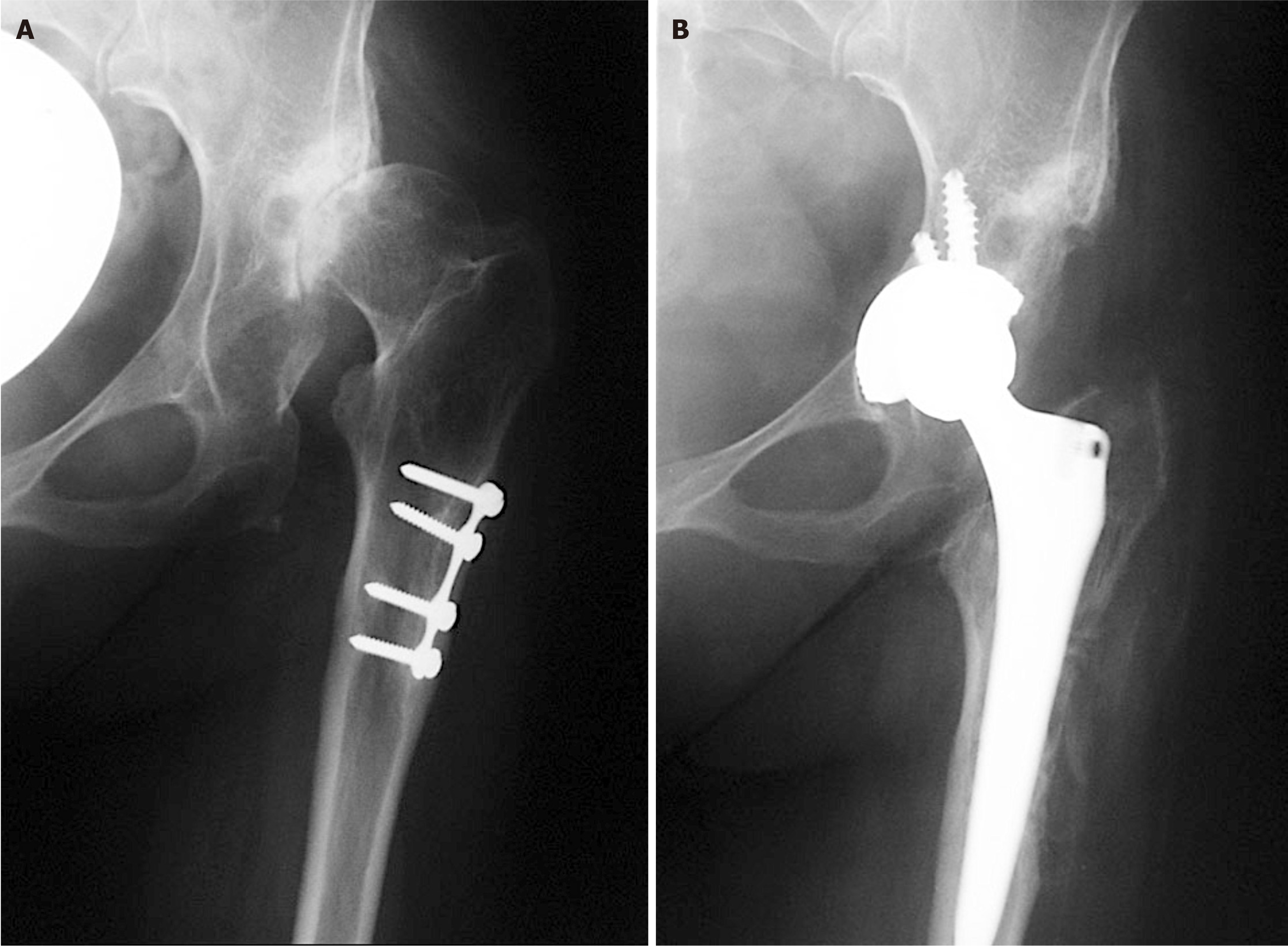
The ideal technique involves medialising the acetabular component as much as possible to reduce the hip load while avoiding complications from superolateral segment coverage. Cotyloplasty is technique that allows us exactly that. It includes deliberate fracture of the medial side (bottom) of acetabulum and placing a cup more medially, beyond the Kohler’s ilioischial line. Although originally performed with cemented cups, uncemented porous acetabular cups have recently shown better outcomes[22-24]. This method includes controlled reaming of the bone from acetabulum until medial wall becomes paper-thin. Any hole in acetabulum, and medial side of acetabulum is jam-packed with cancellous bone, after which the press fit cup is inserted. In this way, acetabular component is medialized and cancellous bone between cup and paper-thin medial wall will allow preserving the bone stock for future revision surgeries, what is quite important for these young patients. Acetabular component is additionally stabilized with screws for better primary stability, and with time, bone ingrowth will provide the secondary stability of acetabular component (Figure 1). In such manner hip biomechanics and leg length are restored, reducing the risk for impingement and dislocation. Potential obstacle of cotyloplasty is the difficulty in controlling the extent of the medial wall fracture, which can lead to complications, for example dislocation of the cup into the pelvis[24]. In an in vitro study we published in 2023, using pig pelvises, we established guidelines for preserving the primary stability of the acetabular component. Our results indicate that, under a load of 700 N, the acetabular component remains stable if the medial wall defect is lower than 68% of its diameter or if the uncovered surface area is no greater than 27%. For higher loads of 1000 N, stability is maintained as long as the defect is less than 45% and uncoverage is less than 18%[25].

Figure 1
Figure 1 X-rays of left hip. A: Preoperative X-ray of a patient with secondary osteoarthritis of the left hip due to dysplasia. Plate and screws in the proximal femur are left from the previous surgery performed in childhood; B: Postoperative X-ray with implanted uncemented acetabular cup and femoral stem. Acetabular cup is protruding beyond the Kohler’s line inside the pelvis and secured with 3 additional screws. Lesser trochanter is brought distally to the normal level so there is no leg length discrepancy postoperatively.

Femoral component placement in patients with paediatric hip sequelae is also complex due to altered anatomy, particularly after corrective osteotomies, osteoporotic or sclerotic bone, and narrow medullary canals. In some cases, a standard femoral stem cannot be used, requiring a modular stem instead[1]. Femur is often too long and must be shortened. Shortening is necessary for achieving position of endoprosthesis in ideal centre of rotation, correction of proximal femoral anteversion and to avoid sciatic nerve stretching. Additionally, the hip’s abductor mechanism is restored, resulting in same leg length[25]. Over time, various types of femoral shortening osteotomies have been developed. These can be classified based on the location of the osteotomy into proximal, distal and femoral shaft osteotomies[26-30].

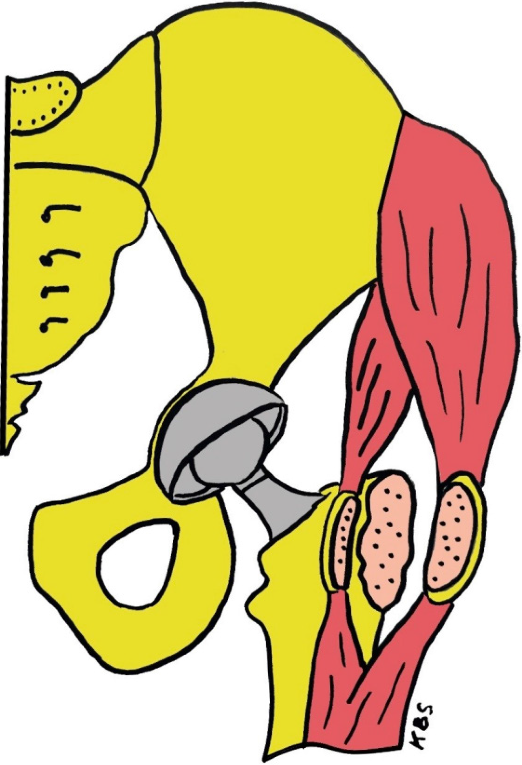
Our preferred approach for THA in patients with paediatric hip sequalae is previously published, a variation of the direct lateral hip approach[9]. During approach, the front half is separated with a flake of bone from the greater trochanter, while remaining connected to the uninterrupted tendons of the gluteus medius and vastus lateralis. The same process is applied to the posterior portion, detaching it with the chisel, leaving a thicker bone flake. This technique strips the muscles from the greater trochanter without performing a trochanteric osteotomy. It preserves the continuity of the abductor muscles, allowing the femur to be shortened without impacting the muscles to much (it splits the continuous tendon only longitudinally) (Figure 2). This approach allows faster recovery and reduces complications related to trochanteric osteotomy[9]. We have used this technique for more than 10 years with good results in more than 65 patients. Initial results for first 12 patients were published with follow up of 2 years[9]. The average Harris hip score was 96.7 (range 92.2-100). The limb lengths were measured before and after the surgery, and after surgery the leg length difference was less than 5 mm in 11 of the 12 patients. Also, medialisation of the cup relative to the ideal centre of rotation according to method of Ranawat et al[31] was on average 8 mm. None of the patients limped or had positive trendelenburg sign, and no one had a peroneal nerve palsy. Additional results for 19 patients were analysed and presented previously. In this cohort some of the results were: Average medialization of the cup (protrusion of the cup bellow the ischio-ileal line) was 6.6 mm ± 2.9 mm (range 2.2-11.5 mm). Average protrusion of the acetabulum in the pelvis was 46% ± 20% (range 7%-77%). In 4 out of 19 cases acetabulum was partially uncovered. In those 4 cases average lateral cup placement was 6.5 mm ± 3.7 mm (range 3.3-10.2 mm). In one case where the cup was medialized 9.6 mm, postoperative instability happened and cup was revised. For revision we used bigger cementless cup and additional grafting of the bottom. Long term follow-up of the patients is underway which mostly focuses on possible long-term deterioration of functional outcome (if any) and Kaplan–Meier survivorship analysis with aseptic loosening and revision as endpoint which is beyond the scope of this paper.

Figure 2
Figure 2 Drawing of our prefer method of treatment. Acetabular cup is medialized (cotyloplasty) so that the dome of the cup is protruding beyond Kohler’s line inside the pelvis. On the femoral side anterior and posterior half of the continuous tendon is mobilized by a chisel. First the anterior part is divided with a thin layer of bone from the greater trochanter but remains attached to the continuous tendon of the gluteus medius and the vastus lateralis. And the same is done with the posterior half of the continuous tendon of the gluteus medius and the vastus lateralis which are detached with the chisel leaving a bone flake of at least 2 mm to 3 mm thickness attached to tendons. With that procedure continuity of abductor muscles is preserved and femur can be shortened without affecting the muscles. KBS: Critical region for definable segment defects.
CONCLUSION

THA in patients with childhood hip diseases sequelae demand individual approach with precise preoperative planning and specific procedures that will allow restoration of ideal biomechanical conditions. It is necessary to place the acetabulum into the ideal centre of rotation, as medially as possible, then to restore the lateral offset with the femoral component and correct the leg length, with necessary preservation of the abductor muscles. Only in that way good functional results in these young patients can be achieved. We suggest modified direct lateral approach on hip which allows good visualization of both, acetabulum and femur, preserving the continuity of abductor muscles without trochanteric osteotomy. And for achieving biomechanically best position of acetabulum cotyloplasty is our method of choice.

Footnotes

Provenance and peer review: Invited article; Externally peer reviewed.

Peer-review model: Single blind

Specialty type: Orthopedics

Country of origin: Croatia

Peer-review report’s classification

Scientific Quality: Grade B

Novelty: Grade B

Creativity or Innovation: Grade B

Scientific Significance: Grade B

P-Reviewer: Zhang L S-Editor: Luo ML L-Editor: A P-Editor: Zhao YQ

References
1.  De Salvo S, Sacco R, Mainard N, Lucenti L, Sapienza M, Dimeglio A, Andreacchio A, Canavese F. Total hip arthroplasty in patients with common pediatric hip orthopedic pathology. J Child Orthop. 2024;18:134-152.  [PubMed]  [DOI]  [Cited in This Article: ]  [Reference Citation Analysis (0)]
2.  Erdemli B, Yilmaz C, Atalar H, Güzel B, Cetin I. Total hip arthroplasty in developmental high dislocation of the hip. J Arthroplasty. 2005;20:1021-1028.  [PubMed]  [DOI]  [Cited in This Article: ]  [Cited by in Crossref: 62]  [Cited by in F6Publishing: 64]  [Article Influence: 3.2]  [Reference Citation Analysis (0)]
3.  Noble PC, Kamaric E, Sugano N, Matsubara M, Harada Y, Ohzono K, Paravic V. Three-dimensional shape of the dysplastic femur: implications for THR. Clin Orthop Relat Res. 2003;27-40.  [PubMed]  [DOI]  [Cited in This Article: ]  [Cited by in Crossref: 5]  [Cited by in F6Publishing: 8]  [Article Influence: 0.4]  [Reference Citation Analysis (0)]
4.  Robertson DD, Essinger JR, Imura S, Kuroki Y, Sakamaki T, Shimizu T, Tanaka S. Femoral deformity in adults with developmental hip dysplasia. Clin Orthop Relat Res. 1996;196-206.  [PubMed]  [DOI]  [Cited in This Article: ]  [Cited by in Crossref: 66]  [Cited by in F6Publishing: 63]  [Article Influence: 2.2]  [Reference Citation Analysis (0)]
5.  Anthony CA, Wasko MK, Pashos GE, Barrack RL, Nunley RM, Clohisy JC. Total Hip Arthroplasty in Patients With Osteoarthritis Associated With Legg-Calve-Perthes Disease: Perioperative Complications and Patient-Reported Outcomes. J Arthroplasty. 2021;36:2518-2522.  [PubMed]  [DOI]  [Cited in This Article: ]  [Cited by in Crossref: 2]  [Cited by in F6Publishing: 2]  [Article Influence: 0.5]  [Reference Citation Analysis (0)]
6.  Te Velde JP, Buijs GS, Schafroth MU, Saouti R, Kerkhoffs GMMJ, Kievit AJ. Total Hip Arthroplasty in Teenagers: A Systematic Literature Review. J Pediatr Orthop. 2024;44:e115-e123.  [PubMed]  [DOI]  [Cited in This Article: ]  [Reference Citation Analysis (0)]
7.  Charnley J, Feagin JA. Low-friction arthroplasty in congenital subluxation of the hip. Clin Orthop Relat Res. 1973;98-113.  [PubMed]  [DOI]  [Cited in This Article: ]  [Cited by in Crossref: 201]  [Cited by in F6Publishing: 195]  [Article Influence: 3.8]  [Reference Citation Analysis (0)]
8.  Kobayashi S, Saito N, Nawata M, Horiuchi H, Iorio R, Takaoka K. Total hip arthroplasty with bulk femoral head autograft for acetabular reconstruction in developmental dysplasia of the hip. J Bone Joint Surg Am. 2003;85:615-621.  [PubMed]  [DOI]  [Cited in This Article: ]  [Cited by in Crossref: 60]  [Cited by in F6Publishing: 59]  [Article Influence: 2.7]  [Reference Citation Analysis (0)]
9.  Delimar D, Bicanic G, Korzinek K. Femoral shortening during hip arthroplasty through a modified lateral approach. Clin Orthop Relat Res. 2008;466:1954-1958.  [PubMed]  [DOI]  [Cited in This Article: ]  [Cited by in Crossref: 12]  [Cited by in F6Publishing: 12]  [Article Influence: 0.7]  [Reference Citation Analysis (0)]
10.  Wu X, Li SH, Lou LM, Cai ZD. The techniques of soft tissue release and true socket reconstruction in total hip arthroplasty for patients with severe developmental dysplasia of the hip. Int Orthop. 2012;36:1795-1801.  [PubMed]  [DOI]  [Cited in This Article: ]  [Cited by in Crossref: 24]  [Cited by in F6Publishing: 25]  [Article Influence: 1.9]  [Reference Citation Analysis (0)]
11.  Shi XT, Li CF, Cheng CM, Feng CY, Li SX, Liu JG. Preoperative Planning for Total Hip Arthroplasty for Neglected Developmental Dysplasia of the Hip. Orthop Surg. 2019;11:348-355.  [PubMed]  [DOI]  [Cited in This Article: ]  [Cited by in Crossref: 17]  [Cited by in F6Publishing: 22]  [Article Influence: 3.7]  [Reference Citation Analysis (0)]
12.  Bicanic G, Barbaric K, Bohacek I, Aljinovic A, Delimar D. Current concept in dysplastic hip arthroplasty: Techniques for acetabular and femoral reconstruction. World J Orthop. 2014;5:412-24.  [PubMed]  [DOI]  [Cited in This Article: ]  [Cited by in CrossRef: 43]  [Cited by in F6Publishing: 39]  [Article Influence: 3.5]  [Reference Citation Analysis (1)]
13.  Shen B, Yang J, Wang L, Zhou ZK, Kang PD, Pei FX. Midterm results of hybrid total hip arthroplasty for treatment of osteoarthritis secondary to developmental dysplasia of the hip-Chinese experience. J Arthroplasty. 2009;24:1157-1163.  [PubMed]  [DOI]  [Cited in This Article: ]  [Cited by in Crossref: 13]  [Cited by in F6Publishing: 11]  [Article Influence: 0.7]  [Reference Citation Analysis (0)]
14.  Haddad FS, Masri BA, Garbuz DS, Duncan CP. Primary total replacement of the dysplastic hip. Instr Course Lect. 2000;49:23-39.  [PubMed]  [DOI]  [Cited in This Article: ]
15.  Delimar D, Cicak N, Klobucar H, Pećina M, Korzinek K. Acetabular roof reconstruction with pedicled iliac graft. Int Orthop. 2002;26:344-348.  [PubMed]  [DOI]  [Cited in This Article: ]  [Cited by in Crossref: 22]  [Cited by in F6Publishing: 23]  [Article Influence: 1.0]  [Reference Citation Analysis (0)]
16.  Fujiwara M, Nishimatsu H, Sano A, Misaki T. Acetabular roof reconstruction using a free vascularized fibular graft. J Reconstr Microsurg. 2006;22:349-352.  [PubMed]  [DOI]  [Cited in This Article: ]  [Cited by in Crossref: 3]  [Cited by in F6Publishing: 2]  [Article Influence: 0.1]  [Reference Citation Analysis (0)]
17.  Inao S, Matsuno T. Cemented total hip arthroplasty with autogenous acetabular bone grafting for hips with developmental dysplasia in adults: the results at a minimum of ten years. J Bone Joint Surg Br. 2000;82:375-377.  [PubMed]  [DOI]  [Cited in This Article: ]  [Cited by in Crossref: 41]  [Cited by in F6Publishing: 42]  [Article Influence: 1.7]  [Reference Citation Analysis (0)]
18.  Kim M, Kadowaki T. High long-term survival of bulk femoral head autograft for acetabular reconstruction in cementless THA for developmental hip dysplasia. Clin Orthop Relat Res. 2010;468:1611-1620.  [PubMed]  [DOI]  [Cited in This Article: ]  [Cited by in Crossref: 78]  [Cited by in F6Publishing: 82]  [Article Influence: 5.5]  [Reference Citation Analysis (0)]
19.  Shinar AA, Harris WH. Bulk structural autogenous grafts and allografts for reconstruction of the acetabulum in total hip arthroplasty. Sixteen-year-average follow-up. J Bone Joint Surg Am. 1997;79:159-168.  [PubMed]  [DOI]  [Cited in This Article: ]  [Cited by in Crossref: 245]  [Cited by in F6Publishing: 234]  [Article Influence: 8.4]  [Reference Citation Analysis (0)]
20.  Siegmeth A, Duncan CP, Masri BA, Kim WY, Garbuz DS. Modular tantalum augments for acetabular defects in revision hip arthroplasty. Clin Orthop Relat Res. 2009;467:199-205.  [PubMed]  [DOI]  [Cited in This Article: ]  [Cited by in Crossref: 99]  [Cited by in F6Publishing: 99]  [Article Influence: 6.2]  [Reference Citation Analysis (0)]
21.  Malizos KN, Bargiotas K, Papatheodorou L, Hantes M, Karachalios T. Survivorship of monoblock trabecular metal cups in primary THA : midterm results. Clin Orthop Relat Res. 2008;466:159-166.  [PubMed]  [DOI]  [Cited in This Article: ]  [Cited by in Crossref: 48]  [Cited by in F6Publishing: 51]  [Article Influence: 3.0]  [Reference Citation Analysis (0)]
22.  Hartofilakidis G, Stamos K, Ioannidis TT. Low friction arthroplasty for old untreated congenital dislocation of the hip. J Bone Joint Surg Br. 1988;70:182-186.  [PubMed]  [DOI]  [Cited in This Article: ]  [Cited by in Crossref: 121]  [Cited by in F6Publishing: 126]  [Article Influence: 3.4]  [Reference Citation Analysis (0)]
23.  Hartofilakidis G, Yiannakopoulos CK, Babis GC. The morphologic variations of low and high hip dislocation. Clin Orthop Relat Res. 2008;466:820-824.  [PubMed]  [DOI]  [Cited in This Article: ]  [Cited by in Crossref: 44]  [Cited by in F6Publishing: 46]  [Article Influence: 2.7]  [Reference Citation Analysis (0)]
24.  Dorr LD, Tawakkol S, Moorthy M, Long W, Wan Z. Medial protrusio technique for placement of a porous-coated, hemispherical acetabular component without cement in a total hip arthroplasty in patients who have acetabular dysplasia. J Bone Joint Surg Am. 1999;81:83-92.  [PubMed]  [DOI]  [Cited in This Article: ]  [Cited by in Crossref: 116]  [Cited by in F6Publishing: 113]  [Article Influence: 4.3]  [Reference Citation Analysis (0)]
25.  Barbaric Starcevic K, Bicanic G, Alar Z, Sakoman M, Starcevic D, Delimar D. Measurement of safe acetabular medial wall defect size in revision hip arthroplasty with a porous cup. Hip Int. 2023;33:478-484.  [PubMed]  [DOI]  [Cited in This Article: ]  [Reference Citation Analysis (0)]
26.  Li X, Sun J, Lin X, Xu S, Tang T. Cementless total hip arthroplasty with a double chevron subtrochanteric shortening osteotomy in patients with Crowe type-IV hip dysplasia. Acta Orthop Belg. 2013;79:287-292.  [PubMed]  [DOI]  [Cited in This Article: ]
27.  Sener N, Tözün IR, Aşik M. Femoral shortening and cementless arthroplasty in high congenital dislocation of the hip. J Arthroplasty. 2002;17:41-48.  [PubMed]  [DOI]  [Cited in This Article: ]  [Cited by in Crossref: 65]  [Cited by in F6Publishing: 80]  [Article Influence: 3.5]  [Reference Citation Analysis (0)]
28.  Kawai T, Tanaka C, Ikenaga M, Kanoe H. Cemented total hip arthroplasty with transverse subtrochanteric shortening osteotomy for Crowe group IV dislocated hip. J Arthroplasty. 2011;26:229-235.  [PubMed]  [DOI]  [Cited in This Article: ]  [Cited by in Crossref: 30]  [Cited by in F6Publishing: 27]  [Article Influence: 1.9]  [Reference Citation Analysis (0)]
29.  Bruce WJ, Rizkallah SM, Kwon YM, Goldberg JA, Walsh WR. A new technique of subtrochanteric shortening in total hip arthroplasty: surgical technique and results of 9 cases. J Arthroplasty. 2000;15:617-626.  [PubMed]  [DOI]  [Cited in This Article: ]  [Cited by in Crossref: 77]  [Cited by in F6Publishing: 76]  [Article Influence: 3.0]  [Reference Citation Analysis (0)]
30.  Togrul E, Ozkan C, Kalaci A, Gülşen M. A new technique of subtrochanteric shortening in total hip replacement for Crowe type 3 to 4 dysplasia of the hip. J Arthroplasty. 2010;25:465-470.  [PubMed]  [DOI]  [Cited in This Article: ]  [Cited by in Crossref: 49]  [Cited by in F6Publishing: 47]  [Article Influence: 3.1]  [Reference Citation Analysis (0)]
31.  Ranawat CS, Dorr LD, Inglis AE. Total hip arthroplasty in protrusio acetabuli of rheumatoid arthritis. J Bone Joint Surg Am. 1980;62:1059-1065.  [PubMed]  [DOI]  [Cited in This Article: ]