Copyright
©The Author(s) 2023.
World J Orthop. Apr 18, 2023; 14(4): 171-185
Published online Apr 18, 2023. doi: 10.5312/wjo.v14.i4.171
Published online Apr 18, 2023. doi: 10.5312/wjo.v14.i4.171
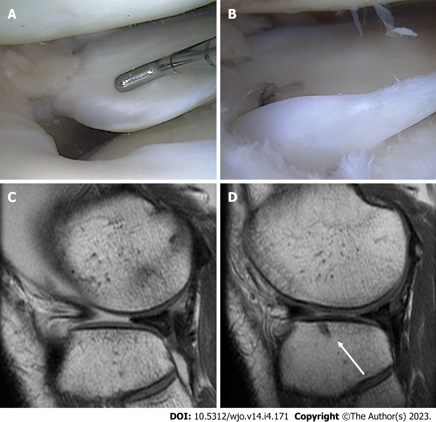
Figure 7 Radial tear of the lateral meniscus.
A: Arthroscopic visualization of the radial tear of the lateral meniscus; B: Tear repair using an all-suture anchor; C: Preoperative T1 sagittal magnetic resonance imaging (MRI) view, showing the extrusion of the anterior portion of the lateral meniscus; D: Six-month follow-up T1 sagittal MRI view, showing the relocation of the anterior portion and the all-suture anchor used for the repair (white arrow).
- Citation: Simonetta R, Russo A, Palco M, Costa GG, Mariani PP. Meniscus tears treatment: The good, the bad and the ugly-patterns classification and practical guide. World J Orthop 2023; 14(4): 171-185
- URL: https://www.wjgnet.com/2218-5836/full/v14/i4/171.htm
- DOI: https://dx.doi.org/10.5312/wjo.v14.i4.171


