Copyright
©The Author(s) 2023.
World J Orthop. Nov 18, 2023; 14(11): 836-842
Published online Nov 18, 2023. doi: 10.5312/wjo.v14.i11.836
Published online Nov 18, 2023. doi: 10.5312/wjo.v14.i11.836
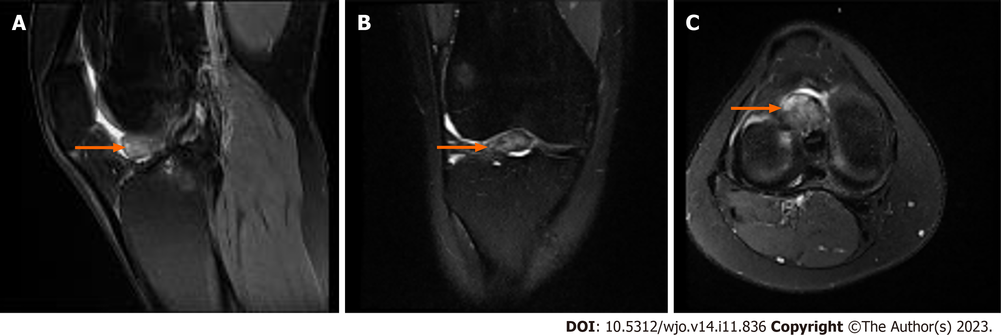
Figure 3 Postoperative magnetic resonance imaging right knee.
A: Magnetic resonance imaging films (3 tesla magnet) in sagittal proton-density fast spin echo with fat saturation (PD FSE FSAT) sequence (orange arrow); B: Coronal T2-weighted FSE FSAT sequence (orange arrow); C: Axial PD FSE FSAT sequence views taken after anterior cruciate ligament reconstruction with cyclops lesion measuring 16 mm × 17 mm × 11 mm (orange arrow).
- Citation: Kelmer G, Johnson AH, Turcotte JJ, Redziniak DE. Recurrent cyclops lesion after primary anterior cruciate ligament reconstruction using bone tendon bone allograft: A case report. World J Orthop 2023; 14(11): 836-842
- URL: https://www.wjgnet.com/2218-5836/full/v14/i11/836.htm
- DOI: https://dx.doi.org/10.5312/wjo.v14.i11.836


