Published online Jun 18, 2021. doi: 10.5312/wjo.v12.i6.423
Peer-review started: December 30, 2020
First decision: January 24, 2021
Revised: February 18, 2021
Accepted: May 7, 2021
Article in press: May 7, 2021
Published online: June 18, 2021
Processing time: 162 Days and 23 Hours
Partial tears of the anterior cruciate ligament (ACL) are frequent, and there is still considerable controversy surrounding their diagnosis, natural history and treatment.
To examine patient-reported outcomes, physical examination and magnetic resonance imaging (MRI) findings of partial ACL tears treated with an intraarticular injection of platelet-rich plasma (PRP) compared to a control group.
From January 2015 to November 2017, consecutive patients from a single institution with partial ACL tears treated nonoperatively were prospectively evaluated. Partial tears were defined as a positive Lachman test with a clear end-point, a negative pivot-shift and less than 3 mm of side-to-side difference using the KT1000 arthrometer. Patients in group 1 were treated with one intraarticular injection of PRP and specific physical therapy protocol. Control group consisted of patients treated only with physical therapy. Prospective analyzed data included physical examination, Tegner activity level and Lysholm and International Knee Documentation Committee scores. Baseline MRI findings and at 6 mo follow-up were reviewed. Failure was defined as those patients with clinical instability at follow-up that required a subsequent ACL reconstruction.
A total of 40 patients where included, 21 treated with PRP injection with a mean follow-up of 25 mo [standard deviation (SD): 3.6] and 19 in the control group with a mean follow-up of 25 mo (SD: 5.68). Overall failure rate was 32.0% (n = 13). No significant differences were observed between groups regarding subjective outcomes, return to sport and failure rate. MRI findings revealed an improvement in the ACL signal in half of the patients of both groups. However, we did not find a significant relationship between MRI findings and clinical outcomes.
Overall, 95.0% of patients returned to sports at a mean follow-up of 25 mo. Mean time to return to sports was 4 mo. Out of these patients, almost 30.0% in each group had a new episode of instability and required surgery at a median time of 5 mo in group 1 and 8 mo in group 2. The addition of PRP alone was not sufficient to enhance any of the outcome measures evaluated, including MRI images, clinical evaluation and failure rate.
Core Tip: This was a prospective comparative study with 40 patients seeking to evaluate the effect of platelet-rich plasma on partial tears of the anterior cruciate ligament. Prospective analyzed data included physical examination, Tegner activity level and Lysholm and International Knee Documentation Committee scores. Baseline magnetic resonance imaging findings and at 6 mo follow-up were also reviewed.
- Citation: Zicaro JP, Garcia-Mansilla I, Zuain A, Yacuzzi C, Costa-Paz M. Has platelet-rich plasma any role in partial tears of the anterior cruciate ligament? Prospective comparative study. World J Orthop 2021; 12(6): 423-432
- URL: https://www.wjgnet.com/2218-5836/full/v12/i6/423.htm
- DOI: https://dx.doi.org/10.5312/wjo.v12.i6.423
Partial tears account for 10% to 30% of all anterior cruciate ligament (ACL) tears[1,2]. There is still considerable controversy surrounding the diagnosis, natural history and treatment of this type of lesion[3,4]. Diverse criteria are often used to define a partial ACL tear. Magnetic resonance imaging (MRI) features include the estimation of the percentage of torn fibers, specific affected bundle (anteromedial or posterolateral) and location of the tear (proximal, middle third or distal). The physical examination is decisive in this type of lesion[1]. Finally, arthroscopy remains the gold standard for the diagnosis of macroscopic integrity of the intact bundle. Intraoperative findings of intact remnant ACL fibers from femur to tibia insertion points confirm the diagnosis[5-7].
Regarding natural history, complete ACL tears have low intrinsic healing capacity, which prevent restoring anatomy and function. However, there has been some disagreement concerning whether partial ACL tears could heal. Though favorable results have been reported with either nonoperative or surgical treatment (such as repair or augmentation) as well as with biological approaches, some authors reported progression to complete deficiency of the ACL and symptomatic knee laxity with conservative treatment[4,7-10].
Platelet-rich plasma (PRP) has received much attention in the last years as a biologic alternative for the treatment of sports-related injuries. Various growth factors and bioactive proteins from the a-granules contained in platelets can potentially enhance tissue healing[11-14]. PRP utilization in ligament injuries has grown remarkably[15-17]. Regarding specifically to ACL injuries, the focus has been mostly on biologic augmentation to improve graft healing after a reconstruction, and only a few studies aimed to improve healing of the native injured ACL[18-22].
The purpose of this study was to examine patient-reported outcomes, physical examination and MRI findings of partial ACL tears treated with an intraarticular injection of PRP compared to a control group.
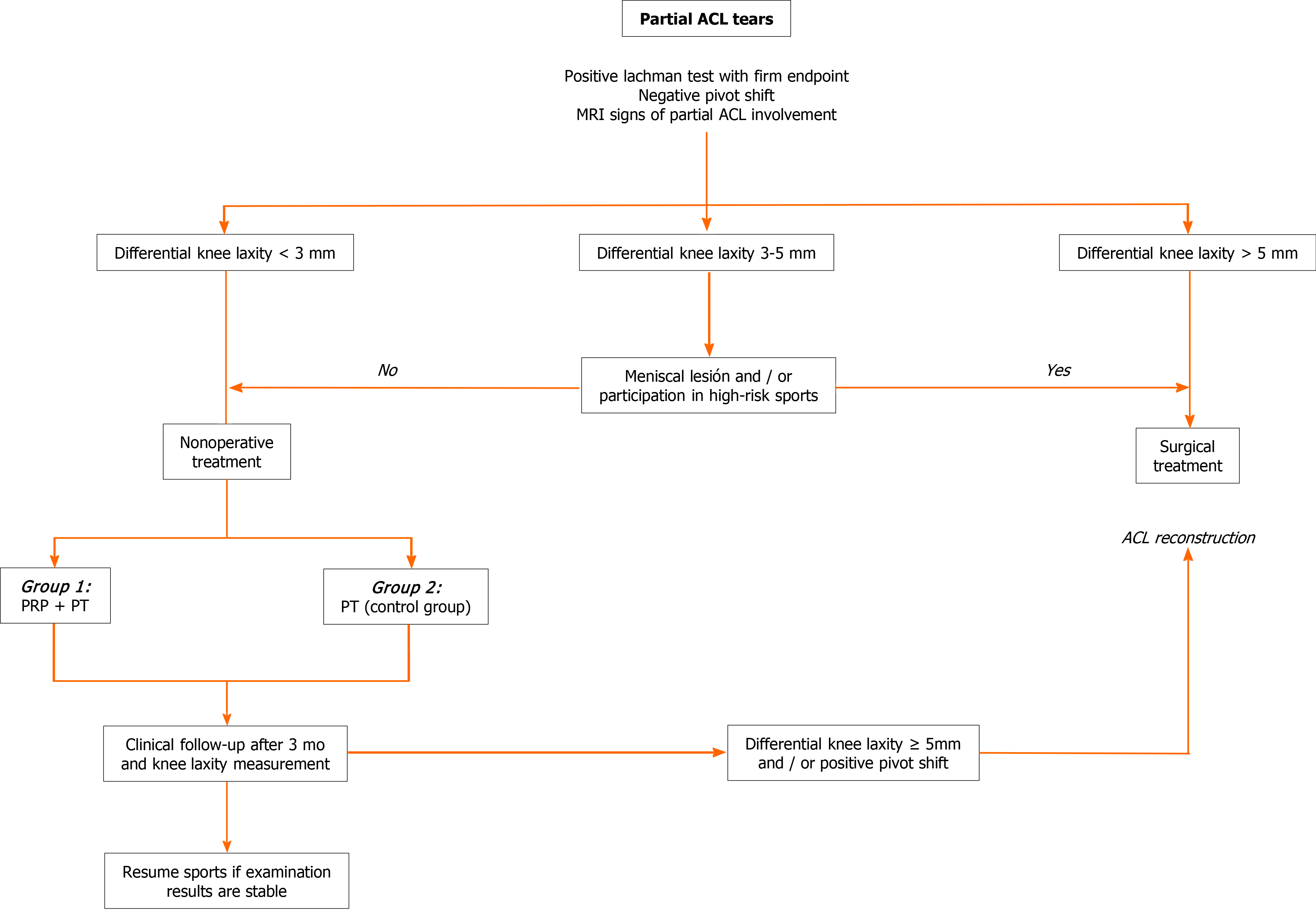
Following Institutional Review Board approval, all patients who were diagnosed with a partial ACL tear and treated nonoperatively between January 2015 and November 2017 were retrospectively selected from a database of prospectively collected data. Figure 1 shows the algorithm used for the diagnosis and treatment of partial ACL tears. It consists of an adaptation of a previously published algorithm by Sonnery-Cottet et al[1]. We included patients with a positive Lachman’s test with a firm endpoint, negative pivot shift, a side-to-side differential laxity less than 3 mm measured by arthrometer (KT-1000 knee arthrometer, MEDmetric Corp.) and MRI signs of partial ACL tear. Patients with a differential laxity between 3 and 5 mm without meniscal lesions and/or participation in high-risk sports were also included.
The exclusion criteria were as follows: patients younger than 18-years-old, patients with less than 6 mo follow-up, patients diagnosed after 1 mo of injury, previous ipsilateral ACL tear or reconstruction, confirmed or suspected contralateral ACL injury, diagnosis of concomitant ipsilateral posterior cruciate ligament, posterolateral corner or grade 3 medial collateral ligament injury and arthritis (International Knee Documentation Committee C or higher).
Nonoperative treatment consisted of one intra-articular injection of leukocyte reduced PRP during the first 4 wk after injury (within the inflammatory phase) and specific physical therapy protocol (group 1). However, not all patients were able to receive the PRP injection, mostly because medical insurance coverage or refusal from the patient to do so. These patients were considered the control group (group 2).
All medical care interventions are centrally registered in a computerized data repository, with only one electronic health record per person. After initial consultation and treatment, patients underwent clinical follow-up at 1, 3, 6 and 12 mo by the same observer. Variables analyzed included patient gender, age, Tegner activity Score both at time of injury and at time of final follow-up, return to sports (RTS) rate, time to RTS and subsequent surgeries. Subjective assessment included Lysholm and International Knee Documentation Committee scores.
Objective stability was tested at the time of injury and at 6 mo follow-up with a knee arthrometer test (KT-1000 knee arthrometer, MEDmetric Corp.), and the manual maximum difference between knees (in mm) was used for analysis of reported mean side-to-side differences. All patients were evaluated by a single orthopedic observer (JPZ-staff member/knee surgeon).
Baseline MRI findings and at 6 mo follow-up were reviewed. Images were analyzed according to a classification published by van Meer et al[23]. Nine features are used to assess the ACL on MRI: fiber continuity, signal intensity, slope of ACL with respect to the Blumensaat line, distance between the Blumensaat line and ACL, tension, thickness, clear boundaries, assessment of original insertions and assessment of intercondylar notch. A total score is determined by summing scores for these 9 features. A score of 10 is maximally abnormal for all features, whereas a score of 0 is normal for all features. Lesion localization was also determined (proximal, middle third and distal). All images were evaluated by a single orthopedic observer (JPZ) who was blinded to the treatment group.
Failure was defined as those patients with clinical instability at follow-up that required a subsequent ACL reconstruction.
On the basis of previously published reports on criteria that influence the composition or biological effect of PRP[24], we included the following information regarding PRP preparation. After harvesting 150 mL of blood, the extracted unit was doubled centrifuged in a Thermo Scientific Sorvall BP-16 Refrigerated Blood Bank Centrifuge. First, a light centrifugation for 4 min at 1400 rpm to separate the PRP from the globular mass was performed. The product obtained was separated into satellite bags without opening the circuit, guaranteeing the sterility of the process. Next, the PRP was centrifuged for 6 min at 3000 rpm to achieve a higher concentration of the product. At the end of the process, quality control was carried out on the product through an XT ROCHE hematological counter. It consists of volume, platelet count, white blood cell count and calculation of product concentration.
Group 1 (PRP group) received a single intra-articular injection of PRP. A median of 8 mL (6-10) was injected. All injections were performed by one of the authors (Zicaro JP). Intra-articular injection aiming at the ACL was performed as a standard sterile procedure. A 25-gauge 3.0-inch needle was inserted through the skin in a similar localization as a medial portal, towards the ACL femoral insertion. No imaging was used as guidance. The median concentrations of platelets and white cells of the product are detailed in Table 1. After the procedure, full weight bearing, cryotherapy and daily life activities was allowed as tolerated. No post treatment bracing was administered. Physical therapy protocol began after 72 h of the injection.
| Initial platelet concentration | PRP | % | |
| Platelets, median (IQR) | 264500 (247000-278000) | 1125500 (1088000-1340500) | 444 (407-519) |
| WC, median (IQR) | 5200 (4790-6480) | 600 (220-810) | 10 (3-15) |
Even though not all patients performed the rehabilitation at the same center, both groups received the same rehabilitation protocol. The goals of the first 4 wk were to recover range of motion, prevent quadriceps inhibition and normalize proximal muscle strength. Nonimpact exercises such as bicycle or swimming were allowed during the first week. Linear impact and strengthening exercises began 6 wk after injection, with progression to multiplanar exercises between 8 to 10 wk. The goal was to return to their previous sport not before 3 to 4 mo after injection.
Descriptive statistics including means, standard deviations (SDs), medians and quartile ranges were applied as appropriate to assess the available demographic, surgical, physical examination and patient-reported outcome data. Statistical hypothesis testing was performed using the Fisher exact test and Wilcoxon rank-sum test. Categorical data was performed using Chi square test. Analysis was performed with 95% confidence interval, and P values < 0.05 were considered statistically significant. All statistical analyses were performed using STATA version 13.
A total of 40 patients where included. In total, 21 treated with PRP injection (group 1) with a mean follow-up of 25 mo (SD: 3.60) and 19 in the control group (group 2) with a mean follow-up of 25 mo (SD: 5.68). The overall median age was 26.0-years-old (IQR 22.5-35.0). Demographic data is shown in Table 2. The only statistically significant difference between groups was gender; no females were included in the control group (P = 0.021).
| Group 1, n = 21 | Group 2, n = 19 | P value | |
| Age in yr, median (IQR) | 25 (22-39) | 31 (26-34) | 0.54 |
| Male, n (%) | 15 (71.4) | 19 (100) | 0.02 |
| FU in mo, median (IQR) | 25 (18-36) | 25 (18-30) | 0.86 |
| MRI tear location: | |||
| Proximal, n (%) | 10 (48) | 12 (63) | |
| Mid-substance, n (%) | 11 (52) | 7 (27) |
Results at final follow-up are shown in Table 3. One patient in each group (5.0%) was unable to RTS due to subjective instability. The other 95.0% in each group were able to return to their previous sports level. Overall mean RTS time was 4 mo (SD: 1.06), without significant differences between groups. We found no significant differences for subjective outcomes between groups.
| Baseline | Group 1, n = 21 | Group 2, n = 19 | P value |
| Lysholm score, median (IQR) | 69.5 (43.0-85.0) | 54.0 (41.0-77.0) | 0.41 |
| IKDC score, median (IQR) | 58.5 (44.0-60.0) | 58.0 (44.0-60.0) | 0.84 |
| TAL, mean ± SD | 6.90 ± 1.07 | 6.70 ± 1.18 | 0.65 |
| At final follow-up | |||
| Lysholm score, median (IQR) | 80 (75-90) | 80 (73-86) | 0.53 |
| IKDC score, median (IQR) | 77 (71-89) | 71 (70-79) | 0.33 |
| TAL, mean ± SD | 6.70 ± 1.52 | 6.50 ± 1.61 | 0.7 |
| RTS rate, n (%) | 20 (95) | 18 (95) | 0.9 |
| Time to RTS in mo, mean ± SD | 3.8 ± 0.8 | 4.3 ± 1.2 | 0.09 |
| Failure rate, n (%) | 7 (33) | 6 (31) | 0.9 |
Regarding objective stability, at 6 mo follow-up in group 1, 13 presented a decrease in the side-to-side difference, 7 remained with the same difference, and 1 had 2 mm more. In group 2, 9 had a decrease in the side-to-side difference, 9 remained with the same difference, and 1 had 1 mm more. None of the patients had a positive pivot shift at final follow-up.
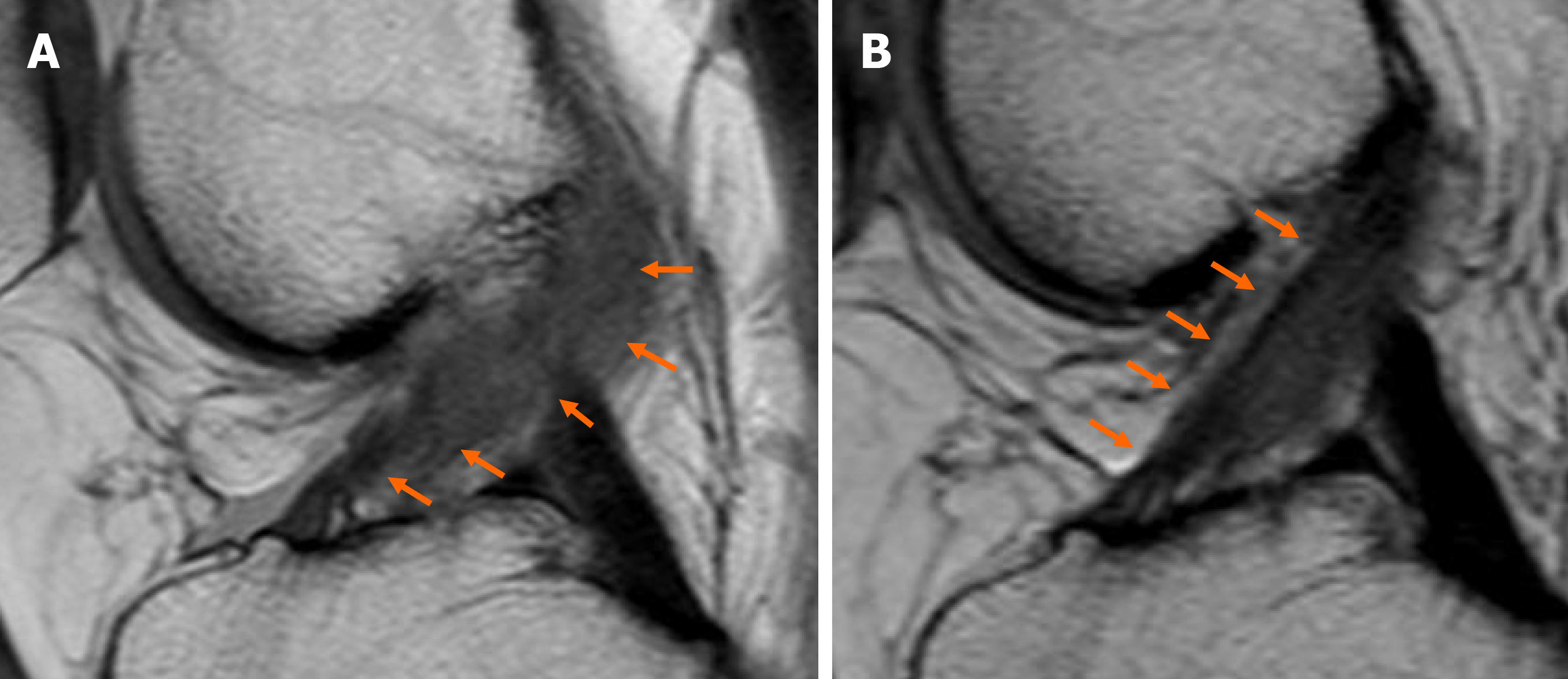
According to van Meer et al[23], MRI classification for partial ACL tears at baseline for most patients were classified between 4 and 10 in both groups (Table 4). At 6 mo follow-up, more than 50.0% of patients in both groups where classified as 0 and 3, showing an improvement in the ACL MRI signal (Figure 2).
| MRI Van Meer classification | |||
| Group 1 | Group 2 | P value | |
| Baseline MRI, mean ± SD | 6.1 ± 1.7 | 6.2 ± 2.4 | 0.97 |
| MRI at 6 mo follow-up, mean ± SD | 3.4 ± 2.9 | 3.6 ± 2.5 | 0.82 |
Overall failure rate was 32.0% (n = 13) with no significant differences between groups (Table 3). Five failures in group 1 where Tegner 7, one Tegner 8 and one Tegner 5. In group 2, four failures where Tegner 7 and two Tegner 6. We found no significant differences when analyzing the location of the lesion and failure in both groups. Two proximal (2/10) and five mid-substance (5/11) tears failed in group 1 (P = 0.21). Four proximal (4/12) and two mid-substance (2/7) tears failed in group 2 (P = 0.83).
The present study evaluated the results of a series of patients with partial ACL tears treated nonoperatively. No significant differences were identified between patients treated or not with a single intraarticular injection of PRP. Overall, 32.0% failures were observed in both groups at a mean follow-up of 25 mo. The remaining 67.0% of patients were able to RTS in a mean of 4 mo.
A key factor in the treatment of partial ACL tears is a correct diagnosis. There has been some disagreement with regard to the definition of this lesion, and most of them are underdiagnosed. Some authors agree that physical examination cannot differentiate a partial ACL tear from an intact ACL[25,26]. On the other hand, MRI has a low level of accuracy for the diagnosis of partial ACL tears (25.0%-50.0%), mainly because of the significant overlap of the imaging findings between partial and complete tears, mucoid degeneration of the ACL and the initial post-traumatic hematoma[27]. Therefore, many surgeons rely on arthroscopy to define the extent of injury. A recently published study analyzed the correlation between preoperative clinical assessment and the arthroscopic examination in patients with ACL tears[26]. While evaluation under anesthesia demonstrated a high sensitivity for the detection of partial tears (100%), it was not necessarily specific (65.5%) and resulted in a high number of false positive partial tears. MRI, on the other hand, demonstrated a relatively high sensitivity (90.9%) and specificity (85.7%). The accuracy of MRI (86.3%) was also greater than that of evaluation under anesthesia (69.5%). These results suggested that MRI is 1.24 times more likely to result in correctly diagnosing a partial tear, which was a statistically significant finding.
Regarding nonsurgical management of partial ACL tears, Pujol et al[8] performed a systematic review and analyzed 12 articles where diagnosis was confirmed on arthroscopy, without ACL surgery. A total of 436 patients were followed up over the period 1976-1997 with a mean follow-up of 5.2 years (range 1.0-15.0). They found good short- and mid-term functional results, especially when patients limited their sports activities. The mean rate of revision ACL was 8.1% (0%-21.0%). RTS rate was 52.0% (21.0%-60.0%), lower than our findings, with 95.0% of patients returning to the same level. Noyes et al[7] reported a 38.0% progression to a complete rupture in a prospective evaluation of 32 patients with a partial ACL tear. Lehnert et al[28] reviewed a series of 39 partial ACL tears 5 years after injury and found that 56.0% had progressed to ACL deficiency. Finally, Fritschy et al[9] reported a rate of 42.0% in 43 patients. and Fruensgaard et al[29] reported 51.0% in a series of 41 patients. Although these results may be comparable to our overall 32.0% failure, these findings are to be viewed in the light of the indications for ACL surgery in vogue 30 years ago. It is important to highlight that in some cases, side-to-side difference in KT-1000 arthrometric evaluation was greater, probably due to a lack of healing of ACL fibers, particularly cases when the anteromedial bundle was affected. Nevertheless, most of these patients were active in their sports practice.
The use of biologic agents, including growth factors, PRP, stem cells and biological scaffolds, has been the focus of current research in ACL repair and healing[3]. In a systematic review analyzing biologic agents for ACL healing, the large majority of articles (21 out of 23) were focused on their application during ACL reconstructive surgery, whereas only two trials, both case series, investigated their potential in partial ACL tears[19]. Centeno et al[30] published a prospective case series of 10 patients treated with percutaneous injection of autologous bone narrow nucleated cells, using fluoroscopic guidance. Patients were included if they had a grade 1, 2 or 3 ACL tear without greater than 1 cm retraction. Treatment protocol consisted of a preinjection of a hypertonic dextrose solution into the ACL followed by a reinjection of 2-3 mL of bone marrow cells, PRP and platelets 2-5 d after, using the same procedure. Seven of ten patients demonstrated improvement in MRI measures of ACL integrity at a mean follow-up of 3 mo. The lack of a control group and the multiple component of the protocol are the main shortcomings of their methods. On the other hand, Seijas et al[20] published a retrospective case series of 19 football players (Tegner 9-10) with a partial ACL tear treated with an arthroscopic intraligamentary application of PRP (leukocyte poor). All cases presented a complete rupture of the anteromedial bundle with an intact posterolateral bundle. RTS rate was 84%. Average RTS in 15 patients Tegner 9 was 16 wk and in 2 patients Tegner 10 was 12 wk. In our study, mean time to return to sport was 4 mo.
Regarding the use of MRI in partial ACL tears, we consider there is an important role in the diagnosis and follow-up. However, these results must not be considered in isolation. Although we thoroughly analyzed nine different imaging parameters, no significant correlation was found between laxity and MRI images. Neither association was identified between lesion localization and treatment failure. These findings might be due to the low number of patients.
The lack of standardization of PRP protocols has been published recently[24]. Chahla et al[24] analyzed 105 studies finding high inconsistences in the way PRP preparation was reported. The majority of studies did not provide sufficient information to allow the protocol to be reproduced, which also prevents comparison of the PRP products. Based on this review and following the proposed guidelines, we included in our study data regarding PRP preparation and composition of the PRP delivered.
It is plausible that a number of limitations may have influenced the results obtained. First, the sample size might be considered to be low. However, a sample size estimation was not possible due to the lack of studies published with the same treatment. Although a control group was established, this group was not randomized, which may raise the possibility of a selection bias. Another possible source of error related to the procedure. The injections were not guided by imaging, and patients underwent only one injection. Nevertheless, there is no standardization in terms of how much and how many PRP injections are required for better results.
Our research provided further evidence about natural history of nonoperative management of partial ACL tears. Overall, 67% of patients with this type of lesion RTS in a mean of 4 mo without clinical instability either with or without an intra-articular PRP injection. The addition of PRP alone was not sufficient to enhance any of the outcome measures evaluated, including MRI images and clinical evaluation.
Platelet-rich plasma (PRP) is being widely used in many orthopedic areas. The use of PRP has for “healing” purposes is still controversial in the field of partial ligamentous lesions.
To our knowledge, there are no comparative series reported in the literature regarding the use of PRP for partial anterior cruciate ligament (ACL) tears.
The aim was to prospectively compare the patient-reported outcomes, rerupture rate and magnetic resonance (MR) findings in patients with partial ACL tears treated with a single PRP intra-articular injection compared to a control group.
Patients who met the inclusion criteria for stable partial ACL tears were divided into two groups. One group received a single intra-articular leukocyte-poor PRP injection within the first 4 wk after the lesion. Both groups received the same rehabilitation protocol. Clinical objective outcomes (KT1000 arthrometric evaluation), subjective outcomes, time to return to sports, rerupture rate and MR findings were evaluated. PRP preparation data was detailed.
Forty patients where included, 21 treated with PRP injection (group 1) (mean follow-up of 25 mo) and 19 in the control group (group 2) (mean follow-up of 25 mo). Overall, 95% of patients in each group returned to their previous sport at a mean time of 4 mo. After 6 mo follow-up, more than 50% of patients improved the ACL signal intensity in the MR. Overall failure rate was 32% (n = 13) with no significant differences between groups.
A single PRP intra-articular injection was not sufficient to enhance any of the outcome measures evaluated, including MR images and clinical evaluation. Overall, 67% of patients returned to sports in a mean of 4 mo without clinical instability either with or without an intra-articular PRP injection.
Further rigorous and objective studies including more patients and different PRP preparations, such as less platelet concentrations or leukocyte-rich preparations, would be useful to determine the true efficacy of PRP for enhancing healing properties of partial ACL lesions.
Manuscript source: Invited manuscript
Specialty type: Orthopedics
Country/Territory of origin: Argentina
Peer-review report’s scientific quality classification
Grade A (Excellent): 0
Grade B (Very good): 0
Grade C (Good): C
Grade D (Fair): 0
Grade E (Poor): 0
P-Reviewer: Schulze-Tanzil G S-Editor: Zhang H L-Editor: Filipodia P-Editor: Xing YX
| 1. | Sonnery-Cottet B, Colombet P. Partial tears of the anterior cruciate ligament. Orthop Traumatol Surg Res. 2016;102:S59-S67. [RCA] [PubMed] [DOI] [Full Text] [Cited by in Crossref: 33] [Cited by in RCA: 33] [Article Influence: 3.7] [Reference Citation Analysis (0)] |
| 2. | Colombet P, Dejour D, Panisset JC, Siebold R; French Arthroscopy Society. Current concept of partial anterior cruciate ligament ruptures. Orthop Traumatol Surg Res. 2010;96:S109-S118. [RCA] [PubMed] [DOI] [Full Text] [Cited by in Crossref: 101] [Cited by in RCA: 80] [Article Influence: 5.3] [Reference Citation Analysis (0)] |
| 3. | Dallo I, Chahla J, Mitchell JJ, Pascual-Garrido C, Feagin JA, LaPrade RF. Biologic Approaches for the Treatment of Partial Tears of the Anterior Cruciate Ligament: A Current Concepts Review. Orthop J Sports Med. 2017;5:2325967116681724. [RCA] [PubMed] [DOI] [Full Text] [Full Text (PDF)] [Cited by in Crossref: 28] [Cited by in RCA: 26] [Article Influence: 3.3] [Reference Citation Analysis (0)] |
| 4. | DeFranco MJ, Bach BR Jr. A comprehensive review of partial anterior cruciate ligament tears. J Bone Joint Surg Am. 2009;91:198-208. [RCA] [PubMed] [DOI] [Full Text] [Cited by in Crossref: 117] [Cited by in RCA: 95] [Article Influence: 5.9] [Reference Citation Analysis (0)] |
| 5. | Zantop T, Brucker PU, Vidal A, Zelle BA, Fu FH. Intraarticular rupture pattern of the ACL. Clin Orthop Relat Res. 2007;454:48-53. [RCA] [PubMed] [DOI] [Full Text] [Cited by in Crossref: 108] [Cited by in RCA: 96] [Article Influence: 5.3] [Reference Citation Analysis (0)] |
| 6. | Crain EH, Fithian DC, Paxton EW, Luetzow WF. Variation in anterior cruciate ligament scar pattern: does the scar pattern affect anterior laxity in anterior cruciate ligament-deficient knees? Arthroscopy. 2005;21:19-24. [RCA] [PubMed] [DOI] [Full Text] [Cited by in Crossref: 189] [Cited by in RCA: 168] [Article Influence: 8.4] [Reference Citation Analysis (0)] |
| 7. | Noyes FR, Mooar LA, Moorman CT 3rd, McGinniss GH. Partial tears of the anterior cruciate ligament. Progression to complete ligament deficiency. J Bone Joint Surg Br. 1989;71:825-833. [RCA] [PubMed] [DOI] [Full Text] [Cited by in Crossref: 171] [Cited by in RCA: 141] [Article Influence: 3.9] [Reference Citation Analysis (0)] |
| 8. | Pujol N, Colombet P, Cucurulo T, Graveleau N, Hulet C, Panisset JC, Potel JF, Servien E, Sonnery-Cottet B, Trojani C, Djian P; French Arthroscopy Society (SFA). Natural history of partial anterior cruciate ligament tears: a systematic literature review. Orthop Traumatol Surg Res. 2012;98:S160-S164. [RCA] [PubMed] [DOI] [Full Text] [Cited by in Crossref: 51] [Cited by in RCA: 46] [Article Influence: 3.5] [Reference Citation Analysis (0)] |
| 9. | Fritschy D, Panoussopoulos A, Wallensten R, Peter R. Can we predict the outcome of a partial rupture of the anterior cruciate ligament? Knee Surg Sports Traumatol Arthrosc. 1997;5:2-5. [RCA] [PubMed] [DOI] [Full Text] [Cited by in Crossref: 39] [Cited by in RCA: 35] [Article Influence: 1.3] [Reference Citation Analysis (0)] |
| 10. | Bak K, Scavenius M, Hansen S, Nørring K, Jensen KH, Jørgensen U. Isolated partial rupture of the anterior cruciate ligament. Long-term follow-up of 56 cases. Knee Surg Sports Traumatol Arthrosc. 1997;5:66-71. [RCA] [PubMed] [DOI] [Full Text] [Cited by in Crossref: 76] [Cited by in RCA: 61] [Article Influence: 2.2] [Reference Citation Analysis (0)] |
| 11. | Foster TE, Puskas BL, Mandelbaum BR, Gerhardt MB, Rodeo SA. Platelet-rich plasma: from basic science to clinical applications. Am J Sports Med. 2009;37:2259-2272. [RCA] [PubMed] [DOI] [Full Text] [Cited by in Crossref: 833] [Cited by in RCA: 803] [Article Influence: 50.2] [Reference Citation Analysis (0)] |
| 12. | Lopez-Vidriero E, Goulding KA, Simon DA, Sanchez M, Johnson DH. The use of platelet-rich plasma in arthroscopy and sports medicine: optimizing the healing environment. Arthroscopy. 2010;26:269-278. [RCA] [PubMed] [DOI] [Full Text] [Cited by in Crossref: 206] [Cited by in RCA: 180] [Article Influence: 12.0] [Reference Citation Analysis (0)] |
| 13. | Taylor DW, Petrera M, Hendry M, Theodoropoulos JS. A systematic review of the use of platelet-rich plasma in sports medicine as a new treatment for tendon and ligament injuries. Clin J Sport Med. 2011;21:344-352. [RCA] [PubMed] [DOI] [Full Text] [Cited by in Crossref: 178] [Cited by in RCA: 138] [Article Influence: 9.9] [Reference Citation Analysis (0)] |
| 14. | Figueroa D, Figueroa F, Calvo R, Vaisman A, Ahumada X, Arellano S. Platelet-rich plasma use in anterior cruciate ligament surgery: systematic review of the literature. Arthroscopy. 2015;31:981-988. [RCA] [PubMed] [DOI] [Full Text] [Cited by in Crossref: 71] [Cited by in RCA: 67] [Article Influence: 6.7] [Reference Citation Analysis (0)] |
| 15. | LaPrade RF, Geeslin AG, Murray IR, Musahl V, Zlotnicki JP, Petrigliano F, Mann BJ. Biologic Treatments for Sports Injuries II Think Tank-Current Concepts, Future Research, and Barriers to Advancement, Part 1: Biologics Overview, Ligament Injury, Tendinopathy. Am J Sports Med. 2016;44:3270-3283. [RCA] [PubMed] [DOI] [Full Text] [Cited by in Crossref: 91] [Cited by in RCA: 83] [Article Influence: 9.2] [Reference Citation Analysis (0)] |
| 16. | LaPrade RF, Goodrich LR, Phillips J, Dornan GJ, Turnbull TL, Hawes ML, Dahl KD, Coggins AN, Kisiday J, Frisbie D, Chahla J. Use of Platelet-Rich Plasma Immediately After an Injury Did Not Improve Ligament Healing, and Increasing Platelet Concentrations Was Detrimental in an In Vivo Animal Model. Am J Sports Med. 2018;46:702-712. [RCA] [PubMed] [DOI] [Full Text] [Cited by in Crossref: 31] [Cited by in RCA: 31] [Article Influence: 4.4] [Reference Citation Analysis (0)] |
| 17. | Cole BJ, Seroyer ST, Filardo G, Bajaj S, Fortier LA. Platelet-rich plasma: where are we now and where are we going? Sports Health. 2010;2:203-210. [RCA] [PubMed] [DOI] [Full Text] [Full Text (PDF)] [Cited by in Crossref: 130] [Cited by in RCA: 148] [Article Influence: 11.4] [Reference Citation Analysis (0)] |
| 18. | Nin JR, Gasque GM, Azcárate AV, Beola JD, Gonzalez MH. Has platelet-rich plasma any role in anterior cruciate ligament allograft healing? Arthroscopy. 2009;25:1206-1213. [RCA] [PubMed] [DOI] [Full Text] [Cited by in Crossref: 147] [Cited by in RCA: 126] [Article Influence: 7.9] [Reference Citation Analysis (0)] |
| 19. | Di Matteo B, Loibl M, Andriolo L, Filardo G, Zellner J, Koch M, Angele P. Biologic agents for anterior cruciate ligament healing: A systematic review. World J Orthop. 2016;7:592-603. [RCA] [PubMed] [DOI] [Full Text] [Full Text (PDF)] [Cited by in CrossRef: 43] [Cited by in RCA: 31] [Article Influence: 3.4] [Reference Citation Analysis (0)] |
| 20. | Seijas R, Ares O, Cuscó X, Alvarez P, Steinbacher G, Cugat R. Partial anterior cruciate ligament tears treated with intraligamentary plasma rich in growth factors. World J Orthop. 2014;5:373-378. [RCA] [PubMed] [DOI] [Full Text] [Full Text (PDF)] [Cited by in CrossRef: 35] [Cited by in RCA: 31] [Article Influence: 2.8] [Reference Citation Analysis (0)] |
| 21. | Murray MM, Palmer M, Abreu E, Spindler KP, Zurakowski D, Fleming BC. Platelet-rich plasma alone is not sufficient to enhance suture repair of the ACL in skeletally immature animals: an in vivo study. J Orthop Res. 2009;27:639-645. [RCA] [PubMed] [DOI] [Full Text] [Full Text (PDF)] [Cited by in Crossref: 118] [Cited by in RCA: 92] [Article Influence: 5.8] [Reference Citation Analysis (0)] |
| 22. | You CK, Chou CL, Wu WT, Hsu YC. Nonoperative Choice of Anterior Cruciate Ligament Partial Tear: Ultrasound-Guided Platelet-Rich Plasma Injection. J Med Ultrasound. 2019;27:148-150. [RCA] [PubMed] [DOI] [Full Text] [Full Text (PDF)] [Cited by in Crossref: 4] [Cited by in RCA: 4] [Article Influence: 0.7] [Reference Citation Analysis (0)] |
| 23. | van Meer BL, Oei EH, Bierma-Zeinstra SM, van Arkel ER, Verhaar JA, Reijman M, Meuffels DE. Are magnetic resonance imaging recovery and laxity improvement possible after anterior cruciate ligament rupture in nonoperative treatment? Arthroscopy. 2014;30:1092-1099. [RCA] [PubMed] [DOI] [Full Text] [Cited by in Crossref: 6] [Cited by in RCA: 6] [Article Influence: 0.5] [Reference Citation Analysis (0)] |
| 24. | Chahla J, Cinque ME, Piuzzi NS, Mannava S, Geeslin AG, Murray IR, Dornan GJ, Muschler GF, LaPrade RF. A Call for Standardization in Platelet-Rich Plasma Preparation Protocols and Composition Reporting: A Systematic Review of the Clinical Orthopaedic Literature. J Bone Joint Surg Am. 2017;99:1769-1779. [RCA] [PubMed] [DOI] [Full Text] [Cited by in Crossref: 301] [Cited by in RCA: 376] [Article Influence: 47.0] [Reference Citation Analysis (0)] |
| 25. | Lintner DM, Kamaric E, Moseley JB, Noble PC. Partial tears of the anterior cruciate ligament. Are they clinically detectable? Am J Sports Med. 1995;23:111-118. [RCA] [PubMed] [DOI] [Full Text] [Cited by in Crossref: 68] [Cited by in RCA: 70] [Article Influence: 2.3] [Reference Citation Analysis (0)] |
| 26. | Jog AV, Smith TJ, Pipitone PS, Toorkey BC, Morgan CD, Bartolozzi AR. Is a Partial Anterior Cruciate Ligament Tear Truly Partial? Arthroscopy. 2020;36:1706-1713. [RCA] [PubMed] [DOI] [Full Text] [Cited by in Crossref: 8] [Cited by in RCA: 10] [Article Influence: 2.0] [Reference Citation Analysis (0)] |
| 27. | Van Dyck P, De Smet E, Veryser J, Lambrecht V, Gielen JL, Vanhoenacker FM, Dossche L, Parizel PM. Partial tear of the anterior cruciate ligament of the knee: injury patterns on MR imaging. Knee Surg Sports Traumatol Arthrosc. 2012;20:256-261. [RCA] [PubMed] [DOI] [Full Text] [Cited by in Crossref: 98] [Cited by in RCA: 86] [Article Influence: 6.6] [Reference Citation Analysis (0)] |
| 28. | Lehnert M, Eisenschenk A, Zellner A. Results of conservative treatment of partial tears of the anterior cruciate ligament. Int Orthop. 1993;17:219-223. [RCA] [PubMed] [DOI] [Full Text] [Cited by in Crossref: 20] [Cited by in RCA: 19] [Article Influence: 0.6] [Reference Citation Analysis (0)] |
| 29. | Fruensgaard S, Johannsen HV. Incomplete ruptures of the anterior cruciate ligament. J Bone Joint Surg Br. 1989;71:526-530. [RCA] [PubMed] [DOI] [Full Text] [Cited by in Crossref: 55] [Cited by in RCA: 57] [Article Influence: 1.6] [Reference Citation Analysis (0)] |
| 30. | Centeno CJ, Pitts J, Al-Sayegh H, Freeman MD. Anterior cruciate ligament tears treated with percutaneous injection of autologous bone marrow nucleated cells: a case series. J Pain Res. 2015;8:437-447. [RCA] [PubMed] [DOI] [Full Text] [Full Text (PDF)] [Cited by in Crossref: 10] [Cited by in RCA: 22] [Article Influence: 2.2] [Reference Citation Analysis (0)] |


