Published online Aug 18, 2020. doi: 10.5312/wjo.v11.i8.357
Peer-review started: April 30, 2020
First decision: July 4, 2020
Revised: July 16, 2020
Accepted: August 1, 2020
Article in press: August 1, 2020
Published online: August 18, 2020
Processing time: 106 Days and 4.8 Hours
Hip avulsion fractures occur mostly during adolescence when actions such as kicking or running cause forceful contraction of attached muscle. Osteochondroma is benign tumor that mostly occurs at the metaphysis of a long bone, being usually asymptomatic.
A 15-year-old patient experienced feeling and sound of a break while kicking a ball in soccer game three years prior to his visit to our hospital. A simple X-ray revealed an avulsion fracture of the apophysis of the anterior inferior iliac spine (AIIS). Later in the follow-up X-ray, a palpable mass was found and demonstrated by magnetic resonance imaging to be a pedunculated osteochondroma in the superolateral aspect of the AIIS. For surgical treatment, we performed osteotomy for surgical excision and excisional biopsy. A mass with smooth surface and an unclear superolateral AIIS border was found intraoperatively. Pathologic exam showed definite diagnosis of osteochondroma. Postoperatively, discomfort during hip flexion was improved, and the hip joint range of motion during walking was recovered at the last follow-up, which was three weeks after the surgery.
This is a rare case to demonstrate relevant previous trauma history prior to the formation of osteochondroma.
Core tip: The patient of this report showed change in the X-rays suggesting bone remodeling due to callus formation of anterior inferior iliac spine apophysis and osteochondroma being caused by anterior inferior iliac spine avulsion fracture. The patient complained of pain, and underwent osteotomy. This case can show the correlation between previous trauma and osteochondroma formation.
- Citation: Cho HG, Kwon HY, Lee YC, Lee YC, Kweon SH. Osteochondroma formation after avulsion fracture of anterior inferior iliac spine: A case report. World J Orthop 2020; 11(8): 357-363
- URL: https://www.wjgnet.com/2218-5836/full/v11/i8/357.htm
- DOI: https://dx.doi.org/10.5312/wjo.v11.i8.357
In adolescence, apophysis avulsion fractures mostly occur in the pelvis and proximal femur. The apophysis, a bony structure to which ligaments or tendons attach, has ossification center related to growth plate and its avulsion fractures are uncommon but can be induced by abrupt muscular contractions[1]. Osteochondroma is benign tumor that mostly occurs at the metaphysis of a long bone, being usually asymptomatic. However, occasionally, it is symptomatic due to complications according to size and location. The precise mechanism is unknown, except for some cases reported of osteochondroma after trauma. We hereby report a case of osteochondroma found after inferior iliac spine (AIIS) apophysis avulsion fracture in a 15-year-old male with a literature review.
A 15-year-old male visited our hospital in August 2019, and a 3 cm × 3 cm mass-like lesion at right AIIS was identified. The area was tender, without limitation in the joint range of motion.
He experienced feeling and sound of a break while kicking a soccer ball in February 2016. He visited a local hospital because the active hip flexion became difficult. A simple X-ray revealed an AIIS apophysis avulsion fracture (Figure 1). He underwent conservative treatment in the hospital and physical therapy for six months. The follow-up X-ray after three months demonstrated callus formation of AIIS apophysis (Figure 2). Later, he felt a blocking sensation in the right hip joint while walking, and a palpable mass was found anterior to the joint.
The patient had no significant previous medical history.
The patient himself and no others in the family suffered from this symptom before.
Tenderness and swelling were evident along the right hip joint. He showed limping gait and reported extensive pain from the right hip joint to thigh. He felt discomfort with full hip flexion in supine position (Figure 3).
Laboratory tests showed no significant result.
Magnetic resonance imaging (MRI) of the superolateral portion of AIIS revealed an ossifying erosive mass with unclear border with the ilium. Lower signal was noted on T1 stress view compared to the adjacent bone versus higher signal around the periphery of the mass on T2 stress view and gadolinium contrast-enhanced T1 view. Due to continuity between the superolateral portion of AIIS and cortical and medullary bone of the mass, which was surrounded by cartilaginous cap, we suspected it to be a pedunculated osteochondroma (Figure 4).
The surgical biopsy sample underwent hematoxylin and eosin staining to check malignancy, and revealed three layers consisting of a perichondrium cartilage cap and bone tissue; giving the patient definite diagnosis of osteochondroma (Figure 5).
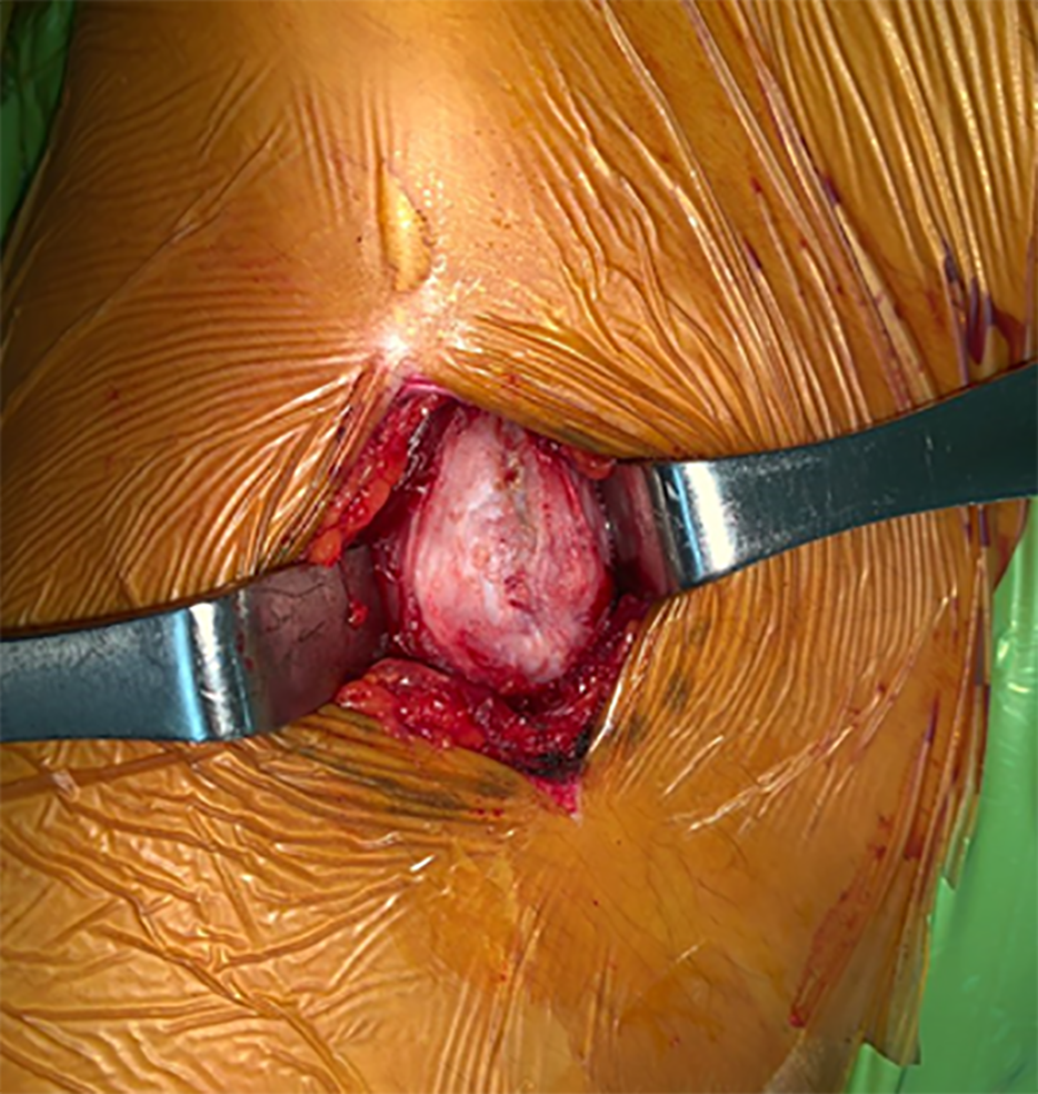
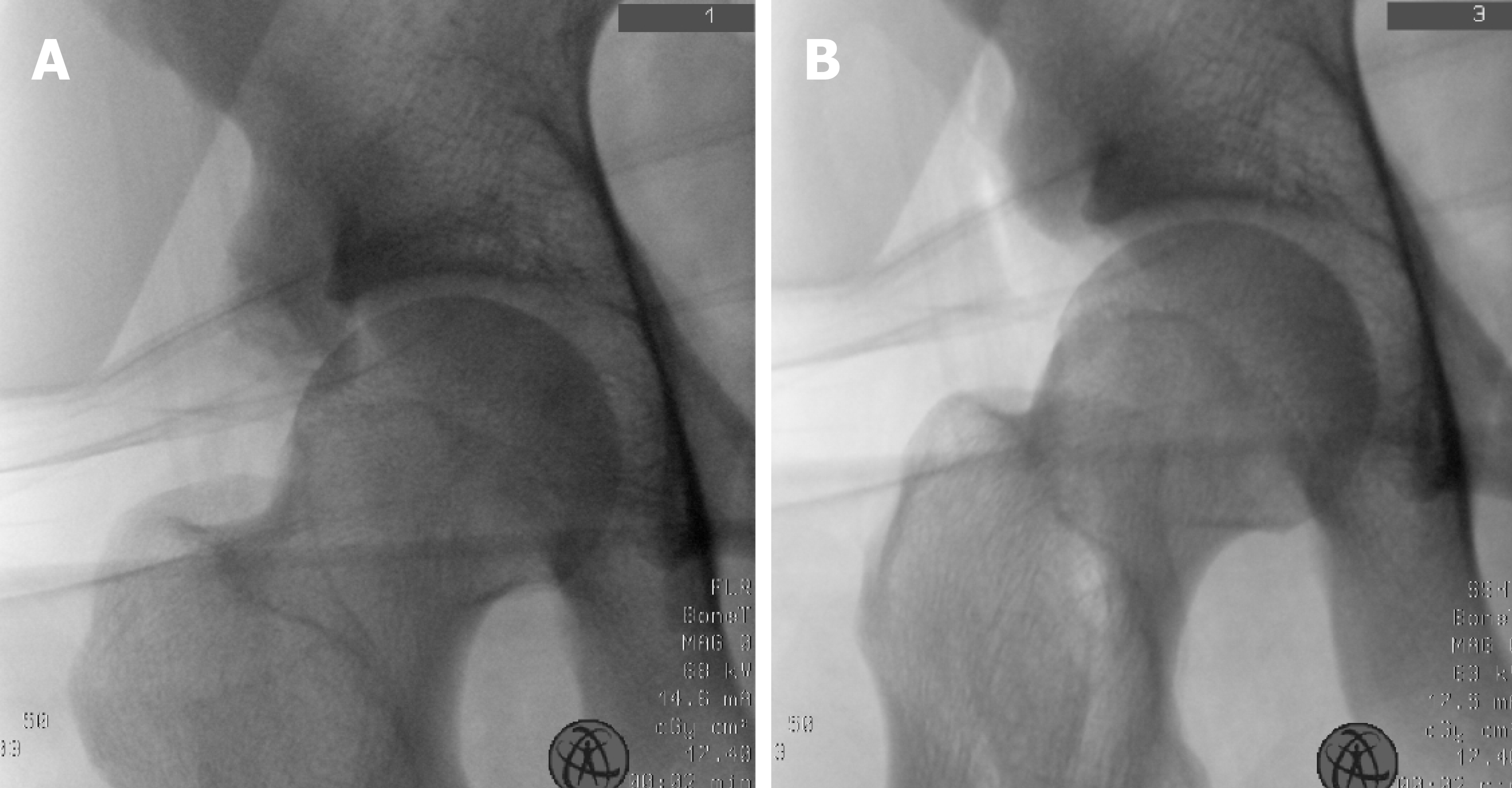
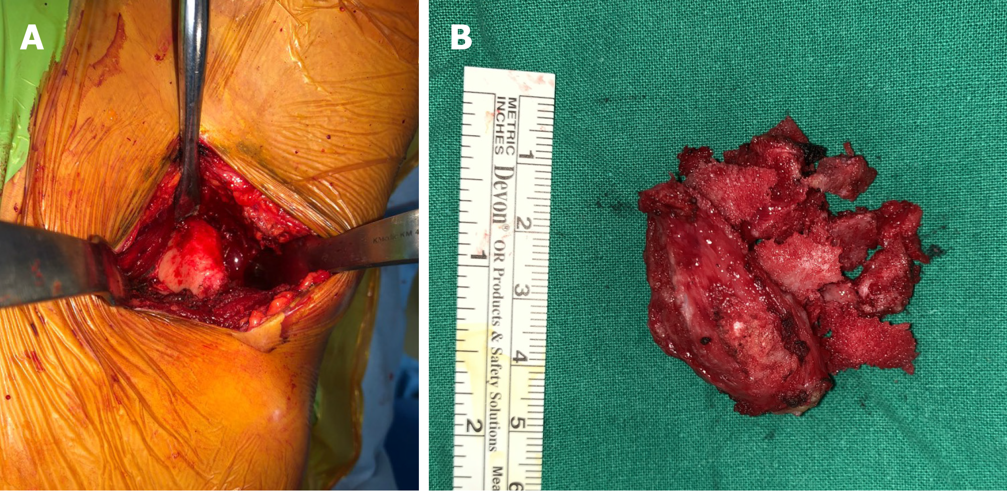
We planned to perform osteotomy for surgical excision and excisional biopsy. Intraoperatively, a mass with smooth surface and an unclear superolateral AIIS border was found (Figure 6). The mass was located anterior to the insertion site of the rectus femoris tendon, not involving it. We used a curved osteotome to expose the fresh bone following the border of ilium. Intraoperative fluoroscopic device showed the bony border being not the same as that of the unaffected side (Figure 7). We could confirm the removal with additional osteotomy by only the surface being peeled without the osteotome internally proceeding (Figure 8). Postoperatively, we found out that border of the bone was different from that of the unaffected side (Figure 9).
Postoperatively, the discomfort during hip flexion was improved, and the hip joint range of motion during walking was recovered at the last follow-up, three weeks after the surgery. He provided informed consent of his data being publicated.
Hip avulsion fractures occur mostly during adolescence when actions such as kicking or running cause forceful contraction of attached muscle. The fracture morphology varies. Ischial tuberosity fractures usually occur during gymnastics, while those of anterior superior iliac spine and AIIS occur during soccer[2]. The symptoms tend to be mild, with localized tenderness and swelling in the fracture site. Primary ossification of the hip joint occurs from the age of 11 to 17 years. Fractures of this period can affect the secondary ossification center; therefore, the unaffected site must be the reference. In AIIS avulsion fractures, fracture displacement is rare, as other connected tendons bind it if proximal rectus femoris is intact. Most hip avulsion fractures can be satisfactorily treated conservatively. Partial weight bearing using crutches are allowed after two weeks, while stretching the muscles needs more care.
Osteochondroma is benign tumor that mostly occurs at the metaphysis of a long bone, being usually asymptomatic. However, occasionally, it is symptomatic due to complications according to size and location. Skeletal complications include deformity, defective function, fracture, and malignant change. Non-skeletal complications can occur because of vessel and nerve compression. Physical examination and simple X-ray are usually used for diagnosis, and computed tomography and MRI are helpful for confirming its exact size and location. MRI discerns surrounding soft tissue and the exact border between osteochondroma and normal bone using signal intensity difference. However, a definite diagnosis requires pathologic biopsy, which shows characteristic features of a well-divided cartilage cap over the bone’s surface. Asymptomatic single osteochondroma does not require treatment, while X-rays are needed to monitor changes. Surgical excision must be considered when pain, limited range of motion, restricted blood supply, nerve symptoms, or malignant changes exist.
The origin of osteochondroma is unclear. In 1891, Virchow reported osteochondroma occurring in cartilage separated from growth plate. In 1920, Keith reported it occurring when a defect in the periosteal bone collar is, causing abnormal proliferation[3]. Geshickter and Copeland mentioned that a cluster of embryonic connective tissue can transform into osteochondroma with repetitive external force at the tendon insertion site[4]. Van Winkle and Mazur reported a case of osteochondroma at surgical drainage site of osteomyelitis in a ten-month-old infant, mentioning that surgical drainage may have damaged the growth plate and induced the osteochondroma, according to Keith’s theory. Some authors have reported that a trauma history prior to osteochondroma formation is relevant[5,6]. Mintzer et al[7] reported a case of osteochondroma after a displaced Salter-Harris type II fracture in the distal femur[7]. Miah et al[8] reported a case of osteochondroma occurring ten years after trauma[8].
The patient of this report underwent osteotomy until fresh bone was exposed. The border was different from that of the unaffected side. Compared to previous X-rays, this suggested bone remodeling due to callus formation of AIIS apophysis and that osteochondroma was caused by AIIS avulsion fracture.
This case can show the correlation between previous trauma and osteochondroma formation. Therefore, the authors found out the importance of history taking when we meet an osteochondroma patient. Previous traumatic history needs to be ruled out. Further case reports regarding this type of history and development of osteochondroma are needed for accumulation of data.
Manuscript source: Unsolicited manuscript
Specialty type: Medicine, research and experimental
Country/Territory of origin: South Korea
Peer-review report’s scientific quality classification
Grade A (Excellent): A
Grade B (Very good): B
Grade C (Good): 0
Grade D (Fair): 0
Grade E (Poor): 0
P-Reviewer: Rodriguez-Merchan EC, Yukata K S-Editor: Zhang L L-Editor: A E-Editor: Xing YX
| 1. | Calderazzi F, Nosenzo A, Galavotti C, Menozzi M, Pogliacomi F, Ceccarelli F. Apophyseal avulsion fractures of the pelvis. A review. Acta Biomed. 2018;89:470-476. [RCA] [PubMed] [DOI] [Full Text] [Full Text (PDF)] [Cited by in RCA: 17] [Reference Citation Analysis (1)] |
| 2. | Rossi F, Dragoni S. Acute avulsion fractures of the pelvis in adolescent competitive athletes: prevalence, location and sports distribution of 203 cases collected. Skeletal Radiol. 2001;30:127-131. [RCA] [PubMed] [DOI] [Full Text] [Cited by in Crossref: 270] [Cited by in RCA: 176] [Article Influence: 7.3] [Reference Citation Analysis (0)] |
| 3. | Brien EW, Mirra JM, Luck JV. Benign and malignant cartilage tumors of bone and joint: their anatomic and theoretical basis with an emphasis on radiology, pathology and clinical biology. II. Juxtacortical cartilage tumors. Skeletal Radiol. 1999;28:1-20. [RCA] [PubMed] [DOI] [Full Text] [Cited by in Crossref: 120] [Cited by in RCA: 84] [Article Influence: 3.2] [Reference Citation Analysis (0)] |
| 4. | Geschickter CF, Copeland MM. Tumors of Bone. Philadelphia: JB Lippincott, 1949. |
| 5. | Bates DL, Osborne WM. Post-traumatic osteochondroma of the calcaneus. J Am Podiatr Med Assoc. 1990;80:606-607. [RCA] [PubMed] [DOI] [Full Text] [Cited by in Crossref: 5] [Cited by in RCA: 5] [Article Influence: 0.1] [Reference Citation Analysis (0)] |
| 6. | Ogden JA. Multiple hereditary osteochondromata. Report of an early case. Clin Orthop Relat Res. 1976;48-60. [PubMed] |
| 7. | Mintzer CM, Klein JD, Kasser JR. Osteochondroma formation after a Salter II fracture. J Orthop Trauma. 1994;8:437-439. [RCA] [PubMed] [DOI] [Full Text] [Cited by in Crossref: 16] [Cited by in RCA: 15] [Article Influence: 0.5] [Reference Citation Analysis (0)] |
| 8. | Miah A, Chu JS, Yegorov A. Post-traumatic osteochondroma of the distal femur. Radiol Case Rep. 2018;13:208-211. [RCA] [PubMed] [DOI] [Full Text] [Full Text (PDF)] [Cited by in Crossref: 4] [Cited by in RCA: 4] [Article Influence: 0.5] [Reference Citation Analysis (0)] |


