Published online Feb 18, 2020. doi: 10.5312/wjo.v11.i2.123
Peer-review started: September 14, 2019
First decision: October 24, 2019
Revised: December 2, 2019
Accepted: December 14, 2019
Article in press: December 14, 2019
Published online: February 18, 2020
Processing time: 158 Days and 0.9 Hours
We report an unusual case of the long head of the biceps brachii tendon rupture near the musculotendinous junction in a young patient. The injury occurred in a young athlete during sports competition. The clinical presentation, surgical treatment, and technique with tenodesis using a unicortical button of the ruptured tendon were presented. The post-surgical recovery was uneventful, and the patient returned to sports in 6 mo. The treatment approach and surgical technique of the long head of biceps brachii rupture was reviewed and discussed. In conclusion, surgical treatment of the long head of the biceps brachii tendon rupture with unicortical button tenodesis resulted in a favorable outcome in a young athlete.
Core tip: We report an unusual case of rupture of the long head of the biceps brachii tendon near the musculotendinous junction in a young atheletc patient. The clinical presentation, surgical treatment, and technique with tenodesis using a unicortical button of the ruptured tendon were presented. The treatment approach and surgical technique of the long head of biceps brachii rupture was reviewed and discussed.
- Citation: Liu X, Tan AHC. Rupture of the long head of the biceps brachii tendon near the musculotendinous junction in a young patient: A case report. World J Orthop 2020; 11(2): 123-128
- URL: https://www.wjgnet.com/2218-5836/full/v11/i2/123.htm
- DOI: https://dx.doi.org/10.5312/wjo.v11.i2.123
Most of the biceps tendon pathologies occur secondary to degeneration and attrition of the rotator cuff[1]. Isolated injury to the long head at the biceps tendon near the musculotendinous junction is uncommon in young patients and it may be associated with overhead sports, weightlifting, or motions that require forceful supination, where an isolated biceps ruptures due to sudden stress[2]. Based on an epidemiological study in the United States, it is uncommon (1.1%) to perform isolated biceps tenodesis in young patients (age < 30 years) and most biceps surgeries were performed in patients with associated diagnoses related to rotator cuff or labral pathologies[3].
Ruptures of the long head of the biceps brachii tendon generally occur in a background of tendon degeneration and are often associated with subscapularis tears, superior labral anterior-posterior lesions or osteophytes[4]. Isolated ruptures are less common and occur more frequently in middle-aged patients[4,5]. They almost always occur near the tendon origin or at the proximal intertubercular groove[4,5]. Tears near the musculotendinous junctions are even rarer. We reported a case of an isolated rupture of the long head of the biceps tendon at the musculotendinous junction in a healthy 22-year-old man. The clinical course, operative management, and post-surgery recovery are presented.
In this case report, we are presenting a young athlete with biceps tear at musculotendinous junction. We used a unicortical biceps tenodesis button instead of the more commonly used bicortical button. The clinical outcome showed that the patient was able to return to professional competition and was satisfied with the outcome. This report provided information on the successful management of patient with high performance demand with a novel technique of using unicortical biceps tenodesis button and a brief literature review of the current practice of the management of biceps tear.
A 22-year-old, right hand-dominant male presented with a chief complaint of swelling, weakness and pain of the right arm following a softball injury.
The patient is a professional softball player. His injury occurred 2 wk before the consultation during a softball competition. He felt a pop sound over the right arm and a snapping sensation when pitching a ball and thereafter, had pain and a noticeable lump over the right anterior arm. The patient did not have prior shoulder or elbow symptoms.
The patient did not have any significant past illness.
The patient did not have any significant personal or family illness.
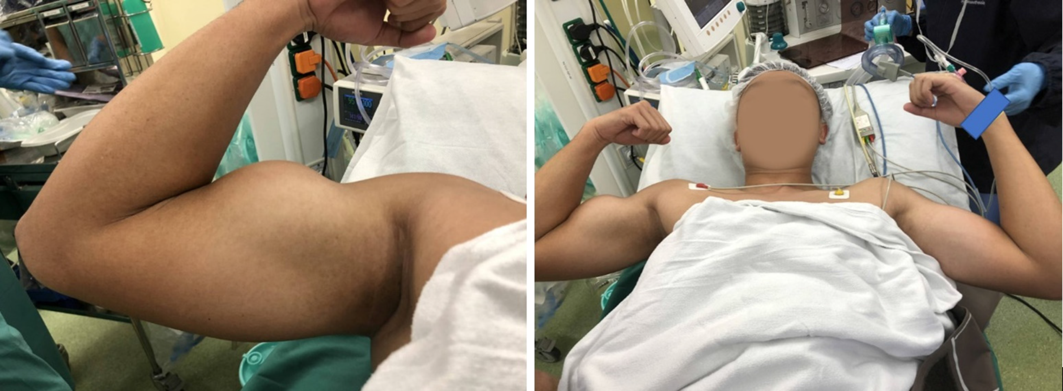
On physical examination, a bulge was seen in the anterior aspect of the right mid-arm that became more pronounced with active flexion of the right supinated forearm, commonly known as “Popeye’s sign” (Figure 1). There was tenderness around the bulge. During resisted elbow flexion, tension was palpable at the biceps tendon insertion but lost over the origin. There was reduced strength of forearm supination and elbow flexion compared to the contralateral side.
The laboratory examination was used for pre-operation evaluation only and included full blood cells counts and renal panel, which are all within normal ranges.
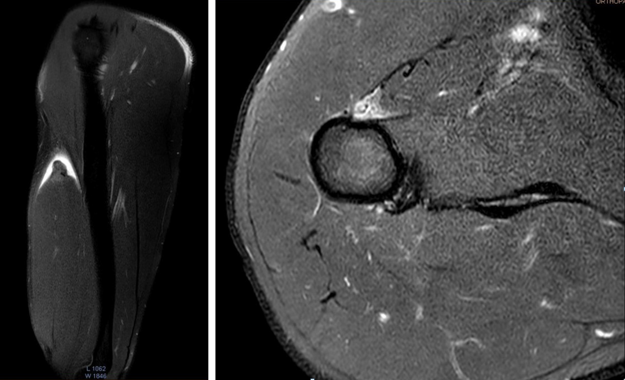
Radiographic examination of the right upper limb did not show any significant soft-tissue or bony abnormalities. Magnetic resonance imaging (MRI) of the right arm revealed a complete rupture of the long head of biceps tendon in the proximal arm near the musculotendinous junction. The proximal portion of the long head of biceps muscle appeared retracted with prominent surrounding fluid/hematoma (Figure 2). This yielded a tendon gap of 6.0 cm. No pathology was identified in the shoulder joint or other parts of the arm.
An isolated rupture of the long head of the biceps tendon.
The patient is a professional athlete and intended to continue his sports career. The treatment options were discussed with the patient and he wanted to maximize shoulder performance and reduce deformity. Surgical treatment was chosen and scheduled one month after the injury. A biceps tenodesis using a unicortical button was performed.
The patient was placed in the beach chair position under general anesthesia. A subpectoral approach with mini-open incision was used. A 3-cm longitudinal incision was made in the anteromedial aspect of the proximal humerus, beginning 1cm proximal to the inferior border of pectoralis major tendon. The dissection was carried on and aimed toward the humerus, avoiding the neurovascular structures over the medial aspect. The biceps which is deep to the pectoralis major tendon was then accessed. During exploration, the short head of the biceps was intact. The long head of the biceps was completely ruptured near the proximal musculotendinous junction.
The musculotendinous stump was then delivered through the wound and a nonabsorbable suture was placed through the remnant tendon, fascia and muscle belly using a whipstitch.
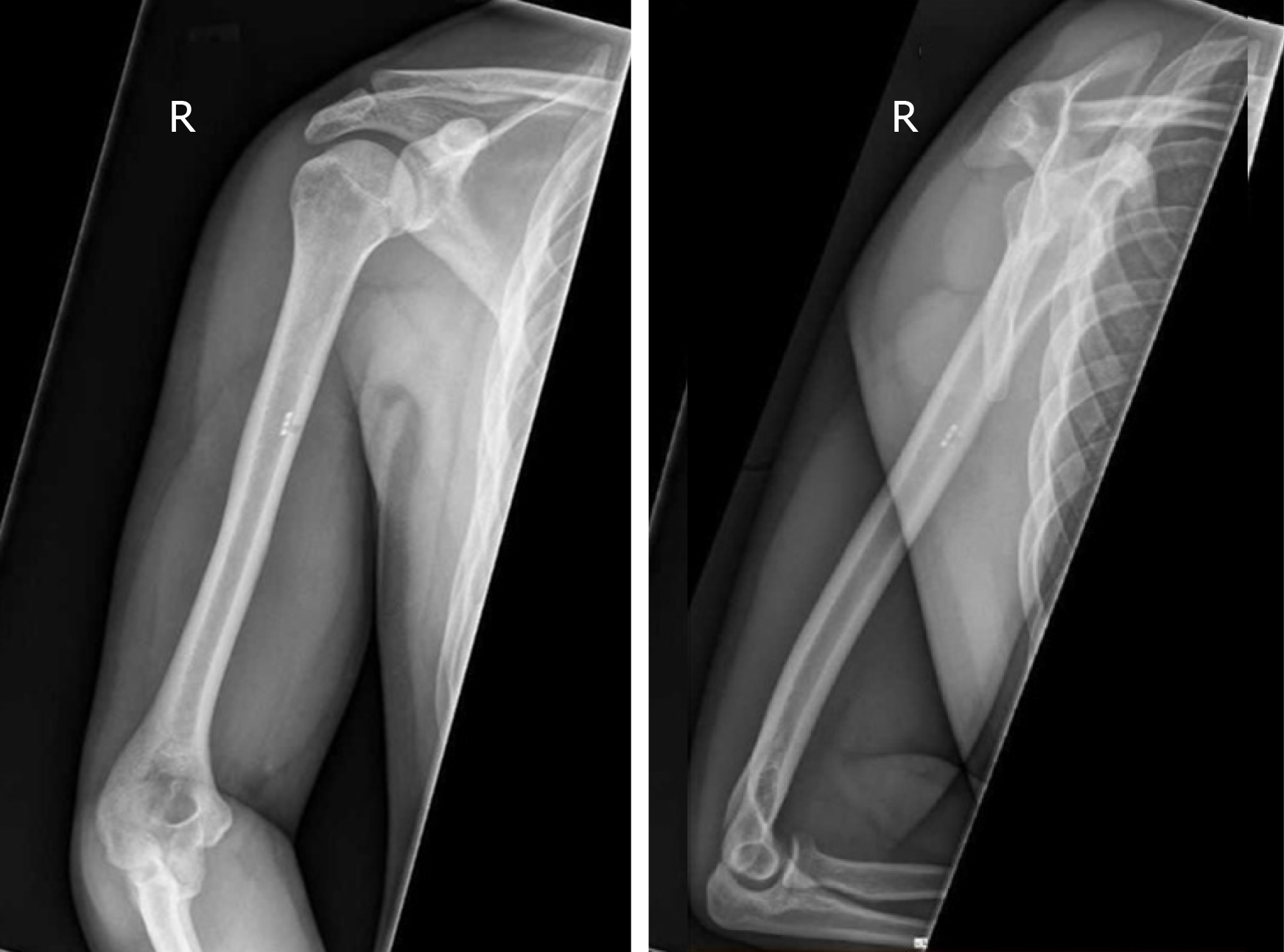
A unicortical metal button (BicepsButtonTM, Arthrex, FL, United States) was used for fixation of the ruptured biceps tendon. A 3.0-mm pin was drilled into the anterior cortex of humerus deep to the inferior border of the pectoralis major tendon. One end of the suture was passed through a hole in the button and then back through the opposite hole. The other end of the sutures was then passed through in the opposite direction. The button was loaded to an inserter and inserted through the pre-drilled 3.0-mm hole. The button was released in the intramedullary canal and retrograde traction was applied to the sutures to toggle the button against the inner anterior cortex. Tension-slip technique was utilized with controlled tension to the two ends of sutures to tighten the button and appose the biceps tendon to the anterior cortex firmly. A free needle attached to one end of the suture was then passed through the biceps, and the two ends were tied to complete the tenodesis. The post-operative radiographs are shown in Figure 3.
Post-operatively, the right arm was placed in a sling at 90 degrees of flexion and followed with passive range of motion exercise in the first four weeks. The patient started gradual active range of motion excise and strengthening from the 5th post-operative week. He regained pre-morbid functional performance and returned to sports 6 months after the surgery and was satisfied with arm strength and cosmetic appearance after surgical treatment.
The biceps brachii muscle functions as a forearm supinator and elbow flexor[1]. The biceps brachii muscle is composed of a short and long head that have different proximal origins on the scapula but share a common distal insertion[1]. The biceps is an important muscle during pitching biomechanics and predominantly activates during cocking to accomplish elbow flexion and then reactivates during follow-through to decelerate the forearm[6]. Rojas et al[7] performed an biomechanics analysis and found that Its activity is higher during windmill pitch than during overhead throw, especially before and after ball release between 9 o’clock and the follow-through phase.
The epidemiology of the pathology varies from degenerative changes in the elderly to traumatic injuries related to weightlifting or throwing in younger patients[8]. The biceps tendon pathologies usually occur due to degeneration of the rotator cuff middle-aged to older patients[1]. Isolated injury to the biceps tendon is uncommon in young patients and it may be associated with overhead sports, weightlifting, or motions that require excessive supination, where a sudden stress may result in an isolated bicep rupture without pre-existing glenohumeral pathology[2]. Barrentine et al[9] reported that forceful overloading of the biceps in throwing athletes, especially baseball pitchers, can result in traction and avulsion of the biceps in the deceleration phase of throwing.
A biceps tendon rupture often associated with a popping sound after traumatic injury. Clinical presentation may include localized sharp pain, ecchymosis and swelling. As shown in Figure 1, a biceps tendon rupture is classically presented with Popeye’s sign, a visible muscle prominence in the mid arm. Radiographic investigation includes magnetic resonance imaging or ultrasound to delineate a complete or partial rupture and investigate associated shoulder pathologies.
Based on an epidemiological study in the United States, it is uncommon (1.1%) to perform isolated biceps tenodesis in young patients (age < 30 years) and most biceps surgeries were performed in patients with associated diagnoses related to rotator cuff or labral pathologies[3]. After rupture of the long head, there may be loss of up to 20% of muscle strength[10]. Surgical treatment of the rupture of the biceps in young or active sportsmen is recommended, which is able to restore both flexion and supination strength and reduce the risk of cosmetic deformity[10].
The treatment of biceps tendon ruptures shoulder be tailored to patients. Factors need to consider include: Age of the patients and their demand, the quality of the tendon to be tenodesed and the position where tenodesis is possible etc.
The patient presented here is a young throwing athlete who places a high demand on his upper limb and who had concerns regarding the cosmetic appearance of the Popeye arm. Surgical treatment with biceps tenodesis was therefore chosen over conservative management.
The ideal location of tenodesis and method of fixation is debated[1-3,5,11]. Tenodesis of the long head of the biceps can be done proximally (suprapectoral) or distally (subpectoral). The proximal fixation site is either within the glenohumeral joint to the intact rotator cuff or just proximal to or within the bicipital groove, and the fixation is typically carried out arthroscopically. Subpectoral biceps tenodesis has emerged as a new technique to treat biceps ruptures. Initially described by Mazzocca et al[12,13], subpectoral biceps tenodesis secures the biceps tendon distal to the bicipital groove through a mini-open incision. There are a few proposed advantages of subpectoral tenodesis[11]. Firstly, the relevant anatomy is can be easily oriented and identified, which aids the consistency of the length-tension relationship. Secondly, as the fixation occurs distally to the bicipital groove, it reduces of risk of pain at this site. Thirdly, subpectoral biceps tenodesis has the versatility of using interference screw and suture anchor fixation with their attendant biomechanical advantages.
The rupture occurred near the proximal musculotendinous junction. There was inadequate tendon length for suprapectoral tenodesis, and it would be technically challenging to achieve a proper length-tension relationship. Apart from its purported advantages as mentioned above, subpectoral tenodesis was chosen based on these considerations. A shoulder arthroscopy was not performed in this case. The patient’s profile, history and clinical examination suggested it was an isolated biceps tendon lesion and the MRI performed confirmed this finding. The complication of leaving the proximal stump in situ is unclear and to the authors’ knowledge, not reported in literature. As additional proximal incision and dissection would be required to retrieve the proximal stump, it was not performed.
In this case, we used a unicortical biceps tenodesis button instead of the more commonly used bicortical button. The benefits of using unicortical button comparing to bicortical or interference screw are reduced risk of iatrogenic brachial plexus injury and humeral fracture[14,15] as only one cortex is drilled under direct visual with a cortical defect of 3-mm which minimizes the risk of humerus fracture[16]. In a biomechanical cadaveric study comparing the unicortical button with interference screw fixation, considerably less displacement in cyclic loading in the unicortical button group was demonstrated with equivalent ultimate load to failure and stiffness[17].
Isolated rupture of the long head of the biceps brachii tendon near the musculotendinous junction is an uncommon injury. In young and active patients, surgery is the preferred treatment choice to restore elbow flexion and forearm supination strength and should be recommended. A unicortical button is effective in achieving a stable repair with favorable surgical outcome.
Manuscript source: Unsolicited manuscript
Specialty type: Orthopedics
Country of origin: Singapore
Peer-review report classification
Grade A (Excellent): 0
Grade B (Very good): 0
Grade C (Good): C
Grade D (Fair): 0
Grade E (Poor): 0
P-Reviewer: Fanter NJ S-Editor: Ma RY L-Editor: A E-Editor: Liu MY
| 1. | Elser F, Braun S, Dewing CB, Giphart JE, Millett PJ. Anatomy, function, injuries, and treatment of the long head of the biceps brachii tendon. Arthroscopy. 2011;27:581-592. [RCA] [PubMed] [DOI] [Full Text] [Cited by in Crossref: 179] [Cited by in RCA: 163] [Article Influence: 11.6] [Reference Citation Analysis (0)] |
| 2. | Rockwood CA, Wirth MA, Fehringer EV, Sperling JW. Editors: Matsen FA, Lippitt SB. Rockwood and Matsen's the shoulder. 5th ed. Philadelphia: Elsevier, 2017: 1-1304. |
| 3. | Werner BC, Brockmeier SF, Gwathmey FW. Trends in long head biceps tenodesis. Am J Sports Med. 2015;43:570-578. [RCA] [PubMed] [DOI] [Full Text] [Cited by in Crossref: 98] [Cited by in RCA: 108] [Article Influence: 10.8] [Reference Citation Analysis (0)] |
| 4. | Geaney LE, Mazzocca AD. Biceps brachii tendon ruptures: a review of diagnosis and treatment of proximal and distal biceps tendon ruptures. Phys Sportsmed. 2010;38:117-125. [RCA] [PubMed] [DOI] [Full Text] [Cited by in Crossref: 16] [Cited by in RCA: 16] [Article Influence: 1.1] [Reference Citation Analysis (0)] |
| 5. | Jayamoorthy T, Field JR, Costi JJ, Martin DK, Stanley RM, Hearn TC. Biceps tenodesis: a biomechanical study of fixation methods. J Shoulder Elbow Surg. 2004;13:160-164. [RCA] [PubMed] [DOI] [Full Text] [Cited by in Crossref: 51] [Cited by in RCA: 50] [Article Influence: 2.4] [Reference Citation Analysis (0)] |
| 6. | Jobe FW, Moynes DR, Tibone JE, Perry J. An EMG analysis of the shoulder in pitching. A second report. Am J Sports Med. 1984;12:218-220. [RCA] [PubMed] [DOI] [Full Text] [Cited by in Crossref: 314] [Cited by in RCA: 265] [Article Influence: 6.5] [Reference Citation Analysis (0)] |
| 7. | Rojas IL, Provencher MT, Bhatia S, Foucher KC, Bach BR, Romeo AA, Wimmer MA, Verma NN. Biceps activity during windmill softball pitching: injury implications and comparison with overhand throwing. Am J Sports Med. 2009;37:558-565. [RCA] [PubMed] [DOI] [Full Text] [Cited by in Crossref: 61] [Cited by in RCA: 59] [Article Influence: 3.7] [Reference Citation Analysis (0)] |
| 8. | Barrentine SW, Fleisig GS, Whiteside JA, Escamilla RF, Andrews JR. Biomechanics of windmill softball pitching with implications about injury mechanisms at the shoulder and elbow. J Orthop Sports Phys Ther. 1998;28:405-415. [RCA] [PubMed] [DOI] [Full Text] [Cited by in Crossref: 93] [Cited by in RCA: 98] [Article Influence: 3.6] [Reference Citation Analysis (0)] |
| 9. | Andrews JR, Carson WG, McLeod WD. Glenoid labrum tears related to the long head of the biceps. Am J Sports Med. 1985;13:337-341. [RCA] [PubMed] [DOI] [Full Text] [Cited by in Crossref: 751] [Cited by in RCA: 627] [Article Influence: 15.7] [Reference Citation Analysis (0)] |
| 10. | Sturzenegger M, Béguin D, Grünig B, Jakob RP. Muscular strength after rupture of the long head of the biceps. Arch Orthop Trauma Surg. 1986;105:18-23. [RCA] [PubMed] [DOI] [Full Text] [Cited by in Crossref: 41] [Cited by in RCA: 45] [Article Influence: 1.2] [Reference Citation Analysis (0)] |
| 11. | Provencher MT, LeClere LE, Romeo AA. Subpectoral biceps tenodesis. Sports Med Arthrosc Rev. 2008;16:170-176. [RCA] [PubMed] [DOI] [Full Text] [Cited by in Crossref: 86] [Cited by in RCA: 92] [Article Influence: 5.4] [Reference Citation Analysis (0)] |
| 12. | Mazzocca AD, Rios CG, Romeo AA, Arciero RA. Subpectoral biceps tenodesis with interference screw fixation. Arthroscopy. 2005;21:896. [RCA] [PubMed] [DOI] [Full Text] [Cited by in Crossref: 167] [Cited by in RCA: 164] [Article Influence: 8.2] [Reference Citation Analysis (0)] |
| 13. | Mazzocca AD, Bicos J, Santangelo S, Romeo AA, Arciero RA. The biomechanical evaluation of four fixation techniques for proximal biceps tenodesis. Arthroscopy. 2005;21:1296-1306. [RCA] [PubMed] [DOI] [Full Text] [Cited by in Crossref: 243] [Cited by in RCA: 218] [Article Influence: 10.9] [Reference Citation Analysis (0)] |
| 14. | Sears BW, Spencer EE, Getz CL. Humeral fracture following subpectoral biceps tenodesis in 2 active, healthy patients. J Shoulder Elbow Surg. 2011;20:e7-11. [RCA] [PubMed] [DOI] [Full Text] [Cited by in Crossref: 105] [Cited by in RCA: 101] [Article Influence: 7.2] [Reference Citation Analysis (0)] |
| 15. | Rhee PC, Spinner RJ, Bishop AT, Shin AY. Iatrogenic brachial plexus injuries associated with open subpectoral biceps tenodesis: a report of 4 cases. Am J Sports Med. 2013;41:2048-2053. [RCA] [PubMed] [DOI] [Full Text] [Cited by in Crossref: 53] [Cited by in RCA: 46] [Article Influence: 3.8] [Reference Citation Analysis (0)] |
| 16. | Hipp JA, Edgerton BC, An KN, Hayes WC. Structural consequences of transcortical holes in long bones loaded in torsion. J Biomech. 1990;23:1261-1268. [RCA] [PubMed] [DOI] [Full Text] [Cited by in Crossref: 60] [Cited by in RCA: 56] [Article Influence: 1.6] [Reference Citation Analysis (0)] |
| 17. | DeAngelis JP, Chen A, Wexler M, Hertz B, Grimaldi Bournissaint L, Nazarian A, Ramappa AJ. Biomechanical characterization of unicortical button fixation: a novel technique for proximal subpectoral biceps tenodesis. Knee Surg Sports Traumatol Arthrosc. 2015;23:1434-1441. [RCA] [PubMed] [DOI] [Full Text] [Cited by in Crossref: 20] [Cited by in RCA: 19] [Article Influence: 1.9] [Reference Citation Analysis (0)] |


