Copyright
©The Author(s) 2018.
World J Orthop. Sep 18, 2018; 9(9): 180-184
Published online Sep 18, 2018. doi: 10.5312/wjo.v9.i9.180
Published online Sep 18, 2018. doi: 10.5312/wjo.v9.i9.180
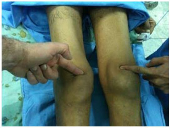
Figure 1 Palpation showing bilateral depression above the patella.
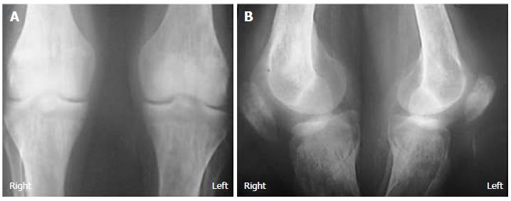
Figure 2 Knee radiographs showing patellar lowering and diffuse bone demineralization.
A: Antero-posterior X-rays; B: Lateral X-rays.
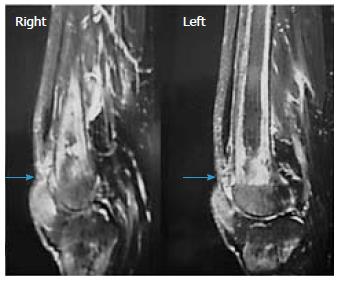
Figure 3 Magnetic resonance imaging of the knee (sagittal view) showing a complete bilateral rupture of the tendon.
Figure 4 Surgical management of the injury through bilateral reinsertion of the tendon.
A: Exposing the bilateral rupture of the quadriceps tendon; B: Quadriceps tendon reinserted through tendon-to-bone repair.
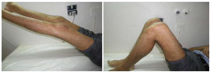
Figure 5 Complete active extensions of the knees.
- Citation: Zribi W, Zribi M, Guidara AR, Ben Jemaa M, Abid A, Krid N, Naceur A, Keskes H. Spontaneous and simultaneous complete bilateral rupture of the quadriceps tendon in a patient receiving hemodialysis: A case report and literature review. World J Orthop 2018; 9(9): 180-184
- URL: https://www.wjgnet.com/2218-5836/full/v9/i9/180.htm
- DOI: https://dx.doi.org/10.5312/wjo.v9.i9.180


