Copyright
©The Author(s) 2018.
World J Orthop. Sep 18, 2018; 9(9): 156-164
Published online Sep 18, 2018. doi: 10.5312/wjo.v9.i9.156
Published online Sep 18, 2018. doi: 10.5312/wjo.v9.i9.156
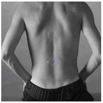
Figure 1 Placement of the electrodes according to SENIAM[27].
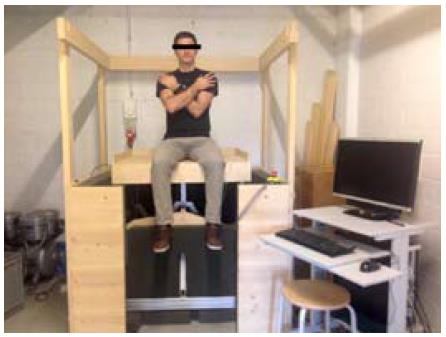
Figure 2 Sitting position on the vibration platform.
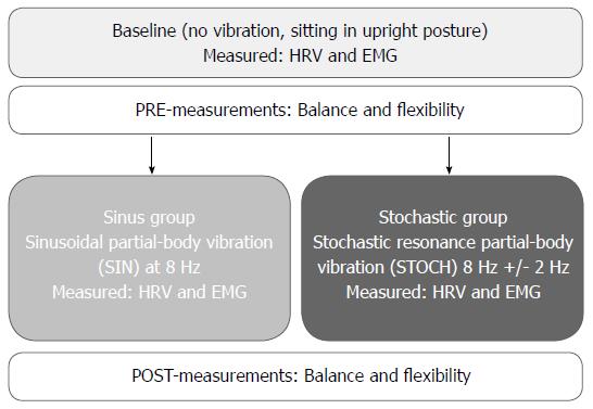
HRV: Heart rate variability; EMG: Electromyography.
Figure 3 Flowchart of the procedure.
EMG: Electromyography, muscular activation of the lower back muscle erector spinae (ES); HRV: Heart rate variability was measured with Polar V800. Balance was measured using the modified star excursion balance test (mSEBT) and expressed in centimeter; Flexibility was assessed with the fingertip-to-floor method (mFTF) and measured in centimeter.
- Citation: Faes Y, Banz N, Buscher N, Blasimann A, Radlinger L, Eichelberger P, Elfering A. Acute effects of partial-body vibration in sitting position. World J Orthop 2018; 9(9): 156-164
- URL: https://www.wjgnet.com/2218-5836/full/v9/i9/156.htm
- DOI: https://dx.doi.org/10.5312/wjo.v9.i9.156


