Copyright
©The Author(s) 2018.
World J Orthop. Sep 18, 2018; 9(9): 130-137
Published online Sep 18, 2018. doi: 10.5312/wjo.v9.i9.130
Published online Sep 18, 2018. doi: 10.5312/wjo.v9.i9.130
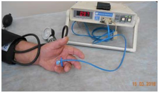
Figure 1 Assessment of skin microcirculation in patient with Dupuytren’s contracture using laser Doppler flowmetry and local ischemic test.
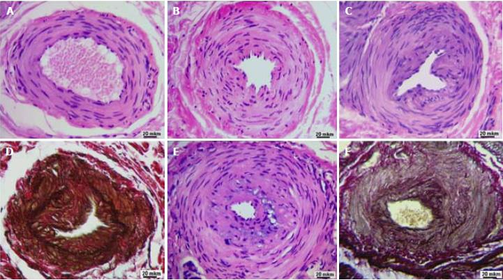
Figure 2 Transverse paraffin sections of palmar fascia perforating arteries from Dupuytren’s contracture patients.
A: Almost normal structure of perforating artery; B: Adventitial fibrosis, constriction of media and deformity of artery lumen; C: Artery with thick neointimal layer; D: The same artery with breakdown of internal elastic laminae evident due to specific elastin staining (dark brown); E: Extreme lumen narrowing due to neointimal thickening; F: Residual fragments of old internal elastic laminae newly formed elastic fibers in the neointimal layer. Staining: Hematoxylin-eosin (A, B, C, E); Weigert - van Gieson (D, E). Magnification 200 ×.
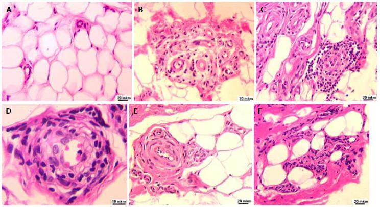
Figure 3 Fragments of hypodermis paraffin sections from Dupuytren’s contracture patients.
A: Thick-walled capillaries in adipose tissue of hypodermis; B: Inflammatory infiltration of vascular pool; C: Multilayered round-cellular cuff outside the narrowed vessel; D: Perivascular and intramural infiltration with lymphocytes and macrophages; E: Pronounced perivascular fibrosis; F: Collagen deposits and perivascular clusters of fibroblasts. Staining: Hematoxylin-eosin. Magnification 200 ×.
- Citation: Shchudlo N, Varsegova T, Stupina T, Dolganova T, Shchudlo M, Shihaleva N, Kostin V. Assessment of palmar subcutaneous tissue vascularization in patients with Dupuytren’s contracture. World J Orthop 2018; 9(9): 130-137
- URL: https://www.wjgnet.com/2218-5836/full/v9/i9/130.htm
- DOI: https://dx.doi.org/10.5312/wjo.v9.i9.130


