Copyright
©The Author(s) 2018.
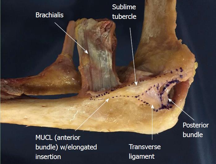
Figure 1 Medial ulnar collateral ligament complex of the elbow with outlined ligaments generated by co-registering the three dimensional digitized anatomy and computed tomography scan of a cadaveric elbow.
Note the tapered and distally elongated insertion of the medial ulnar collateral ligament on the sublime tubercle and ulnar ridge. MUCL: Medial ulnar collateral ligament.
Figure 2 Cadaveric specimen outlining all ligaments of the medial ulnar collateral ligament complex including the medial ulnar collateral ligament/anterior bundle, posterior bundle, and transverse ligament.
MUCL: Medial ulnar collateral ligament.
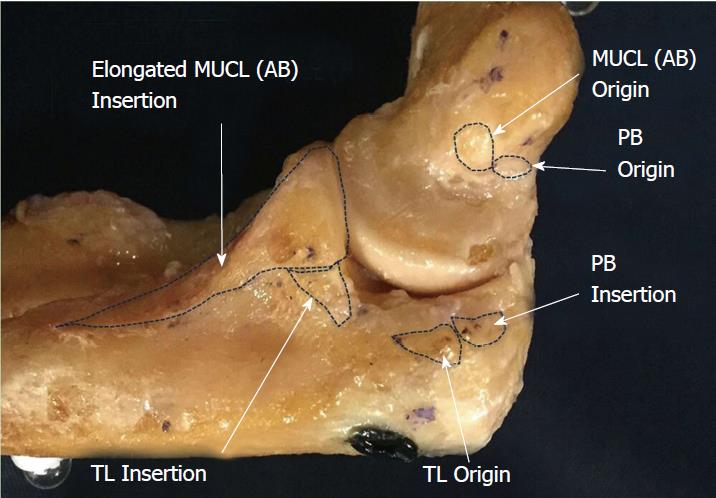
Figure 3 Cadaveric specimen showing origins and insertions of all ligaments of the medial ulnar collateral ligament complex including: Anterior bundle, posterior bundle and transverse ligament.
AB: Anterior bundle; PB: Posterior bundle; TL: Transverse ligament.
- Citation: Labott JR, Aibinder WR, Dines JS, Camp CL. Understanding the medial ulnar collateral ligament of the elbow: Review of native ligament anatomy and function. World J Orthop 2018; 9(6): 78-84
- URL: https://www.wjgnet.com/2218-5836/full/v9/i6/78.htm
- DOI: https://dx.doi.org/10.5312/wjo.v9.i6.78


