Copyright
©The Author(s) 2018.
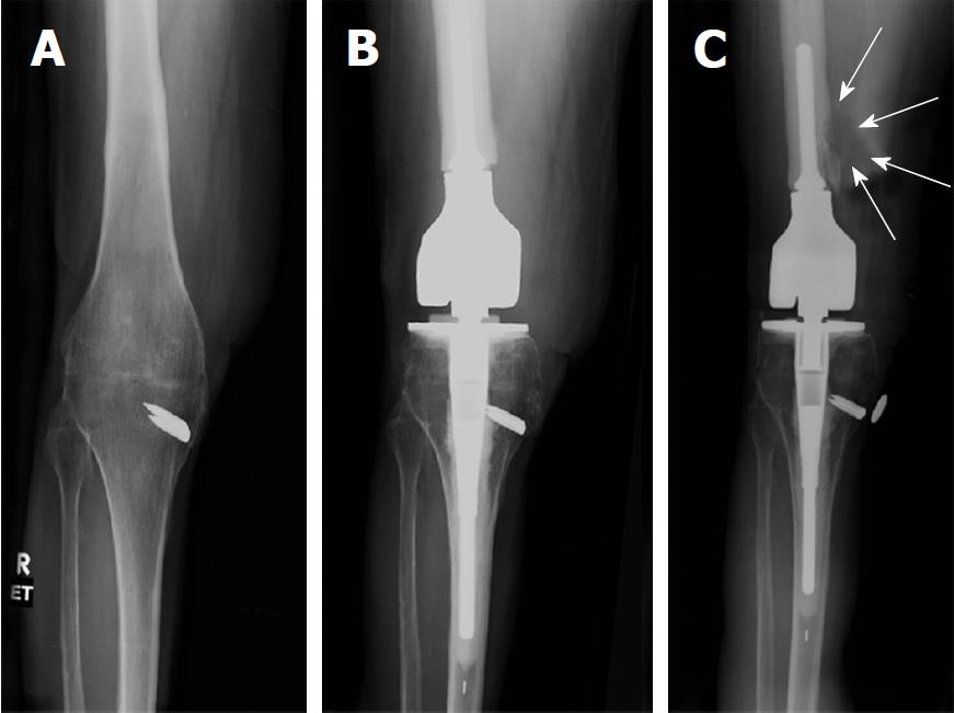
Figure 1 AP radiograph of right knee reviewing life-cycle of right knee reconstruction.
A: Pre-operative radiograph showing successful knee fusion. The area over the medial staple has a split thickness skin graft (STSG) of 5 cm x 7 cm. Patient ambulates with full weight bearing on the right leg. Localized osteopenia is present in the knee region; B: Four months post-operative radiograph of cemented endoprosthetic hinge total knee arthroplasty using a modified Zimmer Biomet RS (Reduced Size) OSS Knee system. Note particularly the placement of the joint line in the cephalad direction. This was purposefully done to avoid dissecting near the area of the tibia with the STSG. Also, note the relatively short cemented femoral stem; C: Radiograph at 6 years. Patient complains of mild pain for the past 5 months. Note the erosive lesion in the distal femoral diaphysis. White arrows outline the soft tissue mass surrounding the erosive lesion. Also note the radiolucent line in the distal femoral diaphysis. This suggests cement-bone separation, likely from repetitive cantilever bend.
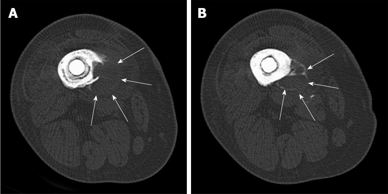
Figure 2 Transverse computed tomography scan images of distal femoral diaphysis centered over lytic lesion.
A: Image shows a surrounding soft tissue mass of 2.8 cm. Erosive lytic lesion extends down to the inner cortex. Also note the radiolucent lines between the prosthetic implant and cement mantle, as well as the radiolucent line between the cement mantle and bone; B: Demonstrates septation of the soft tissue mass with peripheral calcification (arrows). This is typically a worrisome sign for a neoplastic process.
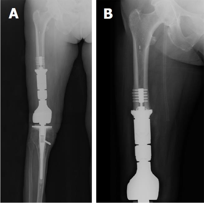
Figure 3 Postoperative radiographs showing the additional bone resection, resection of the soft tissue pseudotumor, and the definitive reconstruction with an extended femoral endoprosthesis.
A: Four week post-operative radiograph showing resection of the distal 9 cm of femoral diaphysis and reconstruction with a 9 cm intercalary segment. The intercalary segment is secured to the femoral diaphysis with a femoral compress device (Zimmer Biomet, Warsaw, IN, United States); B: One-year postoperative radiograph showing distal femoral bone remodeling and bone integration into the porous end of the femoral compress device. The patient’s thigh pain was completely resolved and her gait returned to a reciprocating smooth gait that appeared symmetrical to the contralateral leg.
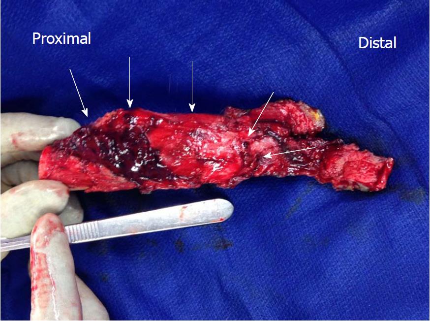
Figure 4 Resected specimen that contains the distal 9 cm of the femoral diaphysis along with the soft tissue tumor.
Specimen is rotated to view the soft tissue mass (arrows). Note the absence of metal debris within the extended joint space.
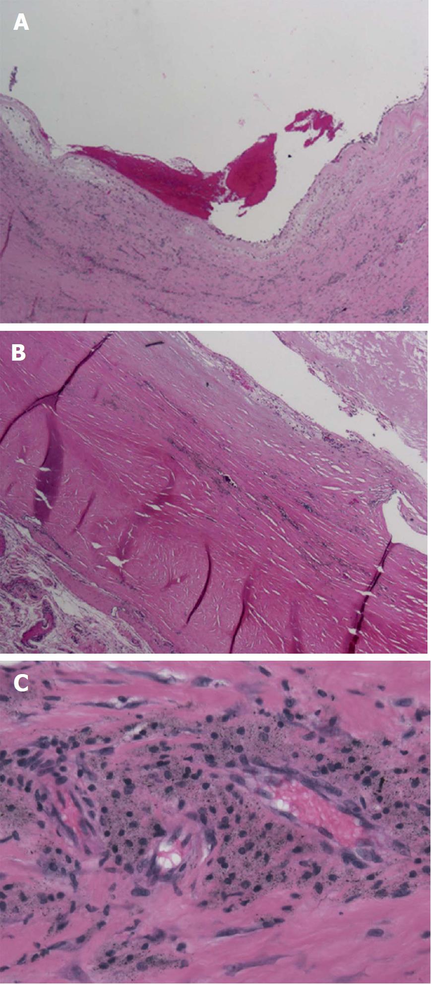
Figure 5 Histologic examination of the pseudotumor with specimen taken in the middle of the cystic mass.
A: Low power (100 ×) view of mass demonstrating its cystic nature with blood and fibrin products within the centre of cystic structure. The wall of the lesion is very thick, studded with islands of metallic particles; B: Intermediate power (200 ×) view of the wall of pseudotumor mass. In the lower left corner, the cellular reaction is primarily lymphocytic and histiocytic with perivascular lymphocytes. The eosinophilic (red-color) area shows the pseudotumor cyst wall with adherent fibrin debris (top right corner) and microscopic metal particles within the wall; C: High power (400 ×) view of the cellular reaction surrounding pseudotumor mass. This shows the "classic" histologic finding of a reactive pseudotumor due to small metallic particles. The micrograph shows perivascular infiltration of histiocytes, containing particles of metallic debris, along with lymphocytes.
- Citation: Chowdhry M, Dipane MV, McPherson EJ. Periosteal pseudotumor in complex total knee arthroplasty resembling a neoplastic process. World J Orthop 2018; 9(5): 72-77
- URL: https://www.wjgnet.com/2218-5836/full/v9/i5/72.htm
- DOI: https://dx.doi.org/10.5312/wjo.v9.i5.72


