Copyright
©The Author(s) 2018.
World J Orthop. Oct 18, 2018; 9(10): 210-219
Published online Oct 18, 2018. doi: 10.5312/wjo.v9.i10.210
Published online Oct 18, 2018. doi: 10.5312/wjo.v9.i10.210
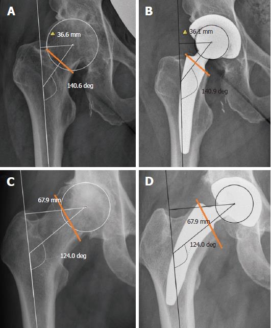
Figure 1 “Top down concept” of the MiniHipTM to restore the individual joint geometry.
The individual femoral neck cut and physiological orientation of the partially retained femoral neck allows the reconstruction of the individual joint geometry. A: Valgus hip deformity; B: A deeper femoral neck cut leads a reduction of the femoral offset with an accurate reconstruction of the joint geometry; C: Varus hip; D: A low femoral neck provides a reconstruction of the geometry with an increased femoral offset with an appropriate successfully reconstructed joint geometry.
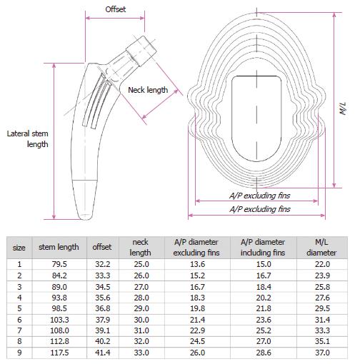
Figure 2 Dimensions (mm) of the different sizes of the MiniHipTM stem.
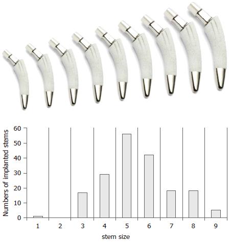
Figure 3 Frequencies of the implanted stem sizes used in this study.
The distribution of sizes used in this study is similar to a Gauss curve. The increasing dimensions of the conus according to the nine different sizes of the implant are depicted. The distal bullet tip of the prosthesis is polished. This design might prevent a fixation in this area and therefore reduce the risk of thigh pain.
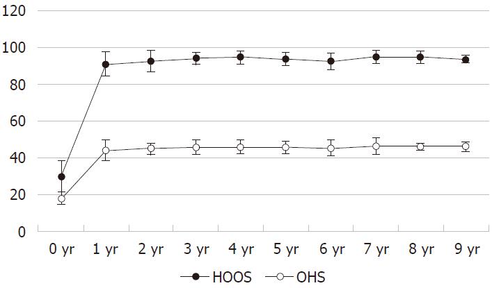
Figure 4 Outcome at the Hip Dysfunction Osteoarthritis and Outcome Score and Oxford Hip Score scoring over ten years.
After the initial improvement after one year, the subsequent scorings at our follow-up investigations two to ten years after the implantation showed only slight increases, which were not significant, P > 0.05 respectively. HOOS: Hip Dysfunction Osteoarthritis and Outcome Score; OHS: Oxford Hip Score.
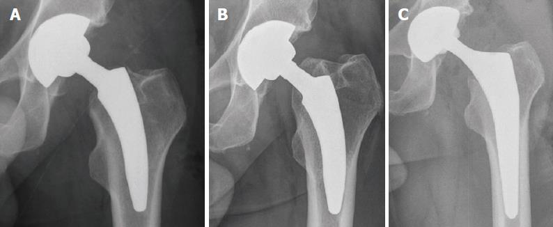
Figure 5 Exemplary X-ray of one of the two cases with a symptomatic subsidence.
A: Postoperative X-ray; B: Subsidence of 15 mm twelve months after surgery; C: X-ray of the one-stage revision to a conventional stem.
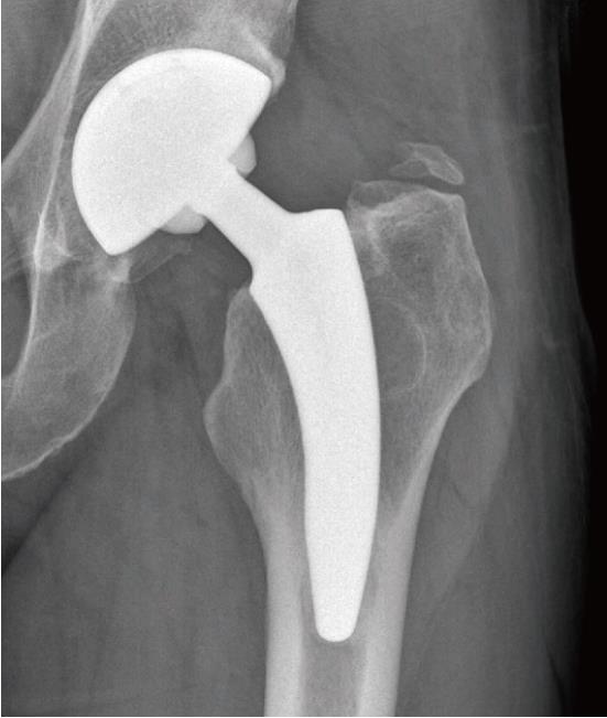
Figure 6 Exemplary X-ray of one of the nine cases with a heterotopic ossifications grade I according to Brooker.
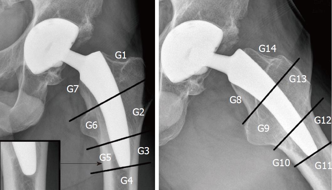
Figure 7 Periprosthetic bone resorptions or bony hypertrophies were assessed within the Gruen zones 1-14.
A small ostolysis of less than 2 mm outlined by a discrete sclerotic margin was detected in a large number of patients around the tip of the stem. Being detected exclusively around the polished tip of the stem, it might be indicative for a fibrous ingrowth at the bullet polished tip of the stem. Further radiological and/or clinical signs of a loosening were not noticed in these cases.
- Citation: von Engelhardt LV, Breil-Wirth A, Kothny C, Seeger JB, Grasselli C, Jerosch J. Long-term results of an anatomically implanted hip arthroplasty with a short stem prosthesis (MiniHipTM). World J Orthop 2018; 9(10): 210-219
- URL: https://www.wjgnet.com/2218-5836/full/v9/i10/210.htm
- DOI: https://dx.doi.org/10.5312/wjo.v9.i10.210


