Copyright
©The Author(s) 2018.
World J Orthop. Oct 18, 2018; 9(10): 185-189
Published online Oct 18, 2018. doi: 10.5312/wjo.v9.i10.185
Published online Oct 18, 2018. doi: 10.5312/wjo.v9.i10.185
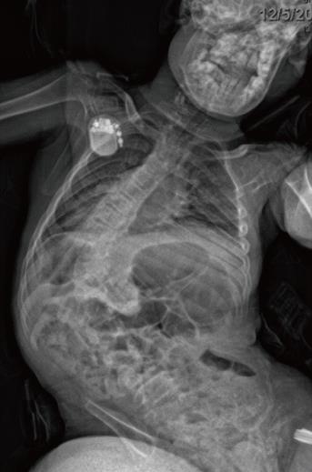
Figure 1 Posteroanterior of a sweeping thoracolumbar curve with pelvic obliquity typical of neuromuscular scoliosis.
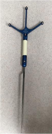
Figure 2 Navigation probe.
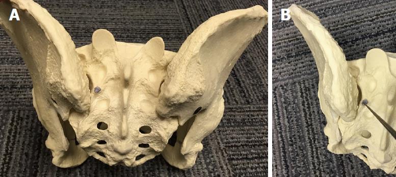
Figure 3 Starting point for the S2-alar-iliac technique screw.
A: On pelvis; B: Navigation probe identifying the appropriate location.
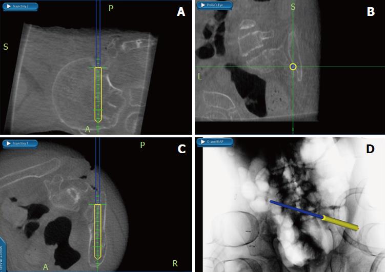
Figure 4 Intra-operative navigation screen depicting safe starting point and projected placement of an S2-alar-iliac technique screw.
A: Sagittal; B: Coronal; C: Axial; D: Current position of navigation probe.
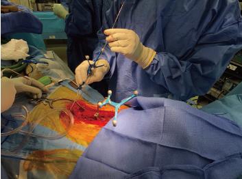
Figure 5 Intraoperative clinical photo depicting the navigation probe placed down the dilated path for the S2-alar-iliac technique screw with a guidewire in place.
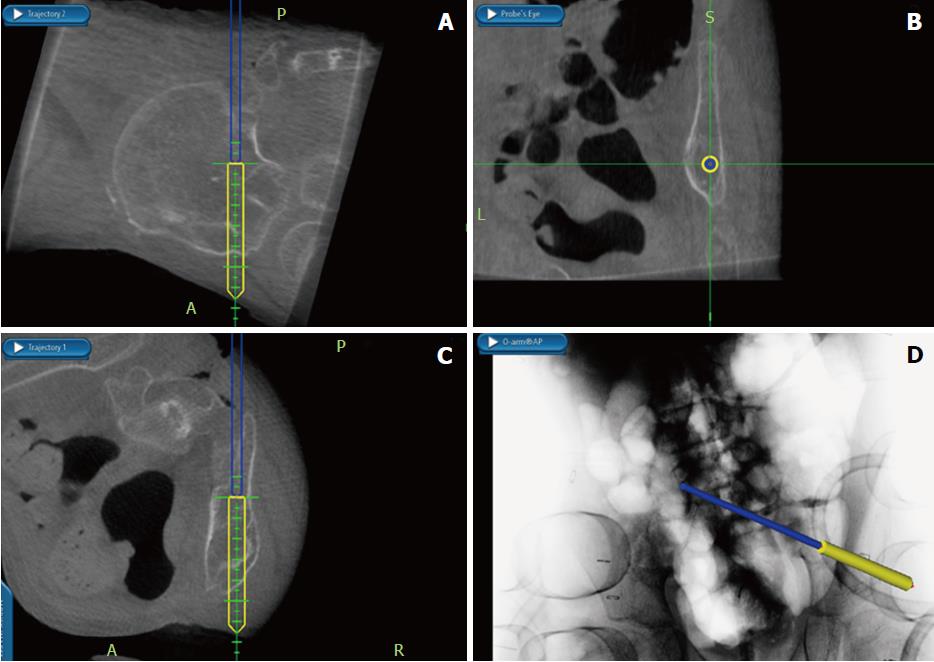
Figure 6 Intra-operative navigation screens depicting a safe trajectory for the S2-alar-iliac technique pelvic screw.
A: Sagittal; B: Coronal; C: Axial; D: Anterior-posterior radiograph showing current position of navigation probe.
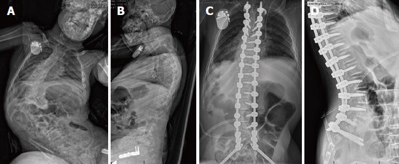
Figure 7 Pre-operative and post-operative radiographs in a patient with neuromuscular scoliosis who underwent T3 to pelvis instrumented posterior spinal fusion using navigation to place pedicle screw and S2-alar-iliac technique instrumentation.
A: Posteroanterior; B: Lateral; C: Anteroposterior; D: Lateral.
- Citation: Anari JB, Cahill PJ, Flynn JM, Spiegel DA, Baldwin KD. Intra-operative computed tomography guided navigation for pediatric pelvic instrumentation: A technique guide. World J Orthop 2018; 9(10): 185-189
- URL: https://www.wjgnet.com/2218-5836/full/v9/i10/185.htm
- DOI: https://dx.doi.org/10.5312/wjo.v9.i10.185


