Copyright
©The Author(s) 2017.
World J Orthop. Sep 18, 2017; 8(9): 681-687
Published online Sep 18, 2017. doi: 10.5312/wjo.v8.i9.681
Published online Sep 18, 2017. doi: 10.5312/wjo.v8.i9.681
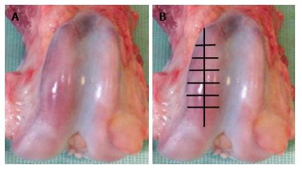
Figure 1 Condyles of canine thigh bone after experimental modelling of osteoarthrosis (A), scheme of articular cartilage harvesting (B).
Figure 2 X-ray electron probe microanalysis of articular cartilage in intact dog.
A: SEM micrograph of articular cartilage and subchondral zone; B: Scanning line regimen, graph shows calcium distribution on line; C: Scanning area regimen shows calcium distribution on the sample surface. Instrumental magnification × 110.
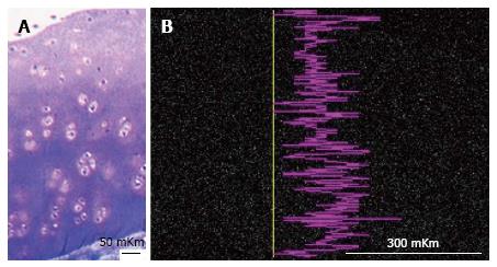
Figure 3 Articular cartilage of intact dog.
A: Semithin section, methylene blue stain. Ob. -6.3 х; oc. -12.5 х; B: Smart map shows sulphur distribution on scanned line. Instrumental magnification 170.
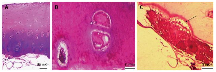
Figure 4 Articular cartilage in dog with experimentally induced osteoarthrosis.
Methylene blue - basic fuchsin staining. A: General appearance. Ob. - 6.3 x, oc. - 12.5 x; B: Focal basophilia of intercellular matrix in deep zone. Ob. - 100 OI, oc. - 12.5 x; C: Osteoclast, resorbing the ground substance of calcified cartilage. Howship lacuna (arrow). Ob. - 100 OI, oc. - 12.5 x.
Figure 5 X-ray electron probe microanalysis of articular cartilage in dog with experimentally induced osteoarthrosis.
A: SEM micrograph of articular cartilage and subchondral zone; B: Scanning line regimen, graph shows calcium distribution on line; C: Scanning area regimen shows calcium distribution on the sample surface. Instrumental magnification 100.
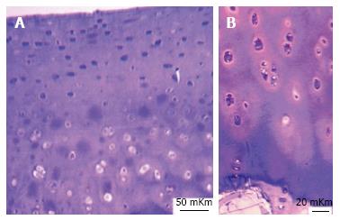
Figure 6 Age-related changes of articular cartilage in intact group.
Semithin section. Methylene blue - basic fuchsin staining. A: Focal metachromasy of articular cartilage in five year old dog. Ob. -6,3 х; oc. - 12,5 х; B: Intensive metachromasy in intermediate and deep zones of articular cartilage in eight year old dog. Ob. -16 х; oc. - 12,5 х.
Figure 7 X-ray electron probe microanalysis of articular cartilage in intact five years old dog.
A: SEM micrograph of articular cartilage and subchondral zone; B: Scanning line regimen, graph shows phosphorus distribution on line; C: Scanning area regimen shows phosphorus distribution on the sample surface. Instrumental magnification 120.
- Citation: Stupina T, Shchudlo M, Stepanov M. Electron probe microanalysis оf experimentally stimulated osteoarthrosis in dogs. World J Orthop 2017; 8(9): 681-687
- URL: https://www.wjgnet.com/2218-5836/full/v8/i9/681.htm
- DOI: https://dx.doi.org/10.5312/wjo.v8.i9.681


