Copyright
©The Author(s) 2017.
World J Orthop. Aug 18, 2017; 8(8): 606-611
Published online Aug 18, 2017. doi: 10.5312/wjo.v8.i8.606
Published online Aug 18, 2017. doi: 10.5312/wjo.v8.i8.606
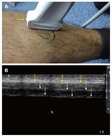
Figure 1 Ultrasound examination of the tibial shaft.
A linear probe having a frequency of 10-12 MHz was used (A). The marker (yellow arrow) is pointing proximally. The plain surface of the tibia makes the examination easy. Normal ultrasound findings (B) include a hyperechoic line (yellow arrows) representing the cortical line of the bone. There are reverberation artifacts deeper to this line (white arrows). These are linear lines parallel to the cortex, having the same distance between them and decreasing in density. A black shadow is located deeper to that. S: Sonographic shadow of the shaft of the tibia. Ultrasound study was performed by Professor Fikri Abu-Zidan, Department of Surgery, Al-Ain Hospital, Al-Ain, UAE.
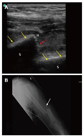
Figure 2 Point-of-care ultrasound of a 42-year-old laborer who fell from 8 meters high during work.
The patient presented with pain, swelling and deformity of the left arm. He had left wrist drop. B mode point-of-care ultrasound of the humeral shaft using a linear probe having a frequency of 10-12 MHz (A) showed that the white cortical line of the humeral shaft (yellow arrows) has been interrupted by a large gap (red arrow) suggesting a displaced fracture at the shaft. X-ray of the humerus (B) confirmed the presence of a displaced fracture (white arrow). S: Sonographic shadow of the humeral shaft; H: Hematoma at the edge of the fracture. Ultrasound study was performed by Professor Fikri Abu-Zidan, Department of Surgery, Al-Ain Hospital, Al-Ain, UAE.
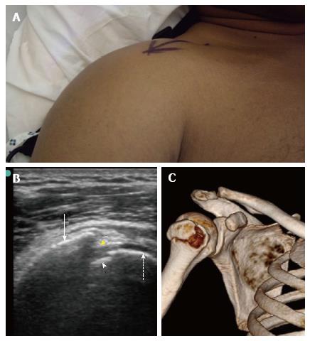
Figure 3 Point-of-care ultrasound of a 40-year-old front seat male passenger who was involved in a front impact collision with a tree.
When presenting to the hospital, he had severe pain, swelling and limitation of the movement of the right shoulder (A). B mode point-of-care ultrasound of the right shoulder using a linear probe having a frequency of 10-12 MHz (B) shows that the humeral head (dashed arrow) and the greater humeral tuberosity (white arrow). There is a discontinuity of the bony line (yellow arrow head) indicating a fracture in the greater tuberosity. A small piece of fractured bone is also seen near the humeral head (white arrow head). Three-dimensional bony reconstruction of the right shoulder confirms the ultrasound findings (C). Ultrasound study was performed by Professor Fikri Abu-Zidan, Department of Surgery, Al-Ain Hospital, Al-Ain, UAE.
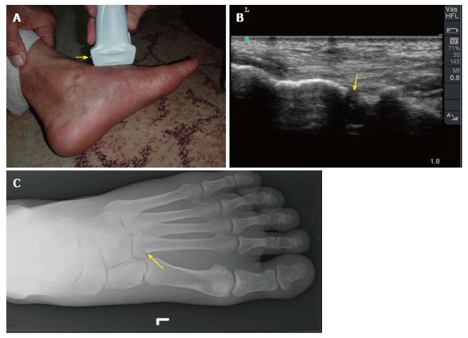
Figure 4 Point-of-care ultrasound of a 60-year-old man who twisted his left ankle and could not walk on it.
He had swelling of the left foot with maximum tenderness on the base of second metatarsal bone (A). The yellow arrow indicates the marker of linear probe which is shown on the left side of the screen while the groove on the other side is shown on the right side of the screen. B mode images of the previous patient showed a cortical defect (yellow arrow) at the base of the second metatarsal bone suggestive of a fracture (B). Plain X-ray of the foot confirmed these findings (C). Ultrasound study was performed by Professor Fikri Abu-Zidan, Department of Surgery, Al-Ain Hospital, Al-Ain, UAE.
- Citation: Abu-Zidan FM. Ultrasound diagnosis of fractures in mass casualty incidents. World J Orthop 2017; 8(8): 606-611
- URL: https://www.wjgnet.com/2218-5836/full/v8/i8/606.htm
- DOI: https://dx.doi.org/10.5312/wjo.v8.i8.606


