Copyright
©The Author(s) 2017.
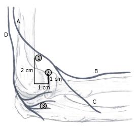
Figure 1 Proximity of nerves and portals: Lateral side view.
A: Radial nerve; B: Superficial branch; C: Deep branch; D: Posterior antebrachial cutaneous nerve; 1: Proximal lateral portal; 2: Anterolateral portal; 3: Midlateral portal.
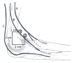
Figure 2 Proximity of nerves and portals: Medial side view.
A: Median nerve; B: Medial antebrachial cutaneous nerve; C: Ulnar nerve; 1: Proximal medial portal; 2: Anteromedial portal.
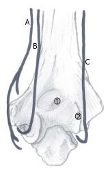
Figure 3 Proximity of nerves and portals: Posterior side view.
A: Medial antebrachial cutaneous nerve; B: Ulnar nerve; C: Posterior antebrachial cutaneous nerve; 1: Transtricipital portal; 2: Posterolateral portal.
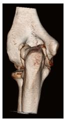
Figure 4 Osteophytes can cause changed anatomy, for example posteromedial osteophytes could push the ulnar nerve out of its groove.
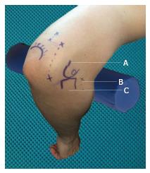
Figure 5 Marking of anatomical structures and portals: Lateral side view.
A: Lateral epicondyle; B: Anterolateral portal; C: Radial head.
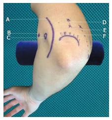
Figure 6 Marking of anatomical structures and portals: Medial side view.
A: Ulnar nerve; B: Proximal medial portal; C: Medial epicondyle; D: Transtricipetal portals; E: Posterolateral portals; F: Olecranon.
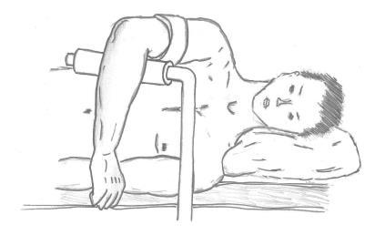
Figure 7 Lateral decubitus position.
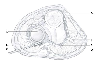
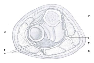
Figure 8 Non-distended joint.
A: Head of radius; B: Radial nerve; C: Lateral antebrachial nerve; D: Cross-section of olecranon; E: Ulnar nerve; F: Capsule; G: Median nerve.
Figure 9 Distended joint joint.
A: Head of radius; B: Radial nerve; C: Lateral antebrachial nerve; D: Cross-section of olecranon; E: Ulnar nerve; F: Capsule; G: Median nerve.
- Citation: Hilgersom NFJ, Oh LS, Flipsen M, Eygendaal D, van den Bekerom MPJ. Tips to avoid nerve injury in elbow arthroscopy. World J Orthop 2017; 8(2): 99-106
- URL: https://www.wjgnet.com/2218-5836/full/v8/i2/99.htm
- DOI: https://dx.doi.org/10.5312/wjo.v8.i2.99


