Copyright
©The Author(s) 2017.
World J Orthop. Feb 18, 2017; 8(2): 156-162
Published online Feb 18, 2017. doi: 10.5312/wjo.v8.i2.156
Published online Feb 18, 2017. doi: 10.5312/wjo.v8.i2.156
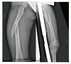
Figure 1 Undisplaced diaphyseal femoral fracture classified as 32-D/5.
1 according to AO pediatrics classification. Associated injuries, such as thoracic or abdominal traumata, often require surgical management of this kind of fracture.
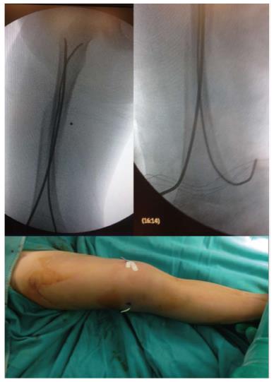
Figure 2 Intraoperative X-rays showing the correct positioning of titanium elastic nail.
Entry points were performed, almost 2.5 cm proximal to the distal physis, one medial and one lateral. To facilitate the removal of the titanium elastic nail, its tail could be left over the skin surface as evident from the clinical intraoperative picture.
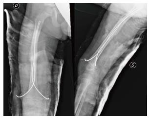
Figure 3 X-ray control at 5 wk of follow-up: Weight-bearing was allowed when advanced consolidation of the fracture with an evident bone callus formation was evident.
Titanium elastic nail was then planned to be removed.
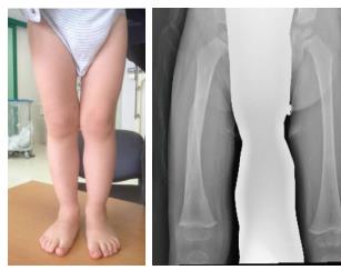
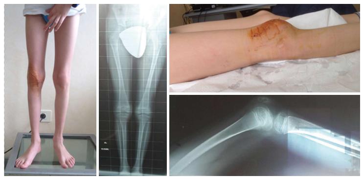
Figure 4 Clinical and radiographic examination 12 mo after fracture with residual varus deformity (< 10°) of the fractured femur.
At longer follow-up, no axial deformities were observed in any patient, while the lengthening of the fractured femur was a common finding, but always < 2 cm.
Figure 5 One patient had a limitation in knee flexion due to associated patellar fracture that was treated for hardware removal three weeks before our evaluation.
- Citation: Donati F, Mazzitelli G, Lillo M, Menghi A, Conti C, Valassina A, Marzetti E, Maccauro G. Titanium elastic nailing in diaphyseal femoral fractures of children below six years of age. World J Orthop 2017; 8(2): 156-162
- URL: https://www.wjgnet.com/2218-5836/full/v8/i2/156.htm
- DOI: https://dx.doi.org/10.5312/wjo.v8.i2.156


