Copyright
©The Author(s) 2017.
World J Orthop. Dec 18, 2017; 8(12): 874-880
Published online Dec 18, 2017. doi: 10.5312/wjo.v8.i12.874
Published online Dec 18, 2017. doi: 10.5312/wjo.v8.i12.874
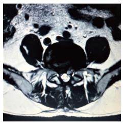
Figure 1 Case of recurrent disc herniation after open discectomy.
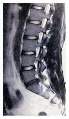
Figure 2 T2 weighted sagittal magnetic resonance imaging demonstrating a disc herniation located at the L5/S1 level.
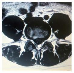
Figure 3 Case of a migrated central hernia.
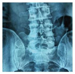
Figure 4 Case of high and steep iliac crests.
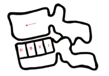
Figure 5 Meyerding’s grading of spondylolisthesis regarding cranial anterior vertebral slippage in accordance with the vertebra below.
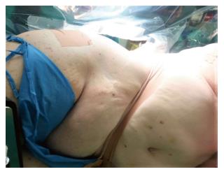
Figure 6 Case of an obese patient to be treated with Transforaminal Percutaneous Endoscopic Discectomy for lumbar disc herniation.
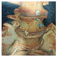
Figure 7 Case of Bertolotti’s syndrome.
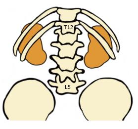
Figure 8 Posterior schematic illustration of the location of kidneys in accordance with lumbar spine levels.
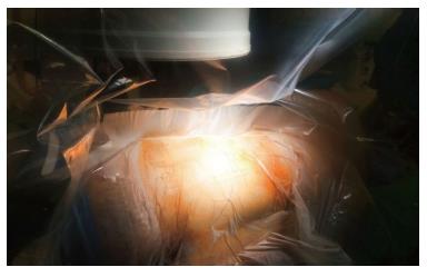
Figure 9 Placement of the patient at the lateral decubitus position and disinfection of the surgical field.
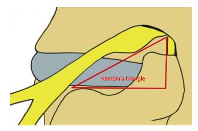
Figure 10 Kambin’s triangle.
The hypotenuse is parallel to the exiting nerve root, the base is according to the superior border of the transverse process of the caudal vertebra, and the height represents the trajectory of the traversing nerve root.
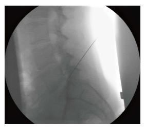
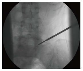
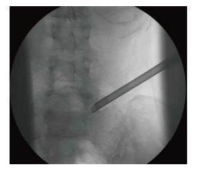
Figure 11 Fluoroscopic verification of the operated level and insertion of the needle.
Figure 12 Sequential transforaminal passage of different size reamers.
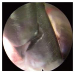
Figure 13 Insertion of the cannula and the endoscope afterwards.
Figure 14 Removal of herniated disc material with a grasper.
- Citation: Kapetanakis S, Gkasdaris G, Angoules AG, Givissis P. Transforaminal Percutaneous Endoscopic Discectomy using Transforaminal Endoscopic Spine System technique: Pitfalls that a beginner should avoid. World J Orthop 2017; 8(12): 874-880
- URL: https://www.wjgnet.com/2218-5836/full/v8/i12/874.htm
- DOI: https://dx.doi.org/10.5312/wjo.v8.i12.874


