Copyright
©The Author(s) 2017.
World J Orthop. Nov 18, 2017; 8(11): 836-845
Published online Nov 18, 2017. doi: 10.5312/wjo.v8.i11.836
Published online Nov 18, 2017. doi: 10.5312/wjo.v8.i11.836
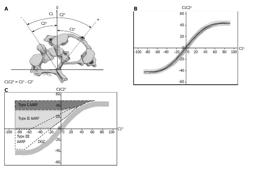
Figure 1 Classification by Pang and Li.
A: Measurements from the dynamic CT scan include the angles between C1 and C2 relative to the vertical axis, and the C1C2 angle is then calculated. By convention, positive values are assigned to the presenting side (side to which the chin points at presentation of torticollis) and negative is assigned to the opposite side (corrected side). Normative data is depicted as motion curves in which the C1 angle is plotted against the C1C2 angle (1B), and the different classes are illustrated as shaded areas in (C) (Figure 1A reprinted with permission from Pang D, Li V, Atlanto-axial rotatory fixation: part 2--new diagnostic paradigm and a new classification based on motion analysis using computed tomographic imaging. Neurosurgery 2005; 57: 941-953. Figures 1B and C reprinted with permission from Pang D, Li V. Atlantoaxial rotatory fixation: Part 1-Biomechanics of normal rotation at the atlantoaxial joint in children. Neurosurgery 2004; 55: 614-625).
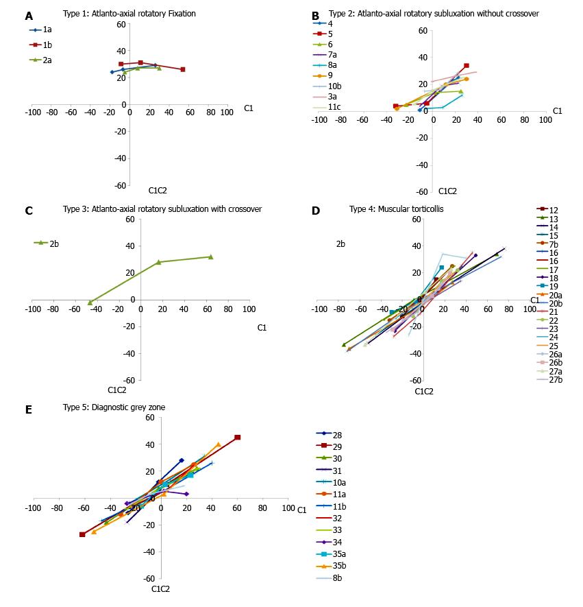
Figure 2 Atlanto-axial rotatory subluxation and fixation.
A: Type 1 (fixation). Three studies (two patients) could be classified as a fixed rotatory subluxation, in which there was less than 20% correction of the C1C2 angle on maximal rotation to the opposite side; B: Type 2 (pathologic stickiness without crossover). Eight studies illustrated an improvement in the angle of divergence of more than 20%, but C1 did not cross over C2; C: Type 3 (pathologic stickiness with crossover). In three studies there was improvement in the C1C2 angle and C1 did cross over C1, but well beyond the null point or midline; D: Type 4 (normal dynamics, muscular torticollis). Twenty-one of our studies exhibited normal dynamic curves and could be classified as muscular torticollis; E: Type 5 (diagnostic grey zone). Ten studies fell into the diagnostic grey zone. In these cases the C1 crossover was delayed and occurred at 8°-20° beyond the midline or null point.
- Citation: Spiegel D, Shrestha S, Sitoula P, Rendon N, Dormans J. Atlantoaxial rotatory displacement in children. World J Orthop 2017; 8(11): 836-845
- URL: https://www.wjgnet.com/2218-5836/full/v8/i11/836.htm
- DOI: https://dx.doi.org/10.5312/wjo.v8.i11.836


