Copyright
©The Author(s) 2017.
World J Orthop. Oct 18, 2017; 8(10): 790-797
Published online Oct 18, 2017. doi: 10.5312/wjo.v8.i10.790
Published online Oct 18, 2017. doi: 10.5312/wjo.v8.i10.790
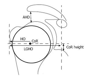
Figure 1 Assessment of the pre- and postoperative joint geometry.
The best-fit circle with the premorbid center of rotation (CoR) is generated with bony landmarks which are not altered by osteoarthritic deformities: The lateral major tubercle border, the medial calcar at the inflection point and the medial edge of the greater tuberosity at the medial supraspinatus insertion. Further parameters were the acromiohumeral distance (AHD), the humeral offset (HO) as the distance between the CoR and the lateral major tubercle border, the lateral glenohumeral offset (LGHO) as the distance between the coracoid and the lateral major tubercle border, and the height of the center of rotation (height CoR) as the distance to the inferior glenoid.
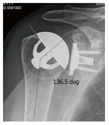
Figure 2 The postoperative center of rotation of the prosthesis shows no deviation compared to the the native one.
The neck shaft angle is defined as the medial angle between the shaft axis and a perpendicular line to the preoperative anatomic neck or the base of the humeral head component.
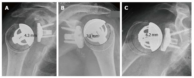
Figure 3 In three cases, a medial deviation of 4.
3 (A), 7.3 (B) and 6.2 (C) mm was caused by an inaccurate humeral neck cut with a resection level which was to high in all cases. These findings were defined as an overstuffing: These patients showed a relatively poor outcome scoring.
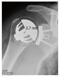
Figure 4 The center of rotation of the prosthesis was 3.
7 mm lateral to the anatomical one and caused by a slightly too small humeral head size. This patient showed a relatively high postoperative constant score.
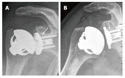
Figure 5 In two cases small radiolucent lines of a maximal thickness of 2 mm were noticed at the upper screw and behind the superior third of the baseplate (white arrows).
- Citation: von Engelhardt LV, Manzke M, Breil-Wirth A, Filler TJ, Jerosch J. Restoration of the joint geometry and outcome after stemless TESS shoulder arthroplasty. World J Orthop 2017; 8(10): 790-797
- URL: https://www.wjgnet.com/2218-5836/full/v8/i10/790.htm
- DOI: https://dx.doi.org/10.5312/wjo.v8.i10.790


